More Related Content

PPT
256865679-bacterial-keratitis-priti-PPT.ppt 
PPTX
Opthalmology BACTERIAL CORNEAL ULCER LUNETA.pptx 
PPTX
Bacterial corneal ulcer (2).pptx CORNEA 
PPTX
BACTERIAL KERATITIS POWERPOINT PRESENTATION 
PPTX
Bacterial corneal ulcer DrBP 
PPTX

PPTX
Ferdous bacterial keratitis copy 
PPTX
Similar to Bacterial_Corneal_Ulcer_notes...PPT.pptx

PPTX

PPT
corneal ulcer.ppt very important topicpics 
PPTX
CORNEAL ULCER types and clinical presentation Ameena C (1).pptx 
PPTX
corneal ulcer (ulcerative keratitis).pptx 
PPTX

PPTX

PPTX

PPTX
Corneal ulcer(bactrial,fungal) 25.02.16, dr.k.n.jha 
PPTX

PPTX
Bacterial and fungal corneal ulcer.pptx for under graduates and postgraduates 
PPTX

PPTX
789_baterial_and_fungal_corneal_ulcers.pptx 
PPTX
Corneal ulcer /corneal opacity and management 
PPTX

PPTX
Corneal ulcer bact & fungal n 
PPTX
CORNEAL ULCER ,DR M SAQUIB , OPHTHALMOLOGY 
PPTX

PPTX
Bacterial corneal ulcers.pptx 
PPT

PPTX
Human being eye CORNEAL ULCER explanation.pptx Recently uploaded

PPTX
ATLS TRAUMA ATLS trauma by Dr.ammar.pptx 
PDF
GLOMERULONEPHRITIS in pathology for Bsc nursing 4th semester 
PDF
50 Re-order Paragraph PTE with Answers – Complete Practice PDF_ Gurully.pdf 
PPTX
PRESENTATION for future educator that will help you 
PDF
SABRE BASIC AIRLINES RESERVATION AND TICKETING SHORT MANUAL BY MD SHAIFULLAR ... 
PDF
Complete List of all 565 CAAHEP & ABHES Accredited Medical Assisting Programs... 
PDF
BCG Attorney Search's Top 24 Law Firm Interview Tips 
PDF
reStartEvents January 22nd TS:SCI & Above Employer Directory.pdf 
PPTX
-Thinking-Skills-Decision-Making-Process-PPT.pptx 
PPTX
VARIOUS FLAP DESIGN in maxillofacial surgery .pptx 
PDF
Explore the Top Platforms for Purchasing Verified PayPal Accounts.pdf 
PDF
A-One Target Institute 9th – 12th Classes.pdf 
PDF
Top 10 Home and Online Tutoring Platforms in Telangana for Teachers.pdf 
PDF
Top 10 Home and Online Tutoring Platforms in Karnataka for Teachers.pdf 
DOCX
OBE-Syllabus in HMPE 7 Catering MNGT. 2023-2024.docx 
PPTX
Brown Vintage Watercolor Creative Portfolio Presentation.pptx 
PPT
Hanry Fayol.pptJBJHFIDHEIRHFIHREHIFHREIFHIOREHIFOH 
PPT
Advantages and disadvantages of the transducer.ppt 
PPTX
Individual_Development_Plan_-_Guide_and_Template.pptx 
PPTX
583395073-Presentation-on-Artificial-intelligence.pptx Bacterial_Corneal_Ulcer_notes...PPT.pptx
- 1.
- 2.
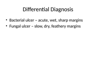
Definition
• Loss ofcorneal epithelium with stromal
infiltration
• Caused by bacterial infection
• Rapid progression with risk of perforation
- 3.
- 4.
Causative Organisms –Gram
Positive
• Staphylococcus aureus
• Streptococcus pneumoniae
• Staphylococcus epidermidis
- 5.
Causative Organisms –Gram
Negative
• Pseudomonas aeruginosa (most dangerous)
• Moraxella
• Haemophilus influenzae
• Neisseria gonorrhoeae
- 6.
Risk Factors
• Cornealtrauma
• Contact lens wear
• Dry eye disease
• Blepharitis
• Chronic dacryocystitis
• Diabetes mellitus
- 7.
Pathogenesis
• Break incorneal epithelium
• Bacterial adherence and multiplication
• Toxin and enzyme release
• Rapid stromal necrosis
• Risk of perforation within 24–48 hours
- 8.
Symptoms
• Severe eyepain
• Redness and watering
• Photophobia
• Rapid decrease in vision
• Purulent discharge
- 9.
- 10.
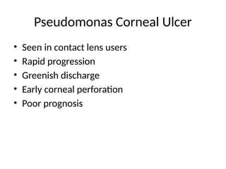
Pseudomonas Corneal Ulcer
•Seen in contact lens users
• Rapid progression
• Greenish discharge
• Early corneal perforation
• Poor prognosis
- 11.
- 12.
- 13.
- 14.
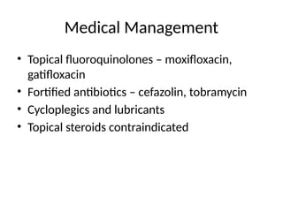
Medical Management
• Topicalfluoroquinolones – moxifloxacin,
gatifloxacin
• Fortified antibiotics – cefazolin, tobramycin
• Cycloplegics and lubricants
• Topical steroids contraindicated
- 15.
- 16.
- 17.
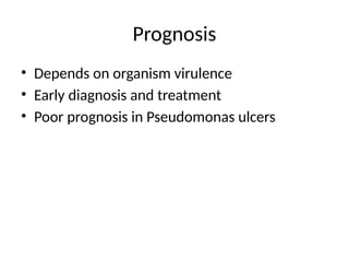
Prognosis
• Depends onorganism virulence
• Early diagnosis and treatment
• Poor prognosis in Pseudomonas ulcers
- 18.
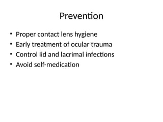
Prevention
• Proper contactlens hygiene
• Early treatment of ocular trauma
• Control lid and lacrimal infections
• Avoid self-medication
- 19.
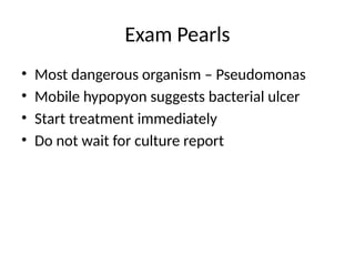
Exam Pearls
• Mostdangerous organism – Pseudomonas
• Mobile hypopyon suggests bacterial ulcer
• Start treatment immediately
• Do not wait for culture report