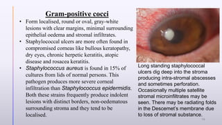
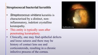
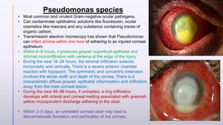
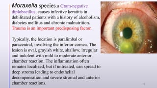
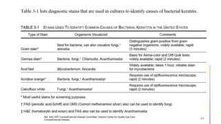
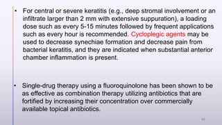
2. Introduction
• A bacterial keratitis was defined as a suppurative corneal
infiltrate and overlying epithelial defect associated with
presence of bacteria on corneal scraping and/or that was cured
with antibiotic therapy.
• Ref. Br J Ophthalmol. 2003 Jul; 87(7): 834–838. doi: 10.1136/bjo.87.7.834
2
3. Demographic Data:
Age: All age group, paediatric microbial keratitis is more likely to be
bacterial etiology1
.
Incidence2,3
:
• USA has an incidence of 11 per 100,000 persons for microbial
keratitis as compared to 799 per 100,000 persons in Nepal and 113
per 100,000 in India.
• Contact lens related corneal ulcers in the general population have
increased from almost 0% in the 1960s to 52% in the 1990s
1. Ref: Microbial Keratitis at Extremes of Age
Parmar, Pragya MS; Salman, Amjad MS; Kalavathy, C M MS; Kaliamurthy, J PhD; Thomas, Philip A MD, PhD; Jesudasan, C A. Nelson MS 3332
2. Ref. AAO PPP Cornea/External Disease Committee, Hoskins Center for Quality Eye Care
Cornea/External Disease
3. Ref. Sultan Qaboos Univ Med J. 2009 Aug; 9(2): 184–195.
Published online 2009 Jun 30.
3
4. Predisposing factor
Ocular Trauma Ocular Surface
Disease
Corneal Foreign Body
Immunosuppressiv
e Agents
Postoperative Extended Contact Lens
Use 4
5. Pathogenesis:
• The break in the continuity of the epithelium that starts the bacteriopathological
process of keratitis.
GENERAL BACTERIOPATHOLOGY IN MICROBIAL KERATITS
• Membrane appendages help these organisms adhere to damaged epithelial cells
and stroma. Pseudomonas aeruginosa and Neisseria gonorrhea utilise
glycocalyx to adhere to each other and to diseased epithelial cells and contact
lenses. Pseudomonas aeruginosa gets attached to both contact lenses and
epithelial breaks due to its biofilm, a coating around the organism.
• During bacterial corneal infections, there is activation of plasminogen to plasmin
which is active proteolytically. Protease, chymase and tryptase cause epithelial
microlesions and healing is delayed due to the degradation of adhesive
glycoproteins by proteolytic enzymes.
Ref. Sultan Qaboos Univ Med J. 2009 Aug; 9(2): 184–195.
Published online 2009 Jun 30.
5
6. Contact Lens Associated Pathogenesis
• The contact lens induces hypoxia, increases corneal temperature
and decreases tear flow over the corneal surface. The adhering of
microorganisms (Staphylococci, Moraxella, Candida) to the contact
lens and epithelium is aided by mucus and proteins.
• N.B: Gram-negative organisms like Pseudomonas, Haemophilus
and Moraxella cause infectious keratitis in extended wear cosmetic
contact lens users, therapeutic soft contact lens wearers on the
other hand are prone to corneal ulcers caused by Gram-positive
bacteria especially Streptococci.
Ref. Sultan Qaboos Univ Med J. 2009 Aug; 9(2): 184–195.
Published online 2009 Jun 30.
6
7. Symptoms: Pain, irritation, redness, photophobia, discharge,
decreased vision, contact lens intolerance.
Ref. Color atlas & synopsis of clinical ophthalmology- Wills eye institute
Associated ocular condition: Gonococcal, Haemophilus and
pneumococcal corneal infections may have associated moderate
to severe conjunctivitis with chemosis and sometimes
pseudomembranes.
Ref. Sultan Qaboos Univ Med J. 2009 Aug; 9(2): 184–195.
Published online 2009 Jun 30.
7
8. H/O use of any immunosuppressive agents should be revealed.
Contact Lens History (e.g., wearing schedule; overnight wear; type of
contact lens; contact lens solution; contact lens hygiene protocol; tap-water
rinsing of contact lenses; swimming, using a hot tub, or showering while
wearing contact lenses; method of purchase, such as over the Internet; and
decorative contact lens use)
Visual acuity depends on the severity of infection, area involved,
duration of infection and the virulence of causative agents.
Ref. AAO PPP Cornea/External Disease Committee, Hoskins Center for Quality Eye Care
Cornea/External Disease
8
9. Clinical presentation based on causative
organism
Ref. Sultan Qaboos Univ Med J. 2009 Aug; 9(2): 184–195.
Published online 2009 Jun 30.
9
10. Gram-positive cocci
• Form localised, round or oval, gray-white
lesions with clear margins, minimal surrounding
epithelial oedema and stromal infiltrates.
• Staphylococcal ulcers are more often found in
compromised corneas like bullous keratopathy,
dry eyes, chronic herpetic keratitis, atopic
disease and rosacea keratitis.
• Staphylococcus aureus is found in 15% of
cultures from lids of normal persons. This
pathogen produces more severe corneal
infiltration than Staphylococcus epidermidis.
Both these strains frequently produce indolent
lesions with distinct borders, non-oedematous
surrounding stroma and they tend to be
localised.
Long standing staphylococcal
ulcers dig deep into the stroma
producing intra-stromal abscesses
and sometimes perforation.
Occasionally multiple satellite
stromal microinfiltrates may be
seen. There may be radiating folds
in the Descemet’s membrane due
to loss of stromal substance.
10
11. Streptococcal bacterial keratitis
• Streptococcus viridans keratitis is
characterised by a distinct, non-
inflammatory, indolent crystalline
keratopathy.
• This entity is typically seen after
penetrating keratoplasty.
• Clinically, one may find epithelial defects
and loose sutures and there may be
history of contact lens use and
corticosteroids, resulting in a chronic
infective process of the cornea. 11
12. • Mycobacterium, Nocardia and Actinomyces
species are Gram-positive branching filamentous bacteria found in soil.
• The keratitis follows soil contaminated corneal injury and produces an
indolent ulcer with elevated hyphae edges, often with satellite lesions,
mimicking a fungal ulcer.
• The cornea has a typical cracked windshield appearance.
12
13. Gram-negative corneal bacterial infections are mostly rapid
in onset and progress fast due to lytic enzymes like protease, lipase
and elastase.
These infections can lead to corneal perforation and the loss of an
eye.
13
14. Pseudomonas species
• Most common and virulent Gram-negative ocular pathogens.
• Can contaminate ophthalmic solutions like fluorescein, ocular
cosmetics like mascara and any substance containing traces of
organic carbon.
• Transmission electron microscopy has shown that Pseudomonas
can infect stroma within one hour of adhering to an injured corneal
epithelium.
• Within 6–8 hours, it produces grayish superficial epithelial and
stromal microinfiltration with oedema at the edge of the injury.
• During the next 18–24 hours, the stromal infiltration extends
horizontally and vertically. There is a severe anterior chamber
reaction with hypopyon. The symmetric and concentric extension
involves the whole width and depth of the cornea. There is a
characteristic diffuse grayish, epithelial inflammation and infiltration
away from the main corneal lesion.
• During the next 48–96 hours, if untreated, a ring infiltration
develops with scleral and corneal melting associated with greenish
yellow mucopurulent discharge adhering to the ulcer.
• Within 2–5 days, an untreated corneal ulcer may lead to
descemetocele formation and perforation of the cornea.
14
15. Moraxella species, a Gram-negative
diplobacillus, causes infective keratitis in
debilitated patients with a history of alcoholism,
diabetes mellitus and chronic malnutrition.
Trauma is an important predisposing factor.
Typically, the location is paralimbal or
paracentral, involving the inferior cornea. The
lesion is oval, grayish white, shallow, irregular
and indolent with mild to moderate anterior
chamber reaction. The inflammation often
remains localized, but if untreated, can spread to
deep stroma leading to endothelial
decompensation and severe stromal and anterior
chamber reactions. 15
16. Grading For Corneal Ulcer:
Mild Reaction: focal, superficial suppuration;
Moderate Reaction: suppuration confined to superficial two-third of the
cornea;
Severe Reaction: suppuration confined to posterior one-third of the cornea
and may present as a ring abscess, scleral suppuration and impending
perforation.
Ref. Sultan Qaboos Univ Med J. 2009 Aug; 9(2): 184–195.
Published online 2009 Jun 30.
16
17. Slit
lamp
examina
tion of
cornea
Epithelium, including defects and punctate keratopathy, oedema, epithelial
movement patterns.
Stroma, including ulceration, thinning, perforation, and infiltrate (location -central,
peripheral, inferior, perineural, surgical, or traumatic wound), density, size, shape
[ring], number [satellite], depth, character of infiltrate margin [suppuration,
necrosis, feathery, soft, crystalline], color), oedema.
Endothelium (endothelial plaque)
Foreign body, including sutures.
Signs of corneal dystrophies (e.g., epithelial basement membrane dystrophy)
Previous corneal inflammation (thinning, scarring, or neovascularization)
Signs of previous corneal or refractive surgery
Ref. AAO PPP Cornea/External
Disease Committee, Hoskins Center
for Quality Eye Care
Cornea/External Disease
17
18. Dyes And Staining
• Fluorescein or rose bengal/lissamine green staining of the
cornea is usually performed and may provide additional
information about other factors, such as the presence of
dendrites, pseudodendrites, loose or exposed sutures, foreign
body, and any epithelial defect.
• Staining of epithelium must be differentiated from pooling of
stain in an area of corneal thinning.
Ref. AAO PPP Cornea/External Disease Committee, Hoskins Center for Quality Eye Care
Cornea/External Disease
18
19. Diagnostic
Test-
Cultures
and
Smears
The majority of community-acquired cases of bacterial keratitis resolve with
empiric therapy and are managed without smears or cultures.
Smears and/or cultures are specifically indicated in the following
circumstances:
A corneal infiltrate is central, large, and/or is associated with significant
stromal involvement or melting;
The infection is chronic or unresponsive to broad-spectrum antibiotic therapy;
There is a history of corneal surgeries; or
Atypical clinical features are present that are suggestive of fungal, amoebic,
or mycobacterial keratitis;
Infiltrates are in multiple locations on the cornea.
Ref. AAO PPP Cornea/External Disease Committee, Hoskins Center for Quality Eye Care
Cornea/External Disease
19
20. Ref. AAO PPP Cornea/External Disease Committee, Hoskins Center for Quality Eye Care
Cornea/External Disease
20
21. Ref. AAO PPP Cornea/External Disease Committee, Hoskins Center for Quality Eye Care
Cornea/External Disease
21
23. MANAGEMENT
Ref. AAO PPP Cornea/External Disease Committee, Hoskins Center for Quality Eye Care
Cornea/External Disease
23
24. PREVENTION
• Avoiding or correcting predisposing factors may reduce the risk of
bacterial keratitis.
• Screening patients for predisposing factors and educating them
about the risks of overnight wear of contact lenses and proper
contact lens care may reduce the incidence of bacterial keratitis in
those who wear contact lenses.
• Ocular surface disease such as corneal epithelial defects, severe
tear deficiency, or lagophthalmos should be treated.
24
26. • For central or severe keratitis (e.g., deep stromal involvement or an
infiltrate larger than 2 mm with extensive suppuration), a loading
dose such as every 5-15 minutes followed by frequent applications
such as every hour is recommended. Cycloplegic agents may be
used to decrease synechiae formation and decrease pain from
bacterial keratitis, and they are indicated when substantial anterior
chamber inflammation is present.
• Single-drug therapy using a fluoroquinolone has been shown to be
as effective as combination therapy utilizing antibiotics that are
fortified by increasing their concentration over commercially
available topical antibiotics.
26
27. • Fortified topical antibiotics should be considered for large and/or
visually significant corneal infiltrates, especially if a hypopyon is
present.
• Ciprofloxacin 0.3%, ofloxacin 0.3%, and levofloxacin 1.5% have
been approved by the Food and Drug Administration (FDA) for the
treatment of bacterial keratitis
• Although widely used, the fourth-generation fluoroquinolones are not
FDA-approved for the treatment of bacterial keratitis.
• In southern India, there has been a sharp increase in resistance of
Pseudomonas aeruginosa to moxifloxacin, from 19% in 2007 to 52%
in 2009.
27
28. • Keratitis from multidrug-resistant Pseudomonas aeruginosa has
been reported, with high morbidity. Topical colistin 0.19% may be
considered in such cases.
• Systemic therapy is necessary in cases of gonococcal keratitis.
• Most antibiotic eye drops should not be tapered below 3 to 4 times a
day because low doses are subtherapeutic and may increase the
risk of developing antibiotic resistance
• Topical povidone-iodine 1.25% has been shown to be as effective as
topical antibiotics for bacterial keratitis in a randomized, controlled
clinical trial performed in Philippines and India.
28
29. CORTICOSTEROID THERAPY
• In a subgroup analysis of SCUT data, there was a potential benefit
for using corticosteroids in Pseudomonas keratitis and in more
severe cases of bacterial keratitis.
• A second subgroup analysis found that the addition of topical
corticosteroids within 2 to 3 days of antibiotic therapy (rather than
after 4 or more days) resulted in a 1-line better visual acuity at 3
months compared with placebo.
• If the ulcer is associated with Nocardia or fungus, the outcomes of
corticosteroid therapy are likely to be poor; for most bacteria other
than Nocardia, the risk is low and may be beneficial.
29
30. Corneal Cross-linking
• It has been used successfully in the treatment of moderate bacterial
ulcers.
• A randomized controlled study with 32 patients found that patients who
received a single cross-linking treatment in addition to standard medical
therapy had faster reepithelialisation and shorter treatment duration than
the control group receiving standard medical therapy alone.
• Cross-linking may be beneficial in cases of bacterial keratitis refractory to
medical therapy alone.
30
31. Corneal Cross-linking cont’d…
• A meta-analysis of 12 articles found that corneal cross-linking is
potentially effective for treatment of bacterial keratitis and can block
corneal melting, especially in bacterial keratitis.
• As ultraviolet energy is absorbed within the first 100 µm, cross-
linking has been proposed to have a greater effect in more shallow
infiltrates.
• One small study found that cross-linking alone, without antibiotic
therapy, can resolve bacterial keratitis in 14 out of 16 cases.
31
32. • In the Bhaktapur Eye Study, patients with corneal abrasions
confirmed by clinical examination who presented within 48 hours of
the injury without signs of corneal infection were enrolled and given
chloramphenicol ointment 1% three times a day for 3 days. Only 18
of 442 patients went on to develop corneal ulcers.
• The WHO applied the Bhaktapur Eye Study model in Bhutan.
Volunteer health workers were trained to follow the inhabitants of 55
villages and to use the same chloramphenicol ointment regimen for
corneal abrasions. There were 115 corneal abrasions during the
study period, and no cases of keratitis developed.
32
33. Ref. AAO PPP Cornea/External
Disease Committee, Hoskins Center
for Quality Eye Care
Cornea/External Disease
33
35. Ref. AAO PPP Cornea/External Disease Committee, Hoskins Center for Quality Eye Care
Cornea/External Disease 35
36. Prognosis of vision:
• On average, patients with mild vision loss (better than 20/40) can expect
about 1 line of improvement, with continuous improvement until 3 months.
• Patients with moderate vision loss (20/40 to 20/800) can expect a rapid
improvement in vision over the first 3 weeks and a cumulative 4- to 5-line
improvement by 3 months, but then little improvement thereafter.
• Patients with severe vision loss (CF or worse) can expect a marked
improvement in vision over the subsequent 12-month period (approximately
11 lines) and can even expect 1 line of this improvement to occur after 3
months.
Ref. Ophthalmology. 2014 Jun; 121(6): 1310–1311.e3. Published online 2014 Mar 5. doi: 10.1016/j.ophtha.2013.12.041
36
37. • Bacterial keratitis carries more favourable outcome measures
than fungal keratitis. Results from the SCUT were compared with
those from the Mycotic Ulcer Treatment Trial (MUTT) and found
that at 3 months, fungal keratitis cases had a larger
infiltrate/scar, a slower re-epithelialization rate, and a higher
perforation rate than bacterial keratitis cases.
Ref. AAO PPP Cornea/External Disease Committee, Hoskins Center for Quality Eye Care
Cornea/External Disease
37