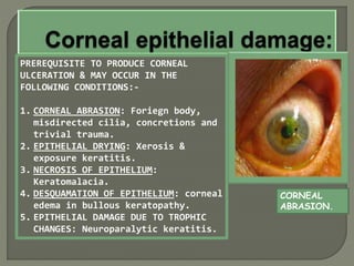
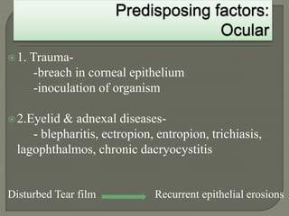
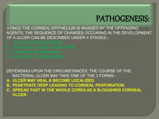
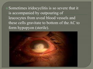
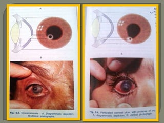
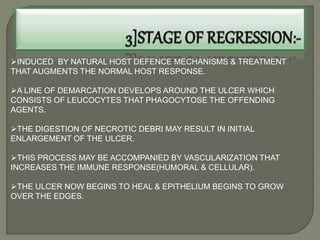
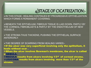
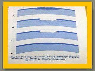
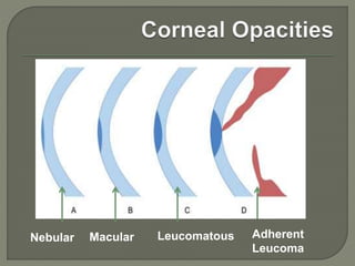
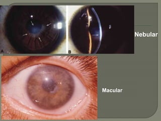
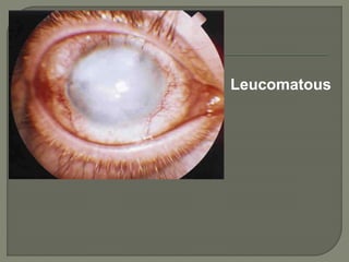
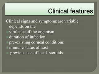
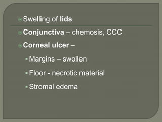
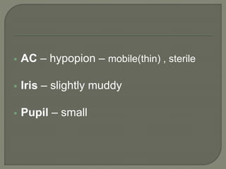
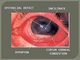
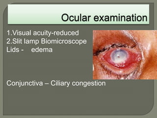
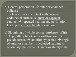
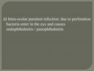
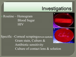
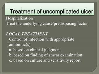
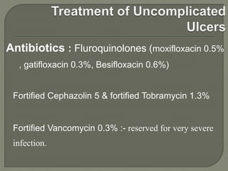
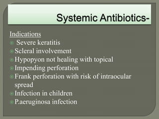
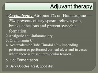
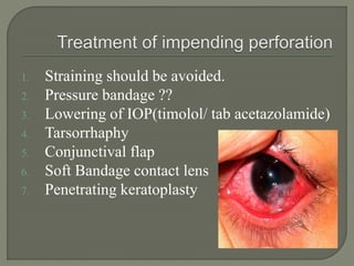
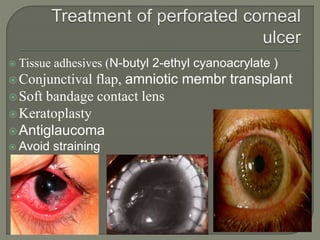
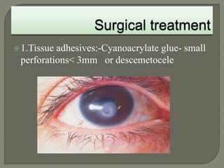
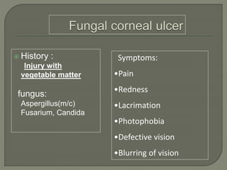
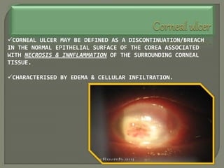
Dr. Atul Kumar Anand
Senior Resident
AIIMS Patna
CORNEAL ULCER MAY BE DEFINED AS A DISCONTINUATION/BREACH
IN THE NORMAL EPITHELIAL SURFACE OF THE COREA ASSOCIATED
WITH NECROSIS & INNFLAMMATION OF THE SURROUNDING CORNEAL
TISSUE.
CHARACTERISED BY EDEMA & CELLULAR INFILTRATION.
BEING THE MOST ANTERIOR PART OF THE EYEBALL,
THE CORNEA IS EXPOSED TO ATMOSPHERE AND HENCE
PRONE TO GET INFECTED EASILY.
AT THE SAME TIME CORNEA IS PROTECTED FROM DAY-
TO-DAY MINOR INFECTIONS BY NORMAL DEFENCE
MECHANISMS PRESENT IN THE TEARS (Present in the
form of lysozyme and other proteins)
THERFORE, INFECTIVE BACTERIAL ULCER MAY
DEVELOP WHEN:-
Either the local defence mechanism is jeopardised.
Presence of local ocular predisposing disease.
Host’s immunity is compromised.
The causative organism is very virulent.
THERE ARE 2 MAJOR FACTORS IN THE PRODUCTION OF A
PURULENT ULCER:-
A]CORNEAL
EPITHELIAL
DAMAGE
B]INFECTION
OF THE
ERODED AREA
HOWEVER, THE FOLLOWING 3
ORGANISMS CAN INVADE AN
INTACT CORNEAL EPITHELIUM AND
PRODUCE ULCERATION....
Neisseria gonorrhoea
N.meingitidis
Corynebacterium diptheriae.
PREREQUISITE TO PRODUCE CORNEAL
ULCERATION & MAY OCCUR IN THE
FOLLOWING CONDITIONS:-
1. CORNEAL ABRASION: Foriegn body,
misdirected cilia, concretions and
trivial trauma.
2. EPITHELIAL DRYING: Xerosis &
exposure keratitis.
3. NECROSIS OF EPITHELIUM:
Keratomalacia.
4. DESQUAMATION OF EPITHELIUM: corneal
edema in bullous keratopathy.
5. EPITHELIAL DAMAGE DUE TO TROPHIC
CHANGES: Neuroparalytic keratitis.
CORNEAL
ABRASION.
1. Trauma-
-breach in corneal epithelium
-inoculation of organism
2.Eyelid & adnexal diseases-
- blepharitis, ectropion, entropion, trichiasis,
lagophthalmos, chronic dacryocystitis
Disturbed Tear film Recurrent epithelial erosions
 3. Ocular surface disorder-
- Dry eye, Steven-Johnson syndrome, ocular
burn, bullous keratopathy.
 4. Contact lens use-
-Increased risk of bacterial keratitis with use of
Extended soft contact lens corneal hypoxia
& decompensation.
- Contamination of CL solution.
 5. Local immune suppression due to topical
corticosteroids(prolong use)
 6.Ocular surgery- cataract , LASIK.
1.Malnutrition
2.Diabetes
3.Immunosupression-Systemic steroids, AIDS
4.Chronic alcoholism
1 EXOGENOUS INFECTION:- Conjuctival sac, lacrimal sac, infected
foriegn bodies, water or air borne infections.
2 FROM THE OCULAR TISSUE: Owing to the direct anatomical
continuity diseases spread from...
•Conjuctiva to the corneal epithelium.
•Sclera to stroma
•Uveal tract to endothelium of cornea.
3 ENDOGENOUS INFECTION: Rare
STAHYLOCOCCUS
AUREUS
PSEUDOONNAS
PYOCYANEA
STREPTOCOCCUS
PNEUMONIAE
E.COLI
PROTEUS
KLEBSIELLA
N.GONORRHOEA
N.MENINGITIDIS
C.DIPTHERIAE
ONCE THE CORNEAL EPITHELIUM IS INVADED BY THE OFFENDING
AGENTS, THE SEQUENCE OF CHANGES OCCURING IN THE DEVELOPMENT
OF A ULCER CAN BE DESCRIBED UNDER 4 STAGES:-
1. STAGE OF INFILTRATION
2. STAGE OF ACTIVE ULCERATION
3. STAGE OF REGRESSION
4. STAGE OF CICATRIZATION.
DEPENDING UPON THE CIRCUMSTANCES, THE COURSE OF THE
BACTERIAL ULCER MAY TAKE ONE OF THE 3 FORMS:-
A. ULCER MAY HEAL & BECOME LOCALIZED.
B. PENETRATE DEEP LEADING TO CORNEAL PERFORATION.
C. SPREAD FAST IN THE WHOLE COREA AS A SLOUGHING CORNEAL
ULCER.
CHARACTERIZED BY INFILTRATION F LYMPHOCYTES INTO THE
EPITHELIUM FROM THE PERIPHERAL CIRCULATION & THE
UNDERLYING STROMA.
SUBSEQUENTLY, NECROSIS OF THE INVOLVED TISSUE MAY
OCCUR.
ACTIVE ULCERATION RESULTS FROM NECROSIS & SLOUGHING OF THE
EPITHELIUM, BOWMAN’S MEMBRANE & THE INVOLVED STROMA.
THE WALLS OF THE ULCER PROJECT OWING TO SWELLING OF THE
LAMELLAE BY IMBIBITION OF FLUID & PACKING OF MASSES OF
LEUCOCYTES BETWEEN THEM.
HYPEREMIA OF CIRCUMCORNEAL VESSELS RESULTING IN ACCUMULATION
OF PURULENT EXUDATES ON THE CORNEA.
EXUDATION INTO THE ANTERIOR CHAMBER FROM VESSELS OF IRIS &
CILIARY BODY LEAD TO HYPOPYON FORMATION.
ULCER MAY FURTHER PROGRESS AS FOLLOWS:
By lateral extension resulting in diffuse superficial ulceration
Or it may progress by deeper penetration leading to decemetocoele fomation
& a possible corneal perforation.
Sometimes iridocyclitis is so severe that it
is accompanied by outpouring of
leucocytes from uveal blood vessels and
these cells gravitate to bottom of the AC to
form hypopyon (sterile).
INDUCED BY NATURAL HOST DEFENCE MECHANISMS & TREATMENT
THAT AUGMENTS THE NORMAL HOST RESPONSE.
A LINE OF DEMARCATION DEVELOPS AROUND THE ULCER WHICH
CONSISTS OF LEUCOCYTES THAT PHAGOCYTOSE THE OFFENDING
AGENTS.
THE DIGESTION OF NECROTIC DEBRI MAY RESULT IN INITIAL
ENLARGEMENT OF THE ULCER.
THIS PROCESS MAY BE ACCOMPANIED BY VASCULARIZATION THAT
INCREASES THE IMMUNE RESPONSE(HUMORAL & CELLULAR).
THE ULCER NOW BEGINS TO HEAL & EPITHELIUM BEGINS TO GROW
OVER THE EDGES.
IN THIS STAGE, HEALING CONTINUES BY PROGRESSIVE EPITHELIZATION
WHICH FORMS A PERMANENT COVERING.
BENEATH THE EPITHELIUM, FIBROUS TISSUE IS LAID DOWN, PARTLY BY
THE CORNEAL FIBROBLASTS & PARTLY BY THE ENDOTHELIUM OF NEW
VESSELS.
THE STROMA THUS THICKENS, PUSHING THE EPITHELIAL SURFACE
ANTERIORLY.
THE DEGREE OF SCARRING FROM HEALING VARIES:-
If the ulcer was very superficial involving only the epithelium, it
heals without scar .
When the ulcer involves Bowman’s membrane, the ulcer is called
a NEBULA.
MACULA results from ulcers involving 1/3rd of corneal stroma.
LEUCOMA results from ulcers involving more than 1/3rd of the
stroma.
The 3 words - all from latin-
NEBULA, MACULA & LEUKOMA,
are the words used to describe the
appearance of a corneal scar.
NEBULA (fog/mist) describes a
hard to see corneal scar- one where
slit lamp detection is required.
MACULA (stain/spot) is typified
by the scar in the photo... It can be
seen by proper illumination.
LEUKOMA (white) is a white scar
that is easily seen just by looking at
the eye.
Nebular Macular Leucomatous Adherent
Leucoma
Nebular
Macular
Leucomatous
Adherent Leucoma
Clinical signs and symptoms are variable
depends on the
virulence of the organism
duration of infection,
pre-existing corneal conditions
immune status of host
 previous use of local steroids
Pain & FB sensation
Lacrimation
Photophobia
Blurred vision
Redness of eyes
Circum corneal
congestion
Swelling of lids
Conjunctiva – chemosis, CCC
Corneal ulcer –
 Margins – swollen
 Floor - necrotic material
 Stromal edema
 AC – hypopion – mobile(thin) , sterile
 Iris – slightly muddy
 Pupil – small
INFILTRATE
EPITHELIAL DEFECT
HYPOPYON
CIRCUM CORNEAL
CONGESTION
Evaluation of predisposing and aggravating
Factors
1. A detailed history.
2. Prior ocular history
3. Review of related medical problems, current
ocular medications and history of systemic
steroids.
1.Visual acuity-reduced
2.Slit lamp Biomicroscope
Lids - edema
Conjunctiva – Ciliary congestion
4. Cornea
-Location of the ulcer- central, paracentral ,
peripheral,total.
-Size , shape, depth, margins & floor-
depends on stage of ulcer.
-Density and extent of stromal infiltration.
5. Anterior chamber
- Cells/flare, mobile Hypopyon.
Iris- muddy
Toxin induced iritis
Pupil – miotic
Other:
-Sac syringing
-corneal sensation
-Fluorescein staining
Pneumococcal C U
 Ulcer serpens is greyish
white or yellowish disc
shaped ulcer occuring
near center of cornea.
 starts at periphery &
spreads towards centre
 Tendency to creep over
the cornea in serpiginous
fashion- Ulcus Serpen.
 Violent iridocyclitis is
often associated with it.
 Hypopyon – Usually
present
 It has great tendency for
PERFORATION.
Pseudomonas C U
 Rapidly spreading.
 Extends periphery &
deep within 24 hrs.
 Stromal necrosis with
shaggy surface
 Spreads concentrically
and symmetrically to
involve whole depth of
cornea-Ring ulcer.
 Greenish-yellow
discharge.
 Hypopyon is present.
 Untreated  corneal
melting.
1. Spread of ulcer horizontally and depth-wise,
leading to thinning of cornea
2. Descemetocele –
This appears as transparent vesicle surrounded
by grayish zone of infiltration.
It represents condition of impending perforation
of cornea
Descemetocele
Descemetocele
3. Perforation of ulcer –
sudden exertion such as coughing, sneezing,
straining at stool or firm closure of eyes increase
in intra-ocular pressure (IOP) perforation
a) Peripheral perforation -
iris prolapse through opening.
Exudation takes place on
prolapsed tissue ->
an adherent leucoma .
Prolapse of iris
Adherent Leucoma
b) Central perforation  anterior chamber
collapse
 lens comes in contact with corneal
endothelial surface  anterior capsular
cataract  repeated healing and perforation
leading to corneal fistula formation
c) Sloughing of whole cornea: prolapse of iris
 pupillary block and exudation on iris 
pseudocornea  anterior synechiae  angle
of anterior chamber is occluded leading to
secondary glaucoma  anterior staphyloma .
Anterior Subcapsular
cataract
Anterior
Synechia
Anterior Staphyloma
d) Intra-ocular purulent infection: due to perforation
bacteria enter in the eye and causes
endophthalmitis / panophthalmitis
Routine – Hemogram
Blood Sugar
HIV
Specific –Corneal scraping(kimura spatula)
Gram stain, Culture &
Antibiotic sensitivity
Culture of contact lens & solution
Hospitalization
Treat the underlying cause/predisposing factor
LOCAL TREATMENT
Control of infection with appropriate
antibiotic(s)
a. based on clinical judgment
b. based on finding of smear examination
c. based on culture and sensitivity report
Antibiotics : Fluroquinolones (moxifloxacin 0.5%
, gatifloxacin 0.3%, Besifloxacin 0.6%)
Fortified Cephazolin 5 & fortified Tobramycin 1.3%
Fortified Vancomycin 0.3% :- reserved for very severe
infection.
Indications
 Severe keratitis
Scleral involvement
Hypopyon not healing with topical
Impending perforation
Frank perforation with risk of intraocular
spread
Infection in children
P.aeruginosa infection
1.Cycloplegic : Atropine 1% or Homatropine
2%- prevents ciliary spasm, relieves pain,
breaks adhesions and prevent synechia
formation.
2.Analgesic anti-inflammatory
3. Oral vitamin C
4. Acetazolamide Tab/ Timolol e/d - impending
perforation or perforated corneal ulcer and in cases
where there is raised intra-ocular tension .
5. Hot Fomentation
6. Dark Goggles, Rest, good diet,
After successful treatment of corneal ulcer:-
CORNEAL SCAR
Dense corneal scars with visual potential-
Lamellar keratoplasty, full thickness graft
Without visual potential –
Cosmetic contact lenses, tattooing.
1. Straining should be avoided.
2. Pressure bandage ??
3. Lowering of IOP(timolol/ tab acetazolamide)
4. Tarsorrhaphy
5. Conjunctival flap
6. Soft Bandage contact lens
7. Penetrating keratoplasty
 Tissue adhesives (N-butyl 2-ethyl cyanoacrylate )
Conjunctival flap, amniotic membr transplant
Soft bandage contact lens
Keratoplasty
Antiglaucoma
 Avoid straining
Signs of healing:-
-resolution of lid edema, congestion
-decreased density of stromal infiltrate
-reduction of corneal oedema
-reduction in AC reaction/hypopyon
-re-epithelization
Signs of non-response
- Increase in infiltration, epithelial defect, height
of hypopyon, Corneal thinning, perforation
Treatment of non healing ulcer
Modification of anti-microbial therapy according
to antimicrobial sensitivity
Removal of any known cause.
-LOCAL
-SYSTEMIC
Scraping of ulcer floor followed by cauterization
with pure (100%) carbolic acid or 10-20%
trichloracetic acid.
Therapeutic keratoplasty
OCULAR /LOCAL
 Secondary glaucoma
 Associated dacryocystitis
 Presence of Foreign body,
trichiasis
 Dry eye, corneal anaesthesia
 Lagophthalmos, lid abnormalities
 Use of topical steroids
 Wrong diagnosis, wrong
treatment, poor compliance with
medications.
SYSTEMIC
 Diabetes mellitus
 Immuno compromised
state
 On systemic steroids or
immuno suppresants.
1.Tissue adhesives:-Cyanoacrylate glue- small
perforations< 3mm or descemetocele
2. Patch graft:- perforation
5mm in diameter
3 . Therapeutic keratoplasty
-large areas of perforation, necrosis
-Non-healing ulcer
Injury with vegetable matter(thorn, leaf or a
wooden stick )
Agricultural occupations.
• use of contact lens, corneal surgery (PK ,LASIK)
Pathogenesis
 History :
Injury with
vegetable matter
fungus:
Aspergillus(m/c)
Fusarium, Candida
Symptoms:
•Pain
•Redness
•Lacrimation
•Photophobia
•Defective vision
•Blurring of vision
 Signs out of proportion
to symptoms
 Dry looking
 Feathery margins
 Immune ring(wesseley
ring)
 Satellite lesions
 Endothelial plaque
 Hypopyon
• Immobile(Thick)
• Infected
Wesseley ring
Hypopyon
History & clinical features
Microbiological investigations of corneal
scrapings:
• KOH, Calcofluor white, Giemsa
• Sabouraud’s dextrose agar
 Specific treatment
 Topical
• Natamycin(5%)
• Amphotericin B(0.15%)
 Systemic
• Ketoconazole
NO CORTICOSTEROID
 Non specific treatment
 Therapeutic Keratoplasty
Corneal  Ulcer.pptx

Corneal Ulcer.pptx

  • 1.
    Dr. Atul KumarAnand Senior Resident AIIMS Patna
  • 2.
    CORNEAL ULCER MAYBE DEFINED AS A DISCONTINUATION/BREACH IN THE NORMAL EPITHELIAL SURFACE OF THE COREA ASSOCIATED WITH NECROSIS & INNFLAMMATION OF THE SURROUNDING CORNEAL TISSUE. CHARACTERISED BY EDEMA & CELLULAR INFILTRATION.
  • 4.
    BEING THE MOSTANTERIOR PART OF THE EYEBALL, THE CORNEA IS EXPOSED TO ATMOSPHERE AND HENCE PRONE TO GET INFECTED EASILY. AT THE SAME TIME CORNEA IS PROTECTED FROM DAY- TO-DAY MINOR INFECTIONS BY NORMAL DEFENCE MECHANISMS PRESENT IN THE TEARS (Present in the form of lysozyme and other proteins) THERFORE, INFECTIVE BACTERIAL ULCER MAY DEVELOP WHEN:- Either the local defence mechanism is jeopardised. Presence of local ocular predisposing disease. Host’s immunity is compromised. The causative organism is very virulent.
  • 5.
    THERE ARE 2MAJOR FACTORS IN THE PRODUCTION OF A PURULENT ULCER:- A]CORNEAL EPITHELIAL DAMAGE B]INFECTION OF THE ERODED AREA HOWEVER, THE FOLLOWING 3 ORGANISMS CAN INVADE AN INTACT CORNEAL EPITHELIUM AND PRODUCE ULCERATION.... Neisseria gonorrhoea N.meingitidis Corynebacterium diptheriae.
  • 6.
    PREREQUISITE TO PRODUCECORNEAL ULCERATION & MAY OCCUR IN THE FOLLOWING CONDITIONS:- 1. CORNEAL ABRASION: Foriegn body, misdirected cilia, concretions and trivial trauma. 2. EPITHELIAL DRYING: Xerosis & exposure keratitis. 3. NECROSIS OF EPITHELIUM: Keratomalacia. 4. DESQUAMATION OF EPITHELIUM: corneal edema in bullous keratopathy. 5. EPITHELIAL DAMAGE DUE TO TROPHIC CHANGES: Neuroparalytic keratitis. CORNEAL ABRASION.
  • 7.
    1. Trauma- -breach incorneal epithelium -inoculation of organism 2.Eyelid & adnexal diseases- - blepharitis, ectropion, entropion, trichiasis, lagophthalmos, chronic dacryocystitis Disturbed Tear film Recurrent epithelial erosions
  • 8.
     3. Ocularsurface disorder- - Dry eye, Steven-Johnson syndrome, ocular burn, bullous keratopathy.  4. Contact lens use- -Increased risk of bacterial keratitis with use of Extended soft contact lens corneal hypoxia & decompensation. - Contamination of CL solution.  5. Local immune suppression due to topical corticosteroids(prolong use)  6.Ocular surgery- cataract , LASIK.
  • 9.
  • 11.
    1 EXOGENOUS INFECTION:-Conjuctival sac, lacrimal sac, infected foriegn bodies, water or air borne infections. 2 FROM THE OCULAR TISSUE: Owing to the direct anatomical continuity diseases spread from... •Conjuctiva to the corneal epithelium. •Sclera to stroma •Uveal tract to endothelium of cornea. 3 ENDOGENOUS INFECTION: Rare
  • 12.
  • 13.
    ONCE THE CORNEALEPITHELIUM IS INVADED BY THE OFFENDING AGENTS, THE SEQUENCE OF CHANGES OCCURING IN THE DEVELOPMENT OF A ULCER CAN BE DESCRIBED UNDER 4 STAGES:- 1. STAGE OF INFILTRATION 2. STAGE OF ACTIVE ULCERATION 3. STAGE OF REGRESSION 4. STAGE OF CICATRIZATION. DEPENDING UPON THE CIRCUMSTANCES, THE COURSE OF THE BACTERIAL ULCER MAY TAKE ONE OF THE 3 FORMS:- A. ULCER MAY HEAL & BECOME LOCALIZED. B. PENETRATE DEEP LEADING TO CORNEAL PERFORATION. C. SPREAD FAST IN THE WHOLE COREA AS A SLOUGHING CORNEAL ULCER.
  • 15.
    CHARACTERIZED BY INFILTRATIONF LYMPHOCYTES INTO THE EPITHELIUM FROM THE PERIPHERAL CIRCULATION & THE UNDERLYING STROMA. SUBSEQUENTLY, NECROSIS OF THE INVOLVED TISSUE MAY OCCUR.
  • 16.
    ACTIVE ULCERATION RESULTSFROM NECROSIS & SLOUGHING OF THE EPITHELIUM, BOWMAN’S MEMBRANE & THE INVOLVED STROMA. THE WALLS OF THE ULCER PROJECT OWING TO SWELLING OF THE LAMELLAE BY IMBIBITION OF FLUID & PACKING OF MASSES OF LEUCOCYTES BETWEEN THEM. HYPEREMIA OF CIRCUMCORNEAL VESSELS RESULTING IN ACCUMULATION OF PURULENT EXUDATES ON THE CORNEA. EXUDATION INTO THE ANTERIOR CHAMBER FROM VESSELS OF IRIS & CILIARY BODY LEAD TO HYPOPYON FORMATION. ULCER MAY FURTHER PROGRESS AS FOLLOWS: By lateral extension resulting in diffuse superficial ulceration Or it may progress by deeper penetration leading to decemetocoele fomation & a possible corneal perforation.
  • 17.
    Sometimes iridocyclitis isso severe that it is accompanied by outpouring of leucocytes from uveal blood vessels and these cells gravitate to bottom of the AC to form hypopyon (sterile).
  • 19.
    INDUCED BY NATURALHOST DEFENCE MECHANISMS & TREATMENT THAT AUGMENTS THE NORMAL HOST RESPONSE. A LINE OF DEMARCATION DEVELOPS AROUND THE ULCER WHICH CONSISTS OF LEUCOCYTES THAT PHAGOCYTOSE THE OFFENDING AGENTS. THE DIGESTION OF NECROTIC DEBRI MAY RESULT IN INITIAL ENLARGEMENT OF THE ULCER. THIS PROCESS MAY BE ACCOMPANIED BY VASCULARIZATION THAT INCREASES THE IMMUNE RESPONSE(HUMORAL & CELLULAR). THE ULCER NOW BEGINS TO HEAL & EPITHELIUM BEGINS TO GROW OVER THE EDGES.
  • 20.
    IN THIS STAGE,HEALING CONTINUES BY PROGRESSIVE EPITHELIZATION WHICH FORMS A PERMANENT COVERING. BENEATH THE EPITHELIUM, FIBROUS TISSUE IS LAID DOWN, PARTLY BY THE CORNEAL FIBROBLASTS & PARTLY BY THE ENDOTHELIUM OF NEW VESSELS. THE STROMA THUS THICKENS, PUSHING THE EPITHELIAL SURFACE ANTERIORLY. THE DEGREE OF SCARRING FROM HEALING VARIES:- If the ulcer was very superficial involving only the epithelium, it heals without scar . When the ulcer involves Bowman’s membrane, the ulcer is called a NEBULA. MACULA results from ulcers involving 1/3rd of corneal stroma. LEUCOMA results from ulcers involving more than 1/3rd of the stroma.
  • 22.
    The 3 words- all from latin- NEBULA, MACULA & LEUKOMA, are the words used to describe the appearance of a corneal scar. NEBULA (fog/mist) describes a hard to see corneal scar- one where slit lamp detection is required. MACULA (stain/spot) is typified by the scar in the photo... It can be seen by proper illumination. LEUKOMA (white) is a white scar that is easily seen just by looking at the eye.
  • 23.
  • 24.
  • 25.
  • 26.
  • 27.
    Clinical signs andsymptoms are variable depends on the virulence of the organism duration of infection, pre-existing corneal conditions immune status of host  previous use of local steroids
  • 29.
    Pain & FBsensation Lacrimation Photophobia Blurred vision Redness of eyes
  • 31.
  • 33.
    Swelling of lids Conjunctiva– chemosis, CCC Corneal ulcer –  Margins – swollen  Floor - necrotic material  Stromal edema
  • 34.
     AC –hypopion – mobile(thin) , sterile  Iris – slightly muddy  Pupil – small
  • 35.
  • 36.
    Evaluation of predisposingand aggravating Factors 1. A detailed history. 2. Prior ocular history 3. Review of related medical problems, current ocular medications and history of systemic steroids.
  • 37.
    1.Visual acuity-reduced 2.Slit lampBiomicroscope Lids - edema Conjunctiva – Ciliary congestion
  • 38.
    4. Cornea -Location ofthe ulcer- central, paracentral , peripheral,total. -Size , shape, depth, margins & floor- depends on stage of ulcer. -Density and extent of stromal infiltration. 5. Anterior chamber - Cells/flare, mobile Hypopyon.
  • 39.
    Iris- muddy Toxin inducediritis Pupil – miotic Other: -Sac syringing -corneal sensation -Fluorescein staining
  • 40.
    Pneumococcal C U Ulcer serpens is greyish white or yellowish disc shaped ulcer occuring near center of cornea.  starts at periphery & spreads towards centre  Tendency to creep over the cornea in serpiginous fashion- Ulcus Serpen.  Violent iridocyclitis is often associated with it.  Hypopyon – Usually present  It has great tendency for PERFORATION.
  • 41.
    Pseudomonas C U Rapidly spreading.  Extends periphery & deep within 24 hrs.  Stromal necrosis with shaggy surface  Spreads concentrically and symmetrically to involve whole depth of cornea-Ring ulcer.  Greenish-yellow discharge.  Hypopyon is present.  Untreated  corneal melting.
  • 42.
    1. Spread ofulcer horizontally and depth-wise, leading to thinning of cornea 2. Descemetocele – This appears as transparent vesicle surrounded by grayish zone of infiltration. It represents condition of impending perforation of cornea
  • 43.
  • 44.
  • 45.
    3. Perforation ofulcer – sudden exertion such as coughing, sneezing, straining at stool or firm closure of eyes increase in intra-ocular pressure (IOP) perforation a) Peripheral perforation - iris prolapse through opening. Exudation takes place on prolapsed tissue -> an adherent leucoma .
  • 46.
  • 47.
    b) Central perforation anterior chamber collapse  lens comes in contact with corneal endothelial surface  anterior capsular cataract  repeated healing and perforation leading to corneal fistula formation c) Sloughing of whole cornea: prolapse of iris  pupillary block and exudation on iris  pseudocornea  anterior synechiae  angle of anterior chamber is occluded leading to secondary glaucoma  anterior staphyloma .
  • 48.
  • 49.
  • 50.
    d) Intra-ocular purulentinfection: due to perforation bacteria enter in the eye and causes endophthalmitis / panophthalmitis
  • 51.
    Routine – Hemogram BloodSugar HIV Specific –Corneal scraping(kimura spatula) Gram stain, Culture & Antibiotic sensitivity Culture of contact lens & solution
  • 52.
    Hospitalization Treat the underlyingcause/predisposing factor LOCAL TREATMENT Control of infection with appropriate antibiotic(s) a. based on clinical judgment b. based on finding of smear examination c. based on culture and sensitivity report
  • 53.
    Antibiotics : Fluroquinolones(moxifloxacin 0.5% , gatifloxacin 0.3%, Besifloxacin 0.6%) Fortified Cephazolin 5 & fortified Tobramycin 1.3% Fortified Vancomycin 0.3% :- reserved for very severe infection.
  • 54.
    Indications  Severe keratitis Scleralinvolvement Hypopyon not healing with topical Impending perforation Frank perforation with risk of intraocular spread Infection in children P.aeruginosa infection
  • 55.
    1.Cycloplegic : Atropine1% or Homatropine 2%- prevents ciliary spasm, relieves pain, breaks adhesions and prevent synechia formation. 2.Analgesic anti-inflammatory 3. Oral vitamin C 4. Acetazolamide Tab/ Timolol e/d - impending perforation or perforated corneal ulcer and in cases where there is raised intra-ocular tension . 5. Hot Fomentation 6. Dark Goggles, Rest, good diet,
  • 56.
    After successful treatmentof corneal ulcer:- CORNEAL SCAR Dense corneal scars with visual potential- Lamellar keratoplasty, full thickness graft Without visual potential – Cosmetic contact lenses, tattooing.
  • 57.
    1. Straining shouldbe avoided. 2. Pressure bandage ?? 3. Lowering of IOP(timolol/ tab acetazolamide) 4. Tarsorrhaphy 5. Conjunctival flap 6. Soft Bandage contact lens 7. Penetrating keratoplasty
  • 58.
     Tissue adhesives(N-butyl 2-ethyl cyanoacrylate ) Conjunctival flap, amniotic membr transplant Soft bandage contact lens Keratoplasty Antiglaucoma  Avoid straining
  • 59.
    Signs of healing:- -resolutionof lid edema, congestion -decreased density of stromal infiltrate -reduction of corneal oedema -reduction in AC reaction/hypopyon -re-epithelization
  • 60.
    Signs of non-response -Increase in infiltration, epithelial defect, height of hypopyon, Corneal thinning, perforation Treatment of non healing ulcer Modification of anti-microbial therapy according to antimicrobial sensitivity Removal of any known cause. -LOCAL -SYSTEMIC Scraping of ulcer floor followed by cauterization with pure (100%) carbolic acid or 10-20% trichloracetic acid. Therapeutic keratoplasty
  • 61.
    OCULAR /LOCAL  Secondaryglaucoma  Associated dacryocystitis  Presence of Foreign body, trichiasis  Dry eye, corneal anaesthesia  Lagophthalmos, lid abnormalities  Use of topical steroids  Wrong diagnosis, wrong treatment, poor compliance with medications. SYSTEMIC  Diabetes mellitus  Immuno compromised state  On systemic steroids or immuno suppresants.
  • 62.
    1.Tissue adhesives:-Cyanoacrylate glue-small perforations< 3mm or descemetocele
  • 63.
    2. Patch graft:-perforation 5mm in diameter 3 . Therapeutic keratoplasty -large areas of perforation, necrosis -Non-healing ulcer
  • 65.
    Injury with vegetablematter(thorn, leaf or a wooden stick ) Agricultural occupations.
  • 66.
    • use ofcontact lens, corneal surgery (PK ,LASIK)
  • 67.
  • 68.
     History : Injurywith vegetable matter fungus: Aspergillus(m/c) Fusarium, Candida Symptoms: •Pain •Redness •Lacrimation •Photophobia •Defective vision •Blurring of vision
  • 69.
     Signs outof proportion to symptoms  Dry looking  Feathery margins  Immune ring(wesseley ring)  Satellite lesions  Endothelial plaque  Hypopyon • Immobile(Thick) • Infected
  • 70.
  • 71.
    History & clinicalfeatures Microbiological investigations of corneal scrapings: • KOH, Calcofluor white, Giemsa • Sabouraud’s dextrose agar
  • 72.
     Specific treatment Topical • Natamycin(5%) • Amphotericin B(0.15%)  Systemic • Ketoconazole NO CORTICOSTEROID  Non specific treatment  Therapeutic Keratoplasty