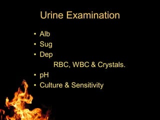
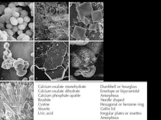
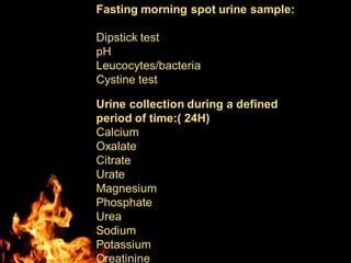
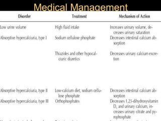
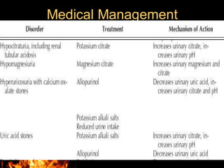
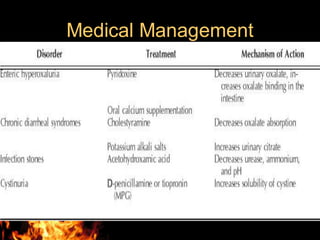
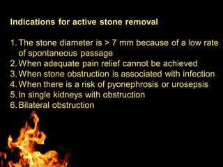
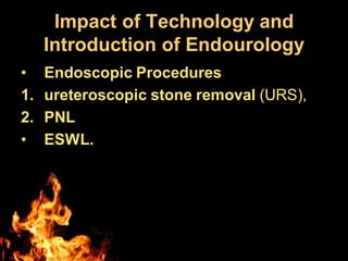
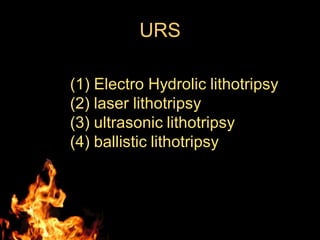
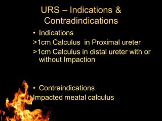
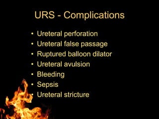
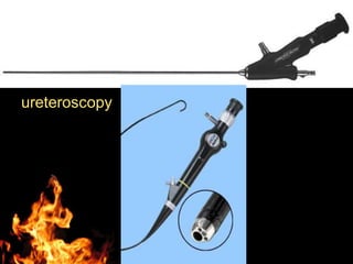
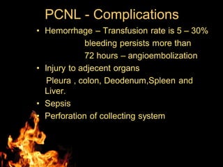
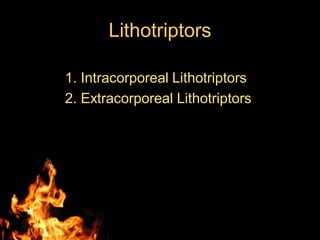
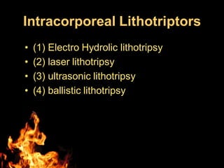
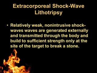
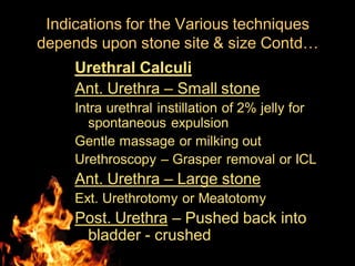
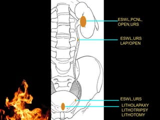
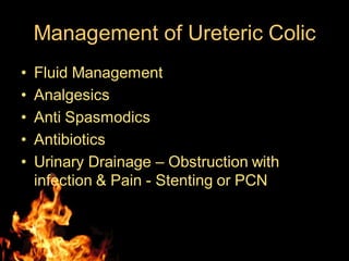
Evaluation & Management
of Urolithiasis
Dept of Urology
Govt Royapettah Hospital and Kilpauk
Medical College
Chennai
Moderators:
Professors:
• Prof. Dr. G. Sivasankar, M.S., M.Ch.,
• Prof. Dr. A. Senthilvel, M.S., M.Ch.,
Asst Professors:
• Dr. J. Sivabalan, M.S., M.Ch.,
• Dr. R. Bhargavi, M.S., M.Ch.,
• Dr. S. Raju, M.S., M.Ch.,
• Dr. K. Muthurathinam, M.S., M.Ch.,
• Dr. D. Tamilselvan, M.S., M.Ch.,
• Dr. K. Senthilkumar, M.S., M.Ch.
Dept of Urology, GRH and KMC, Chennai.
2
Evaluation
History
Diet and fluid intake –
1. half the increased levels of urinary
calcium, oxalate, and uric acid seen in
stone-forming patients may be attributed
to a diet rich in animal protein
2. Milk ingestion can cause hypercalciuria
3. salt
Evaluation Contd…
Medications –
Corticosteroids , aluminum-containing
antacids, loop diuretics, and vitamin D.
Chemotherapeutic agents colchicine or
probenecid
Infection. -Proteus, Klebsiella, Serratia, and
Enterobacter species, & E. coli
Activity level - immobilization
Evaluation Contd…
Systemic disease.
primary hyperparathyroidism, RTA, gout, and sarcoidosis
can cause urolithiasis.
Genetics.
A family history of stones may suggest certain causes, such
as RTA, cystinuria, or absorptive hypercalciuria
Anatomy.
Urinary tract obstruction—congenital (ureteropelvic junction
obstruction or horseshoe kidney) or acquired (benign
prostatic hypertrophy, urethral stricture)—leads to urinary
stasis and stone formation
Previous surgery
Urine Examination
• Alb
• Sug
• Dep
RBC, WBC & Crystals.
• pH
• Culture & Sensitivity
Fasting morning spot urine sample:
Dipstick test
pH
Leucocytes/bacteria
Cystine test
Urine collection during a defined
period of time:( 24H)
Calcium
Oxalate
Citrate
Urate
Magnesium
Phosphate
Urea
Sodium
Potassium
Creatinine
• Blood investigations
pH
Bicorbonate
K+
Chloride.
Calcium
Albumin
Creatine
Urate.
USG - KUBU
USG – Contd…
Plain Abdominal Films
• 90% of urological stones are radio – opaque.
• Radiolucent stones are
1. Pure uric acid
2. Xanthine, hypoxanthine
3. Triamterine & Indinavir
4. Matrix
5. silicates.
6. Dihydroxyadenine.
A – calcium oxalate
B – calcium phosphate
C – uric acid
D – cystine
E - struvite
IVU
• Usually 76% Urograffin , dose - 1ml/kg
• Obstruction by stone is confirmed by delay
in the appearance of contrast medium or
dilated PCS and ureter proximal to stone.
• usual 5-, 10-, and 20-minute urographic
films
• Delayed films may be obtained several
hours or even 1 day after the injection of
contrast material.
Computed Tomography
• Non enhanced spiral CT – investigation of
choice.
• Risk of allergy to contrast agents
• Radiolucent Stones
• accurately measure the size of the stone
• An additional advantage - detect nonurologic
abnormalities
• Useful signs of ureteral obstruction—
hydroureter, stranding of perinephric fat,
hydronephrosis, and nephromegaly
• characterizing the composition of these stones
Magnetic Resonance Imaging
• Contrast to CT, MRI unable to visualize
most stones clinically, this modality is not
useful for characterizing the composition
of these stones
• Renal impairment or allergy to
intravenous contrast agents and when x-
rays are contraindicated .
• Magnetic resonance (MR) urography has
been reported to be effective in detecting
urinary tract dilatation
Management
• General Measures
• Specific Medical Management
• ESWL
• Minimally invasive surgeries
• Open Surgeries.
Management
General Measures of Prevention
1. Drink sufficient water to keep the urine
volume above 3 L/day,
2. Limit their daily meat intake to 8 ounces or
less,
3. Substitute whole wheat bread for white bread
4. Eat natural fiber cereals.
5. Limit their intake of oxalate-rich foods
6. Do not add salt at the table.
7. Not to restrict dairy products but to avoid
overindulgence—no more than three glasses
of milk a day.
8. Increase citrus fruit intake
Medical Management
Medical Management
Medical Management
Indications for active stone removal
1.The stone diameter is > 7 mm because of a low rate
of spontaneous passage
2.When adequate pain relief cannot be achieved
3.When stone obstruction is associated with infection
4.When there is a risk of pyonephrosis or urosepsis
5.In single kidneys with obstruction
6.Bilateral obstruction
Impact of Technology and
Introduction of Endourology
• Endoscopic Procedures
1. ureteroscopic stone removal (URS),
2. PNL
• ESWL.
URS
(1) Electro Hydrolic lithotripsy
(2) laser lithotripsy
(3) ultrasonic lithotripsy
(4) ballistic lithotripsy
URS – Indications &
Contradindications
• Indications
>1cm Calculus in Proximal ureter
>1cm Calculus in distal ureter with or
without Impaction
• Contraindications
Impacted meatal calculus
URS - Complications
• Ureteral perforation
• Ureteral false passage
• Ruptured balloon dilator
• Ureteral avulsion
• Bleeding
• Sepsis
• Ureteral stricture
ureteroscopy
PERCUTANOUS
NEPHROLITHOTOMY
• Requires through understanding of
collecting system
• Access is made with fluoroscopy
after retrograde ureteral catheter
insertion or by USG
• Tract is created by dilatation.
• Nephroscopy is performed and
stone fragmented with ICL.
• Nephrostomy kept.
PCNL Indications &
Contraindications
• Indications
Renal Stones > 2 cm in size.
< 2cm stones where ESWL contraindicated
Failed ESWL
Proximal ureteric stone >1cm
Obstruction distal to stone
Associated upper tract foreign bodies
Cystine stones
• ContraIndications
Irreversible Coagulopathy
PCNL - Complications
• Hemorrhage – Transfusion rate is 5 – 30%
bleeding persists more than
72 hours – angioembolization
• Injury to adjecent organs
Pleura , colon, Deodenum,Spleen and
Liver.
• Sepsis
• Perforation of collecting system
Lithotriptors
1. Intracorporeal Lithotriptors
2. Extracorporeal Lithotriptors
Intracorporeal Lithotriptors
• (1) Electro Hydrolic lithotripsy
• (2) laser lithotripsy
• (3) ultrasonic lithotripsy
• (4) ballistic lithotripsy
Extracorporeal Shock-Wave
Lithotripsy
• Relatively weak, nonintrusive shock-
waves waves are generated externally
and transmitted through the body and
build to sufficient strength only at the
site of the target to break a stone.
Generator Types
• Electrohydraulic (spark gap),
• Electromagnetic,
• Piezoelectric.
Four potential mechanisms for ESWL
stone breakage have been described:
(1) compression fracture,
(2) spallation,
(3) acoustic cavitation, and
(4) dynamic fatigue
ESWL – Indications
• Renal stones up to 2 cm in size
• Ureteric stones up to 1 cm in size
ESWL – Contraindications
• Impacted ureteric stones.
• > 2cm sized stones
• Calcium oxalate > 1.5 cm stones.
• Significant dilatation of collecting system
• Distal ureteric obstruction
• Untreated coagulopathy
• Active infection
• Renal or Aortic aneurysm
• Pregnancy
• Patient with defibrillator or pacemaker
Complications of ESWL
• Hematuria - perinephric and renal
hematomas - 0.2% to 0.6%
• GIT – Pancreatitis, Elevation of
Liver enzyme, Billiary colic,
Mucosal erosion of colon.
• Ureteric obstruction – Steinstrasse
– due to accumulation of multiple
stone fragment in ureter.
• Hypertension.
Open Surgery
• Incidence of open procedures – 3%
• Indications
1. Complex stone burden
2. Failure of ESWL/PCNL/URS
3. Concomitent open surgery – eg PUJ
4. Nephrectomy for poor functioning
kidney
5. Patient preference.
Open Surgeries
• Pyelolithotomy
• Extended Pyelolithotomy
• Coagulum Pyelolithotomy
• Calyceal diverticulolithotomy
• Anatrophic Nephrolithotomy
• Ureterolithotomy
Indications for the Various techniques
depends upon stone site & size
Renal stones – non Staghorn
• for stones less than 10 mm- ESWL
• For stones between 10 and 20 mm -
ESWL &PNL
• Stones greater than 20 mm should
primarily be treated by PNL
Indications for the Various techniques
depends upon stone site & size
Contd…
Staghorn Stones –
• PNL monotheraphy,
• PNL – ESWL – PNL – Sandwitch therapy
• Anatrophic Nephrolithotomy
Ureteric Calculus
Proximal Ureteric Calculi
< 1 cm – ESWL
>1 cm - Ureteroscopy with ICL
Lap. Ureterolithotomy.
Indications for the Various techniques
depends upon stone site & size
Contd…
Ureteric Calculus
Distal Ureteric Calculi
< 1 cm, non impacted – ESWL
>1 cm, impacted - URS.
> 2cm impacted or failed other modality –
Lap or Open.
Indications for the Various techniques
depends upon stone site & size
Contd…
Vesical Calculus
Cystolitholapaxy – upto 2 cms.
cystolithotripsy with electrohydraulic, ultrasonic,
laser, or pneumatic lithotripsy .
Percutaneous cystolithotomy - patients with
narrow urethras
Open cystolithotomy - large stone burdens or
hard stones refractory to an endoscopic
approach and concomitant open prostatectomy
or diverticulectomy
ESWL
Indications for the Various techniques
depends upon stone site & size
Contd…
Urethral Calculi
Ant. Urethra – Small stone
Intra urethral instillation of 2% jelly for
spontaneous expulsion
Gentle massage or milking out
Urethroscopy – Grasper removal or ICL
Ant. Urethra – Large stone
Ext. Urethrotomy or Meatotomy
Post. Urethra – Pushed back into
bladder - crushed
Indications for the Various techniques
depends upon stone site & size Contd…
ESWL,PCNL,
OPEN,URS
ESWL,URS
LAP/OPEN
ESWL,URS
LITHOLAPAXY
LITHOTRIPSY
LITHOTOMY
Management of Ureteric Colic
• Fluid Management
• Analgesics
• Anti Spasmodics
• Antibiotics
• Urinary Drainage – Obstruction with
infection & Pain - Stenting or PCN
Thank You

Urolithiasis evaluation and management brief

  • 1.
    Evaluation & Management ofUrolithiasis Dept of Urology Govt Royapettah Hospital and Kilpauk Medical College Chennai
  • 2.
    Moderators: Professors: • Prof. Dr.G. Sivasankar, M.S., M.Ch., • Prof. Dr. A. Senthilvel, M.S., M.Ch., Asst Professors: • Dr. J. Sivabalan, M.S., M.Ch., • Dr. R. Bhargavi, M.S., M.Ch., • Dr. S. Raju, M.S., M.Ch., • Dr. K. Muthurathinam, M.S., M.Ch., • Dr. D. Tamilselvan, M.S., M.Ch., • Dr. K. Senthilkumar, M.S., M.Ch. Dept of Urology, GRH and KMC, Chennai. 2
  • 3.
    Evaluation History Diet and fluidintake – 1. half the increased levels of urinary calcium, oxalate, and uric acid seen in stone-forming patients may be attributed to a diet rich in animal protein 2. Milk ingestion can cause hypercalciuria 3. salt
  • 4.
    Evaluation Contd… Medications – Corticosteroids, aluminum-containing antacids, loop diuretics, and vitamin D. Chemotherapeutic agents colchicine or probenecid Infection. -Proteus, Klebsiella, Serratia, and Enterobacter species, & E. coli Activity level - immobilization
  • 5.
    Evaluation Contd… Systemic disease. primaryhyperparathyroidism, RTA, gout, and sarcoidosis can cause urolithiasis. Genetics. A family history of stones may suggest certain causes, such as RTA, cystinuria, or absorptive hypercalciuria Anatomy. Urinary tract obstruction—congenital (ureteropelvic junction obstruction or horseshoe kidney) or acquired (benign prostatic hypertrophy, urethral stricture)—leads to urinary stasis and stone formation Previous surgery
  • 6.
    Urine Examination • Alb •Sug • Dep RBC, WBC & Crystals. • pH • Culture & Sensitivity
  • 8.
    Fasting morning spoturine sample: Dipstick test pH Leucocytes/bacteria Cystine test Urine collection during a defined period of time:( 24H) Calcium Oxalate Citrate Urate Magnesium Phosphate Urea Sodium Potassium Creatinine
  • 9.
  • 10.
  • 11.
  • 12.
    Plain Abdominal Films •90% of urological stones are radio – opaque. • Radiolucent stones are 1. Pure uric acid 2. Xanthine, hypoxanthine 3. Triamterine & Indinavir 4. Matrix 5. silicates. 6. Dihydroxyadenine.
  • 13.
    A – calciumoxalate B – calcium phosphate C – uric acid D – cystine E - struvite
  • 15.
    IVU • Usually 76%Urograffin , dose - 1ml/kg • Obstruction by stone is confirmed by delay in the appearance of contrast medium or dilated PCS and ureter proximal to stone. • usual 5-, 10-, and 20-minute urographic films • Delayed films may be obtained several hours or even 1 day after the injection of contrast material.
  • 17.
    Computed Tomography • Nonenhanced spiral CT – investigation of choice. • Risk of allergy to contrast agents • Radiolucent Stones • accurately measure the size of the stone • An additional advantage - detect nonurologic abnormalities • Useful signs of ureteral obstruction— hydroureter, stranding of perinephric fat, hydronephrosis, and nephromegaly • characterizing the composition of these stones
  • 19.
    Magnetic Resonance Imaging •Contrast to CT, MRI unable to visualize most stones clinically, this modality is not useful for characterizing the composition of these stones • Renal impairment or allergy to intravenous contrast agents and when x- rays are contraindicated . • Magnetic resonance (MR) urography has been reported to be effective in detecting urinary tract dilatation
  • 20.
    Management • General Measures •Specific Medical Management • ESWL • Minimally invasive surgeries • Open Surgeries.
  • 21.
    Management General Measures ofPrevention 1. Drink sufficient water to keep the urine volume above 3 L/day, 2. Limit their daily meat intake to 8 ounces or less, 3. Substitute whole wheat bread for white bread 4. Eat natural fiber cereals. 5. Limit their intake of oxalate-rich foods 6. Do not add salt at the table. 7. Not to restrict dairy products but to avoid overindulgence—no more than three glasses of milk a day. 8. Increase citrus fruit intake
  • 22.
  • 23.
  • 24.
  • 25.
    Indications for activestone removal 1.The stone diameter is > 7 mm because of a low rate of spontaneous passage 2.When adequate pain relief cannot be achieved 3.When stone obstruction is associated with infection 4.When there is a risk of pyonephrosis or urosepsis 5.In single kidneys with obstruction 6.Bilateral obstruction
  • 26.
    Impact of Technologyand Introduction of Endourology • Endoscopic Procedures 1. ureteroscopic stone removal (URS), 2. PNL • ESWL.
  • 27.
    URS (1) Electro Hydroliclithotripsy (2) laser lithotripsy (3) ultrasonic lithotripsy (4) ballistic lithotripsy
  • 28.
    URS – Indications& Contradindications • Indications >1cm Calculus in Proximal ureter >1cm Calculus in distal ureter with or without Impaction • Contraindications Impacted meatal calculus
  • 29.
    URS - Complications •Ureteral perforation • Ureteral false passage • Ruptured balloon dilator • Ureteral avulsion • Bleeding • Sepsis • Ureteral stricture
  • 30.
  • 31.
    PERCUTANOUS NEPHROLITHOTOMY • Requires throughunderstanding of collecting system • Access is made with fluoroscopy after retrograde ureteral catheter insertion or by USG • Tract is created by dilatation. • Nephroscopy is performed and stone fragmented with ICL. • Nephrostomy kept.
  • 33.
    PCNL Indications & Contraindications •Indications Renal Stones > 2 cm in size. < 2cm stones where ESWL contraindicated Failed ESWL Proximal ureteric stone >1cm Obstruction distal to stone Associated upper tract foreign bodies Cystine stones • ContraIndications Irreversible Coagulopathy
  • 34.
    PCNL - Complications •Hemorrhage – Transfusion rate is 5 – 30% bleeding persists more than 72 hours – angioembolization • Injury to adjecent organs Pleura , colon, Deodenum,Spleen and Liver. • Sepsis • Perforation of collecting system
  • 35.
  • 36.
    Intracorporeal Lithotriptors • (1)Electro Hydrolic lithotripsy • (2) laser lithotripsy • (3) ultrasonic lithotripsy • (4) ballistic lithotripsy
  • 37.
    Extracorporeal Shock-Wave Lithotripsy • Relativelyweak, nonintrusive shock- waves waves are generated externally and transmitted through the body and build to sufficient strength only at the site of the target to break a stone.
  • 38.
    Generator Types • Electrohydraulic(spark gap), • Electromagnetic, • Piezoelectric.
  • 39.
    Four potential mechanismsfor ESWL stone breakage have been described: (1) compression fracture, (2) spallation, (3) acoustic cavitation, and (4) dynamic fatigue
  • 40.
    ESWL – Indications •Renal stones up to 2 cm in size • Ureteric stones up to 1 cm in size
  • 41.
    ESWL – Contraindications •Impacted ureteric stones. • > 2cm sized stones • Calcium oxalate > 1.5 cm stones. • Significant dilatation of collecting system • Distal ureteric obstruction • Untreated coagulopathy • Active infection • Renal or Aortic aneurysm • Pregnancy • Patient with defibrillator or pacemaker
  • 42.
    Complications of ESWL •Hematuria - perinephric and renal hematomas - 0.2% to 0.6% • GIT – Pancreatitis, Elevation of Liver enzyme, Billiary colic, Mucosal erosion of colon. • Ureteric obstruction – Steinstrasse – due to accumulation of multiple stone fragment in ureter. • Hypertension.
  • 43.
    Open Surgery • Incidenceof open procedures – 3% • Indications 1. Complex stone burden 2. Failure of ESWL/PCNL/URS 3. Concomitent open surgery – eg PUJ 4. Nephrectomy for poor functioning kidney 5. Patient preference.
  • 44.
    Open Surgeries • Pyelolithotomy •Extended Pyelolithotomy • Coagulum Pyelolithotomy • Calyceal diverticulolithotomy • Anatrophic Nephrolithotomy • Ureterolithotomy
  • 45.
    Indications for theVarious techniques depends upon stone site & size Renal stones – non Staghorn • for stones less than 10 mm- ESWL • For stones between 10 and 20 mm - ESWL &PNL • Stones greater than 20 mm should primarily be treated by PNL
  • 46.
    Indications for theVarious techniques depends upon stone site & size Contd… Staghorn Stones – • PNL monotheraphy, • PNL – ESWL – PNL – Sandwitch therapy • Anatrophic Nephrolithotomy
  • 47.
    Ureteric Calculus Proximal UretericCalculi < 1 cm – ESWL >1 cm - Ureteroscopy with ICL Lap. Ureterolithotomy. Indications for the Various techniques depends upon stone site & size Contd…
  • 48.
    Ureteric Calculus Distal UretericCalculi < 1 cm, non impacted – ESWL >1 cm, impacted - URS. > 2cm impacted or failed other modality – Lap or Open. Indications for the Various techniques depends upon stone site & size Contd…
  • 49.
    Vesical Calculus Cystolitholapaxy –upto 2 cms. cystolithotripsy with electrohydraulic, ultrasonic, laser, or pneumatic lithotripsy . Percutaneous cystolithotomy - patients with narrow urethras Open cystolithotomy - large stone burdens or hard stones refractory to an endoscopic approach and concomitant open prostatectomy or diverticulectomy ESWL Indications for the Various techniques depends upon stone site & size Contd…
  • 50.
    Urethral Calculi Ant. Urethra– Small stone Intra urethral instillation of 2% jelly for spontaneous expulsion Gentle massage or milking out Urethroscopy – Grasper removal or ICL Ant. Urethra – Large stone Ext. Urethrotomy or Meatotomy Post. Urethra – Pushed back into bladder - crushed Indications for the Various techniques depends upon stone site & size Contd…
  • 51.
  • 52.
    Management of UretericColic • Fluid Management • Analgesics • Anti Spasmodics • Antibiotics • Urinary Drainage – Obstruction with infection & Pain - Stenting or PCN
  • 53.