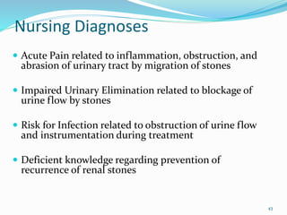
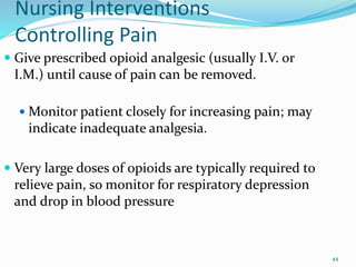
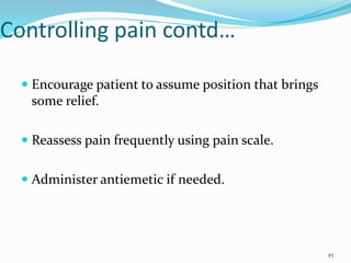
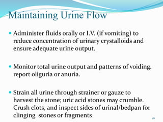
1
NEPHROLITHIASIS AND UROLITHIASIS
 Nephrolithiasis refers to renal stone .
 Urolithiasis refers to the presence of stones in the
urinary system.
 Stones, or calculi, are formed in the urinary tract
from the kidney to bladder by the crystallization of
substances excreted in the urine.
2
Causes and predisposing factors of
kidney stones
Hypercalcemia and hypercalciuria
Excessive intake of vitamin D, milk, and alkali
 Chronic dehydration
 Fluid intake,
 Immobility
Diet high in purines and abnormal purine
metabolism (hyperuricemia and gout)
3
Contd…
 Genetic predisposition (cystinuria)
 Chronic urinary tract infection (Struvite stone)
 Chronic obstruction with stasis of urine, foreign
bodies within the urinary tract
 Excessive oxalate absorption in inflammatory
bowel disease and bowel resection or ileostomy
 Living in mountainous, desert, or tropical areas
4
Contd…
Some medications
some diuretics
calcium-containing antacids
Protease inhibitors: Indinavir sulfate
Steroids:
Dietary factors:
 inadequate fluid intake predisposes to dehydration
 high intake of animal protein,
 a high-salt diet,
 excessive vitamin D supplementation,
 excessive intake of oxalate-containing foods such as
spinach
5
Medical conditions leading kidney
stone
 Gout (uric acid stones)
 Hyperparathyroidism
 kidney diseases such as renal tubular acidosis
 Chronic diseases such as diabetes and
hypertension.
 Inflammatory bowel disease
 Those who have undergone intestinal bypass or
ostomy surgery
6
Pathophysiology
Inhibitors of stone formation:
 Stones can form when there is a deficiency of
substances that normally prevent crystallization
in the urine, such as Citrate,magnesium,
nephrocalcin, and uropontin.
(that inhibit the nucleation, growth, and
aggregation of calcium-containing crystals)
7
Supersaturation of urine:
 When the urine becomes supersaturated with one
or more calculogenic (crystal-forming) substances,
a seed crystal may form through the process of
nucleation.
8
Different types of kidney stones
 Calcium stones: Calcium stones are most common
substance and found in up to 90% of stone.
 They are more common in men between age 20 - 30.
 The peak onset is during a person’s 20.
 They may range from very small particles, often called
SAND or GRAVEL to giant staghorn calculi.
 Calcium can combine with other substances, such as
oxalate (the most common substance), phosphate, or
carbonate, to form the stone.
9
10
Causes of hypercalciuria
 A high rate of bone reabsorption E.g Paget’s disease,
hyperparathyroidism, cushing’s syndrome, immobility etc
 Increase calcium reabsorption in gut as in milk-alkali
syndrome,& excessive intake of vitamin D.
 Impaired renal tubular absorption of filtered calcium as
in renal tubular acidosis.
11
Contd….
 Oxalate: It is the second most common stone,
which is relatively insoluble in urine.
 The mechanism of the oxalate availability is
unclear but may be closely related to diet.
 Oxalate is present in certain foods such as spinach.
It's also found in vitamin C supplements.
 Diseases of the small intestine increase risk of
these stones.
12
13
 Cystine stones: Cystine stones can form in people
who have cystinuria. This disorder runs in families
and affects both men and women.
 Uric acid stones: Uric acid stones are more common
in men than in women. They can occur with gout or
chemotherapy.
 Struvite stones: Struvite stones are mostly found in
women who have a urinary tract infection.
 These stones can grow very large and can block the
kidney, ureter, or bladder.
14
15
Uric acid stones:
16
17
18
Signs and symptoms
 While some kidney stones may not produce
symptoms (known as "silent" stones)
 The main symptom is severe pain that starts
suddenly and may go away suddenly:
 Pain may move to groin area (groin pain) or
testicles (testicle pain)
19
Pain pattern depends on site of
obstruction
 Calyx: Flank or costovertebral angle
pain,hematuria, abdominal distention
 Ureteropelvic junction: pain at flank or
costovertebral junction, migrating to groin and
testicle/libia minora
 Ureterovesical junction: Urgency, frequency,
genital pain
20
Contd..
 Renal colic pain
 Pain relief is immediate after stone passage.
 Large ureteral stones produce symptoms of
obstruction as they pass down the ureter (ureteral
colic).
 Bladder stones produce symptoms similar to cystitis.
21
Others symptoms
 Blood in the urine
 Chills
 Fever
 Nausea
 Vomiting
22
Diagnostic Evaluation
History taking:
 Dietary and medication histories and family history of
renal stones
 Ultrasonography
 X-ray KUB (Kidney, Ureter & Bladder)
 IVU ( Intra Venous Urography) to determine site
and evaluate degree of obstruction
 Spiral CT scan
23
Contd…
 Urinalysis :hematuria and pyuria; pH < 5.5
indicates uric acid stone; > 7.5 indicates struvite
stone; urine culture and drug sensitivity studies to
detect infection.
 Serum kidney function tests, electrolytes,
calcium, phosphorus, uric acid, and magnesium
levels; serum parathyroid hormone may also be
evaluated
24
Management
Non operative treatment
 1. Conservative
 2. Extracorporeal shock wave Lithotripsy ( ESWL)
Operative treatments
 Endoscopic procedures
 Open- rocedures
25
Management
Non - Operative
General Principles
 If small stone (< 5 mm) and able to treat as outpatient,
80% will pass stone spontaneously with hydration,
pain control, and reassurance.
 Intravenous hydrations followed by intravenous
frusemide
 Hospitalized for intractable pain, persistent vomiting,
high-grade fever, obstruction with infection, and
solitary kidney with obstruction.
26
Extracorporeal Shock Wave
Lithotripsy ( ESWL)
27
ESWL contd..
 Noninvasive technique and treatment of choice for
stones less than 2 cm in diameter (80% of stones
fall into this category) and located in the ureter
above the iliac crest.
28
ESWL contd…
 High-energy shock waves are
directed at the kidney stone (
500-1500 shock wave)
 Position of the kidney stone
is located by fluoroscopy, and
the shock waves are targeted
directly at the stone.
 The shock waves do not
affect soft tissue.
29
ESWL contd…
 ESWL eliminates need for
surgery in majority of patients
and can be repeated for
recurrent stones with no
apparent risk to kidney
structure or function.
 Complications include pain,
urinary infection, and
temporary bleeding around
kidney.
30
Endoscopic procedures
 Percutaneous Nephrostolithotomy
 Uretero-renoscopic Lithotripsy (URSL)
31
Percutaneous Nephrostolithotomy
 Percutaneous
nephrolithotomy (PCNL) is a
surgical procedure to remove
stones from the kidney by a small
puncture wound (up to about 1
cm) through the skin.
 It is most suitable to remove
stones of more than 2 cm in size
and which are present near the
Pelvic region.
 It is usually done under general
anesthesia or spinal anesthesia.
32
Contd….
 For stones in renal collecting
system or upper portion of ureter
and larger than 2.5 cm in diameter.
 Under ultrasound guidance, a
needle is advanced into collecting
system; guide wire is advanced
into renal pelvis or ureter.
33
Contd…
 Tract is dilated with mechanical dilators or
high-pressure balloon dilator until
nephroscope can be inserted up against
stone.
 Stones can be broken apart with hydraulic
shock waves or a laser beam administered
by way of nephroscope fragments are
removed using forceps, graspers, or basket
34
Complications of PCNL
 Injury to the colon sepsis
 Injury to the blood vessels
 Urinary leak may persist for a few days.
35
Uretero-renoscopic Lithotripsy (URSL)
 Ureterorenoscopy is a procedure ,where the kidney
stones are removed mechanically using a thin
telescope called ureteroscope is passed through the
urethra into the bladder, through to the ureter or to
the kidneys where the stone I s stuck .
The stones are broken down into smaller pieces using
lithotripsy.
36
Contd….
 This is performed under general anesthetic and
sometimes a plastic tube called a stent is inserted
temporarily to facilitate the movement of the stone
particles into the bladder.
 This is most effective for stones having size of 15 mm
or more.
37
Open Surgical Procedures
Indicated for only 1% to 2% of all stones.
 Pyelolithotomy removal of stones from kidney
pelvis
 Nephrolithotomy a incision into kidney for
removal of stone
38
Contd…
 Nephrectomy a removal of kidney; indicated
when kidney is extensively and irreparably
damaged and is no longer a functioning organ;
partial nephrectomy sometimes done.
 Ureterolithotomy a removal of stone in ureter
 Cystolithotomy a removal of stone from bladder
39
Complications of renal stone
 Calculous hydronephrosis
 Calculous pyonephrosis
 Renal failure
 Squamous cell carcinoma
40
Nursing Assessment
 Obtain history focusing on family history of
calculi, episodes of dehydration, prolonged
immobility, UTI, dietary and medication history.
 Assess pain location and radiation; assess level of
pain using a scale of 1 to 10.
 Observe for presence of associated symptoms:
nausea, vomiting, diarrhea, abdominal distention.
41
Contd…
 Monitor for signs and symptoms of UTI, such as
chills, fever, dysuria, frequency. Examine urine for
hematuria.
 Observe for signs and symptoms of obstruction,
such as frequent urination of small amounts,
oliguria, anuria.
42
Nursing Diagnoses
 Acute Pain related to inflammation, obstruction, and
abrasion of urinary tract by migration of stones
 Impaired Urinary Elimination related to blockage of
urine flow by stones
 Risk for Infection related to obstruction of urine flow
and instrumentation during treatment
 Deficient knowledge regarding prevention of
recurrence of renal stones
43
Nursing Interventions
Controlling Pain
 Give prescribed opioid analgesic (usually I.V. or
I.M.) until cause of pain can be removed.
 Monitor patient closely for increasing pain; may
indicate inadequate analgesia.
 Very large doses of opioids are typically required to
relieve pain, so monitor for respiratory depression
and drop in blood pressure
44
Controlling pain contd…
 Encourage patient to assume position that brings
some relief.
 Reassess pain frequently using pain scale.
 Administer antiemetic if needed.
45
Maintaining Urine Flow
 Administer fluids orally or I.V. (if vomiting) to
reduce concentration of urinary crystalloids and
ensure adequate urine output.
 Monitor total urine output and patterns of voiding.
report oliguria or anuria.
 Strain all urine through strainer or gauze to
harvest the stone; uric acid stones may crumble.
Crush clots, and inspect sides of urinal/bedpan for
clinging stones or fragments 46
Contd..
 For outpatient treatment, the patient may use a
coffee filter to strain urine.
 Help patient to walk, if possible, because
ambulation may help move the stone through the
urinary tract.
47
Controlling Infection
 Administer parenteral or oral antibiotics as
prescribed during treatment, and monitor for
adverse effects.
 Assess urine for color, cloudiness, and odor.
 Obtain vital signs, and monitor for fever and
symptoms of impending sepsis (tachycardia,
hypotension).
48
Prevention of Recurrent Stone
Formation
 For patients with calcium oxalate stones
 Instruct on diet avoid excesses of calcium and
phosphorus; maintain a low-sodium diet
 Teach purpose of drug therapy allopurinol
therapy to reduce uric acid concentration.
49
Contd…
 For patients with uric acid stones
 Teach methods to alkalinize urine to enhance
urate solubility.
 Instruct on testing urine pH.
 Teach purpose of taking allopurinol to lower
uric acid concentration.
 Provide information about reduction of dietary
purine intake (low protein red meat, fish, fowl).
50
 For patients with infection (struvite) stone
 Teach signs and symptoms of urinary infection
(in patients with neurologic or spinal cord
disease, teach use of dipsticks to evaluate urine
for nitrites and leukocytes); encourage him to
report infection immediately; must be treated
vigorously.
 Try to avoid prolonged periods of recumbency
slows renal drainage and alters calcium
metabolism.
51
 For patients with cystine stones(occur in
cystinuria, a hereditary disorder of amino acid
transport).
 Teach patient to alkalinize urine by taking
sodium bicarbonate tablets (Soda Mint) to
increase cystine solubility; instruct patient how
to test urine pH with a pH indicator.
 Teach patient about drug therapy with D-
penicillamine (Depen) to lower cystine
concentration
 Explain importance of maintaining drug therapy
consistently.
52
For all patients with stone disease
 Explain need for consistently increased fluid
intake (24-hour urinary output greater than 2 L)
lowers the concentration of substances involved
in stone formation.
 Drink enough fluids to achieve a urinary
volume of 2,000 to 3,000 mL or more every 24
hours.
 Drink larger amounts of fluid during periods
of strenuous exercise, if patient perspires
freely.
 Take fluids in evening to guarantee a high
urine flow during the night.
53
 Encourage a diet low in sugar and animal
proteins refined carbohydrates appear to lead to
hypercalciuria and urolithiasis; animal proteins
increase urine excretion of calcium, uric acid,
and oxalate.
 Increase consumption of fiber inhibits calcium
and oxalate absorption.
 Save any stone passed for analysis. (Only
patients with more than one episode of
urolithiasis are advised to have a metabolic
evaluation.)
54
References
 http://en.wikipedia.org/wiki/Kidney_stone retrieved
on 2012/08/08
 http://www.medicinenet.com/kidney_stone/article.ht
m retrieved on 2012/08/08
 Nettina, Sandra , M., & Elizabeth ,J. (2006).
Lippincott Manual of Nursing Practice. (8th
ed.).published by Lippincott Williams & Wilkins.
 Black, J.M., & Hawks, J.H.(2009).Medical Surgical
Nursing.(8th ed.). Published by Elsevier India.
56

Nephrolithiasis

  • 1.
  • 2.
    NEPHROLITHIASIS AND UROLITHIASIS Nephrolithiasis refers to renal stone .  Urolithiasis refers to the presence of stones in the urinary system.  Stones, or calculi, are formed in the urinary tract from the kidney to bladder by the crystallization of substances excreted in the urine. 2
  • 3.
    Causes and predisposingfactors of kidney stones Hypercalcemia and hypercalciuria Excessive intake of vitamin D, milk, and alkali  Chronic dehydration  Fluid intake,  Immobility Diet high in purines and abnormal purine metabolism (hyperuricemia and gout) 3
  • 4.
    Contd…  Genetic predisposition(cystinuria)  Chronic urinary tract infection (Struvite stone)  Chronic obstruction with stasis of urine, foreign bodies within the urinary tract  Excessive oxalate absorption in inflammatory bowel disease and bowel resection or ileostomy  Living in mountainous, desert, or tropical areas 4
  • 5.
    Contd… Some medications some diuretics calcium-containingantacids Protease inhibitors: Indinavir sulfate Steroids: Dietary factors:  inadequate fluid intake predisposes to dehydration  high intake of animal protein,  a high-salt diet,  excessive vitamin D supplementation,  excessive intake of oxalate-containing foods such as spinach 5
  • 6.
    Medical conditions leadingkidney stone  Gout (uric acid stones)  Hyperparathyroidism  kidney diseases such as renal tubular acidosis  Chronic diseases such as diabetes and hypertension.  Inflammatory bowel disease  Those who have undergone intestinal bypass or ostomy surgery 6
  • 7.
    Pathophysiology Inhibitors of stoneformation:  Stones can form when there is a deficiency of substances that normally prevent crystallization in the urine, such as Citrate,magnesium, nephrocalcin, and uropontin. (that inhibit the nucleation, growth, and aggregation of calcium-containing crystals) 7
  • 8.
    Supersaturation of urine: When the urine becomes supersaturated with one or more calculogenic (crystal-forming) substances, a seed crystal may form through the process of nucleation. 8
  • 9.
    Different types ofkidney stones  Calcium stones: Calcium stones are most common substance and found in up to 90% of stone.  They are more common in men between age 20 - 30.  The peak onset is during a person’s 20.  They may range from very small particles, often called SAND or GRAVEL to giant staghorn calculi.  Calcium can combine with other substances, such as oxalate (the most common substance), phosphate, or carbonate, to form the stone. 9
  • 10.
  • 11.
    Causes of hypercalciuria A high rate of bone reabsorption E.g Paget’s disease, hyperparathyroidism, cushing’s syndrome, immobility etc  Increase calcium reabsorption in gut as in milk-alkali syndrome,& excessive intake of vitamin D.  Impaired renal tubular absorption of filtered calcium as in renal tubular acidosis. 11
  • 12.
    Contd….  Oxalate: Itis the second most common stone, which is relatively insoluble in urine.  The mechanism of the oxalate availability is unclear but may be closely related to diet.  Oxalate is present in certain foods such as spinach. It's also found in vitamin C supplements.  Diseases of the small intestine increase risk of these stones. 12
  • 13.
  • 14.
     Cystine stones:Cystine stones can form in people who have cystinuria. This disorder runs in families and affects both men and women.  Uric acid stones: Uric acid stones are more common in men than in women. They can occur with gout or chemotherapy.  Struvite stones: Struvite stones are mostly found in women who have a urinary tract infection.  These stones can grow very large and can block the kidney, ureter, or bladder. 14
  • 15.
  • 16.
  • 17.
  • 18.
  • 19.
    Signs and symptoms While some kidney stones may not produce symptoms (known as "silent" stones)  The main symptom is severe pain that starts suddenly and may go away suddenly:  Pain may move to groin area (groin pain) or testicles (testicle pain) 19
  • 20.
    Pain pattern dependson site of obstruction  Calyx: Flank or costovertebral angle pain,hematuria, abdominal distention  Ureteropelvic junction: pain at flank or costovertebral junction, migrating to groin and testicle/libia minora  Ureterovesical junction: Urgency, frequency, genital pain 20
  • 21.
    Contd..  Renal colicpain  Pain relief is immediate after stone passage.  Large ureteral stones produce symptoms of obstruction as they pass down the ureter (ureteral colic).  Bladder stones produce symptoms similar to cystitis. 21
  • 22.
    Others symptoms  Bloodin the urine  Chills  Fever  Nausea  Vomiting 22
  • 23.
    Diagnostic Evaluation History taking: Dietary and medication histories and family history of renal stones  Ultrasonography  X-ray KUB (Kidney, Ureter & Bladder)  IVU ( Intra Venous Urography) to determine site and evaluate degree of obstruction  Spiral CT scan 23
  • 24.
    Contd…  Urinalysis :hematuriaand pyuria; pH < 5.5 indicates uric acid stone; > 7.5 indicates struvite stone; urine culture and drug sensitivity studies to detect infection.  Serum kidney function tests, electrolytes, calcium, phosphorus, uric acid, and magnesium levels; serum parathyroid hormone may also be evaluated 24
  • 25.
    Management Non operative treatment 1. Conservative  2. Extracorporeal shock wave Lithotripsy ( ESWL) Operative treatments  Endoscopic procedures  Open- rocedures 25
  • 26.
    Management Non - Operative GeneralPrinciples  If small stone (< 5 mm) and able to treat as outpatient, 80% will pass stone spontaneously with hydration, pain control, and reassurance.  Intravenous hydrations followed by intravenous frusemide  Hospitalized for intractable pain, persistent vomiting, high-grade fever, obstruction with infection, and solitary kidney with obstruction. 26
  • 27.
  • 28.
    ESWL contd..  Noninvasivetechnique and treatment of choice for stones less than 2 cm in diameter (80% of stones fall into this category) and located in the ureter above the iliac crest. 28
  • 29.
    ESWL contd…  High-energyshock waves are directed at the kidney stone ( 500-1500 shock wave)  Position of the kidney stone is located by fluoroscopy, and the shock waves are targeted directly at the stone.  The shock waves do not affect soft tissue. 29
  • 30.
    ESWL contd…  ESWLeliminates need for surgery in majority of patients and can be repeated for recurrent stones with no apparent risk to kidney structure or function.  Complications include pain, urinary infection, and temporary bleeding around kidney. 30
  • 31.
    Endoscopic procedures  PercutaneousNephrostolithotomy  Uretero-renoscopic Lithotripsy (URSL) 31
  • 32.
    Percutaneous Nephrostolithotomy  Percutaneous nephrolithotomy(PCNL) is a surgical procedure to remove stones from the kidney by a small puncture wound (up to about 1 cm) through the skin.  It is most suitable to remove stones of more than 2 cm in size and which are present near the Pelvic region.  It is usually done under general anesthesia or spinal anesthesia. 32
  • 33.
    Contd….  For stonesin renal collecting system or upper portion of ureter and larger than 2.5 cm in diameter.  Under ultrasound guidance, a needle is advanced into collecting system; guide wire is advanced into renal pelvis or ureter. 33
  • 34.
    Contd…  Tract isdilated with mechanical dilators or high-pressure balloon dilator until nephroscope can be inserted up against stone.  Stones can be broken apart with hydraulic shock waves or a laser beam administered by way of nephroscope fragments are removed using forceps, graspers, or basket 34
  • 35.
    Complications of PCNL Injury to the colon sepsis  Injury to the blood vessels  Urinary leak may persist for a few days. 35
  • 36.
    Uretero-renoscopic Lithotripsy (URSL) Ureterorenoscopy is a procedure ,where the kidney stones are removed mechanically using a thin telescope called ureteroscope is passed through the urethra into the bladder, through to the ureter or to the kidneys where the stone I s stuck . The stones are broken down into smaller pieces using lithotripsy. 36
  • 37.
    Contd….  This isperformed under general anesthetic and sometimes a plastic tube called a stent is inserted temporarily to facilitate the movement of the stone particles into the bladder.  This is most effective for stones having size of 15 mm or more. 37
  • 38.
    Open Surgical Procedures Indicatedfor only 1% to 2% of all stones.  Pyelolithotomy removal of stones from kidney pelvis  Nephrolithotomy a incision into kidney for removal of stone 38
  • 39.
    Contd…  Nephrectomy aremoval of kidney; indicated when kidney is extensively and irreparably damaged and is no longer a functioning organ; partial nephrectomy sometimes done.  Ureterolithotomy a removal of stone in ureter  Cystolithotomy a removal of stone from bladder 39
  • 40.
    Complications of renalstone  Calculous hydronephrosis  Calculous pyonephrosis  Renal failure  Squamous cell carcinoma 40
  • 41.
    Nursing Assessment  Obtainhistory focusing on family history of calculi, episodes of dehydration, prolonged immobility, UTI, dietary and medication history.  Assess pain location and radiation; assess level of pain using a scale of 1 to 10.  Observe for presence of associated symptoms: nausea, vomiting, diarrhea, abdominal distention. 41
  • 42.
    Contd…  Monitor forsigns and symptoms of UTI, such as chills, fever, dysuria, frequency. Examine urine for hematuria.  Observe for signs and symptoms of obstruction, such as frequent urination of small amounts, oliguria, anuria. 42
  • 43.
    Nursing Diagnoses  AcutePain related to inflammation, obstruction, and abrasion of urinary tract by migration of stones  Impaired Urinary Elimination related to blockage of urine flow by stones  Risk for Infection related to obstruction of urine flow and instrumentation during treatment  Deficient knowledge regarding prevention of recurrence of renal stones 43
  • 44.
    Nursing Interventions Controlling Pain Give prescribed opioid analgesic (usually I.V. or I.M.) until cause of pain can be removed.  Monitor patient closely for increasing pain; may indicate inadequate analgesia.  Very large doses of opioids are typically required to relieve pain, so monitor for respiratory depression and drop in blood pressure 44
  • 45.
    Controlling pain contd… Encourage patient to assume position that brings some relief.  Reassess pain frequently using pain scale.  Administer antiemetic if needed. 45
  • 46.
    Maintaining Urine Flow Administer fluids orally or I.V. (if vomiting) to reduce concentration of urinary crystalloids and ensure adequate urine output.  Monitor total urine output and patterns of voiding. report oliguria or anuria.  Strain all urine through strainer or gauze to harvest the stone; uric acid stones may crumble. Crush clots, and inspect sides of urinal/bedpan for clinging stones or fragments 46
  • 47.
    Contd..  For outpatienttreatment, the patient may use a coffee filter to strain urine.  Help patient to walk, if possible, because ambulation may help move the stone through the urinary tract. 47
  • 48.
    Controlling Infection  Administerparenteral or oral antibiotics as prescribed during treatment, and monitor for adverse effects.  Assess urine for color, cloudiness, and odor.  Obtain vital signs, and monitor for fever and symptoms of impending sepsis (tachycardia, hypotension). 48
  • 49.
    Prevention of RecurrentStone Formation  For patients with calcium oxalate stones  Instruct on diet avoid excesses of calcium and phosphorus; maintain a low-sodium diet  Teach purpose of drug therapy allopurinol therapy to reduce uric acid concentration. 49
  • 50.
    Contd…  For patientswith uric acid stones  Teach methods to alkalinize urine to enhance urate solubility.  Instruct on testing urine pH.  Teach purpose of taking allopurinol to lower uric acid concentration.  Provide information about reduction of dietary purine intake (low protein red meat, fish, fowl). 50
  • 51.
     For patientswith infection (struvite) stone  Teach signs and symptoms of urinary infection (in patients with neurologic or spinal cord disease, teach use of dipsticks to evaluate urine for nitrites and leukocytes); encourage him to report infection immediately; must be treated vigorously.  Try to avoid prolonged periods of recumbency slows renal drainage and alters calcium metabolism. 51
  • 52.
     For patientswith cystine stones(occur in cystinuria, a hereditary disorder of amino acid transport).  Teach patient to alkalinize urine by taking sodium bicarbonate tablets (Soda Mint) to increase cystine solubility; instruct patient how to test urine pH with a pH indicator.  Teach patient about drug therapy with D- penicillamine (Depen) to lower cystine concentration  Explain importance of maintaining drug therapy consistently. 52
  • 53.
    For all patientswith stone disease  Explain need for consistently increased fluid intake (24-hour urinary output greater than 2 L) lowers the concentration of substances involved in stone formation.  Drink enough fluids to achieve a urinary volume of 2,000 to 3,000 mL or more every 24 hours.  Drink larger amounts of fluid during periods of strenuous exercise, if patient perspires freely.  Take fluids in evening to guarantee a high urine flow during the night. 53
  • 54.
     Encourage adiet low in sugar and animal proteins refined carbohydrates appear to lead to hypercalciuria and urolithiasis; animal proteins increase urine excretion of calcium, uric acid, and oxalate.  Increase consumption of fiber inhibits calcium and oxalate absorption.  Save any stone passed for analysis. (Only patients with more than one episode of urolithiasis are advised to have a metabolic evaluation.) 54
  • 55.
    References  http://en.wikipedia.org/wiki/Kidney_stone retrieved on2012/08/08  http://www.medicinenet.com/kidney_stone/article.ht m retrieved on 2012/08/08  Nettina, Sandra , M., & Elizabeth ,J. (2006). Lippincott Manual of Nursing Practice. (8th ed.).published by Lippincott Williams & Wilkins.  Black, J.M., & Hawks, J.H.(2009).Medical Surgical Nursing.(8th ed.). Published by Elsevier India. 56