FilariasisDr.T.V.Rao MD
WHAT IS Filariasis
•Filariasis (or philariasis) is a parasitic disease
caused by an infection with roundworms of
the Filarioidea type. These are spread by
blood-feeding black flies and mosquitoes.
This disease belongs to the group of diseases
called helminthiasis.
•Eight known filarial nematodes use humans
as their definitive hosts.
Epidemiology-
International
•120 million in 80 countries
•1 billion at risk
•90% - Wucheraria
Bancrofti
•Remainder – Brugia
Malayi
Parasites
• White, slender roundworms
• Three types: Wuchereria bancrofti,
Brugia malayi, Brugia timori
• Live for 5-7 years, produce millions
of offspring
•Block the lymphatic system
• Network of channels and
lymph nodes that help
maintain fluid levels in the
body
• Blockage leads to edema
(collection of fluid in tissues)
Mosquitos are Vectors and
spread the Infection
•A mosquito is the intermediate host and
carrier. The most common
vectors/carriers are:
•in Africa: Anopheles species
•in the Americas: Culex
quinquefasciatus
•in the Pacific and in Asia: Mansonia and
Aedes species.
Millions are Infected with
filariasis
•One hundred and twenty million people in at
least 80 nations of the world have lymphatic
filariasis One billion people are at risk of
getting infected. Ninety percent of these
infections are caused by Wuchereria
bancrofti, and most of the remainder by
Brugia malayi. For W. bancrofti, humans are
the exclusive host, and even though certain
strains of B. malayi can also infect some
felines and monkeys.
Wucheraria bancrofti
• Primary causative agent of lymphatic
filariasis
• Overt bancroftian filariasis : 115 million
cases worldwide (45.5 million India, 40
million sub-Saharan Africa)
• Widespread throughout the subtropics and
tropics (for e.g. Central Africa, India,
Thailand, Malaysia, Phillipines, Pacific
Islands, Haiti, coastal Brazil)
Microfilaria of Mansonella ozzardi.
Apparently, it died out of Campeche, Mexico.
Stain: methylene blue.
Characters of the Adult
Parasites
•An Adult female
Wuchereria
bancrofti is about
80–100 mm long and
0.24–0.30 mm in
diameter, whereas a
male is about 40 mm
long and 0.1 mm in
diameter.
How the Larva Appear
•A microfilaria is about 240–300 µm
(micrometers) long and 7.5–10 µm
thick. It is sheathed and has
nocturnal periodicity, except the
South Pacific microfilaria which
does not have marked periodicity. It
has a gently curved body, and a tail
that is tapered to a point.
How the Larva Appear
•The nuclear column
(the cells that
constitute its body)
is loosely packed.
The cells can be seen
individually under a
microscope and do
not extend to the tip
of the tail.
Filarial Larvae
Impact of Filariasis
•with the disease can suffer from
lymphedema and elephantiasis and in men,
swelling of the scrotum, called hydrocele.
Lymphatic filariasis is a leading cause of
permanent disability worldwide.
Communities frequently shun and reject
women and men disfigured by the disease.
Affected people frequently are unable to
work because of their disability, and this
harms their families and their communities.
Life Cycle:
•Infective larvae are transmitted
by infected biting mosquitoes
during a blood meal. The larvae
migrate to lymphatic vessels and
lymph nodes, where they
develop into microfilariae-
producing adults.
Life Cycle:
•The adults dwell in lymphatic
vessels and lymph nodes
where they can live for
several years. The female
worms produce microfilariae
which circulate in the blood
Microfilariae
•The microfilariae infect biting mosquitoes.
Inside the mosquito, the microfilariae
develop in 1 to 2 weeks into infective
filariform (third-stage) larvae. During a
subsequent blood meal by the mosquito,
the larvae infect the human host. They
migrate to the lymphatic vessels and lymph
nodes of the human host, where they
develop into adults.
Life cycle of Brugia that also applies to
Wuchereria by CDC
Multiplication and Life
Cycle
•Adult female worms produce
microfilariae. Feeding vector
mosquitoes ingest microfilariae from
the bloodstream. In the mosquito the
microfilariae mature to infective larvae,
which migrate to the mosquito's mouth-
parts, enter a new host via the vector's
puncture wound, migrate to the
lymphatics, mature, and mate.
Why Clinical Manifestations
•Disease manifestations are due
to lymphatic dysfunction
resulting from the presence of
living and dead worms, lymph
thrombi, inflammation, and
immune reactions to worms and
worm products.
Pathogenesis and
Pathology
•Complex interplay of the pathogenic
potential of the parasite, the immune
response of the host, and external
('complicating') bacterial and fungal
infections.
•Most recognizable – Genital damage
( Hydroceles ) and
Lymphoedema/elephantiasis
Clinical features.
There are chronic, acute and asymptomatic
presentations of lymphatic filarial disease, as
well as some syndromes associated with these
infections. Among chronic manifestations,
hydrele, even though found only with W.
bancrofti infections not in Brugia infections is
the most common clinical manifestation of
lymphatic filariasis.
Disease Manifestations
•Although the parasite damages the
lymph system, most infected people
have no symptoms and will never
develop clinical symptoms. These
people do not know they have
lymphatic filariasis unless tested. A
small percentage of persons will
develop lymphedema.
Disease Manifestations
•This is caused by fluid collection
because of improper functioning of
the lymph system resulting in
swelling. This mostly affects the
legs, but can also occur in the arms,
breasts, and genitalia. Most people
develop these symptoms years after
being infected.
What is elephantiasis
characterized by?
•Thickening and hardening of the skin
•Correct.
•+ B) Increased body size due to masses
of worms all over the body, especially in
the nose
•+ C) Eosinophilia, heart failure and
breathing difficulty
What causes
elephantiasis?
•A) Decrease of blood flow due to
worms inside blood vessels
•+ B) Blockage of lymph fluid due
to worms inside lymph vessels
•+ C) Masses of microfilaria in
skin tissue
Tropical pulmonary
eosinophilia (TPE)
• Distinct syndrome in some individuals
• Paroxysmal cough and wheezing
• Weight loss, low grade fever,
pronounced blood eosinophilia
• Total serum IgE and antifilarial Ab titres
raised
• Responds well to treatment but in its
absence progressive pulmonary
damage
Symptoms
Fever
Kidney damage
Skin abnormalities due to
bacterial infection.
Elephantiasis
Swelling of limbs and genitalia
Male: Enlargement of scrotum,
penis retracted under skin,
spermatic cords thickened
Female: Long tumorous mass
covered by thickened ulcerated
skin develops on the vulva
Social Impact of Disease
Sexual Disability
Communities frequently
shun those disfigured.
Inability to work
Women with visible signs
may never marry or spouses
and families will reject
them.
Clinical features.
There are chronic, acute and asymptomatic
presentations of lymphatic filarial disease, as
well as some syndromes associated with these
infections. Among chronic manifestations,
hydrele, even though found only with W.
bancrofti infections not in Brugia infections is
the most common clinical manifestation of
lymphatic filariasis.
Pathogenesis
•Men can develop hydrocele or swelling of the
scrotum due to infection with one of the parasites
that causes LF specifically W. bancrofti.
•Filarial infection can also cause tropical pulmonary
eosinophilia syndrome, although this syndrome is
typically found in persons living with the disease in
Asia. Symptoms of tropical pulmonary eosinophilia
syndrome include cough, shortness of breath, and
wheezing. The eosinophilia is often accompanied by
high levels of IgE (Immunoglobulin E) and antifilarial
antibodies.
•Histologically - dilatation and
proliferation of lymphatic
endothelium & abnormal lymphatic
function
•'non-inflammatory pathway'
•‘inflammatory pathway‘ - adenitis
and retrograde lymphangitis
•bacterial and fungal superinfections
Clinical Features
•Chronic manifestations: Hydrocoele (most common ),
elephantiasis, Chyluria
•Acute manifestations: Acute inflammatory episodes
'DLA'(dermatolymphangioadenitis) , 'filarial fever' ,
tropical pulmonary eosinophilia, acute inflammatory
reaction
•Asymptomatic Presentations
•Other Syndromes: arthritis (typically monoarticular),
endomyocardial fibrosis, tenosynovitis,
thrombophlebitis, glomerulonephritis, lateral popliteal
nerve palsy, and others.
While lymphedema can develop in the absence of
overt inflammatory reactions and in the early
stages be associated with microfilaremia, the
development of elephantiasis (either of the limbs
or the genitals) is most often associated with a
history of recurrent inflammation. The early
pitting edema gives rise to a stronger edema with
the hardening of the tissues.
Diagnosis.
Until very recently, diagnosing lymphatic
filariasis had been extremely difficult, since
parasites had to be detected microscopically in
the blood, and in most parts of the world, the
parasites have a nocturnal periodicity that
restricts their appearance in the blood to only
the hours around midnight.
Diagnosis
Until recently, very difficult to
diagnose
Nocturnal periodicity: The
worms can only be detected in
the blood of those infected
around the hour of midnight.
New specific card test: Detects
parasites using only finger prick
blood tests any time of day.
Ultrasound can identify rapidly
moving adult worms.
Diagnosis
Until recently, diagnosis depended
on the direct demonstration of the
parasite
Antigen detection: Circulating
filarial antigen (CFA) - 'gold
standard' for diagnosing Wuchereria
bancrofti infections.
Clinical Diagnosis
Serology
•Serologic techniques provide an
alternative to microscopic detection of
microfilariae for the diagnosis of
lymphatic filariasis. Patients with active
filarial infection typically have elevated
levels of ant filarial IgG4 in the blood
and these can be detected using routine
assays.
Treatment.
Communities where filariasis is endemic.
The primary goal of treating the
affected community is to eliminate
microfilariae from the blood of
infected individuals so that
transmission of the infection by the
mosquito can be interrupted.
Management
•Treating the infection: DEC (6 mg/kg per
day) for 12 days in bancroftian filariasis and
for 6 days in brugian filariasis, repeated at
1-6 monthly intervals if necessary
•Ivermectin
•Albendazole
•Side effects : headaches, fever, myalgia,
lymphadenopathy and occasionally rash,
itching
Treatment and Management of
Elephantiasis
Prevention
Mosquito nets, insect repellents
Voodoo healing techniques
Elevate and exercise affected body part
Skin treatment
Wash area twice daily
Antibacterial cream
CDP (Complex decongestive physiotherapy)
Lymph drainage, massage, compressive
bandages
Management and Treatment
of Lymphatic Filariasis
•Currently Used:
• Antifilarial drugs (DEC and ivermectin) are
useful against larval offspring
•Testing:
• Doxycycline
• Tested on a Tanzanian village.
• Found to almost completely eliminate adult worms
14 months after treatment.
• Sustained loss of larval offspring for 8-14 months
after treatment.
• Albendazole and DEC
• Given together once a year
• Found to be 99% effective in removing microfilariae
from blood for full year after treatment
Prevention
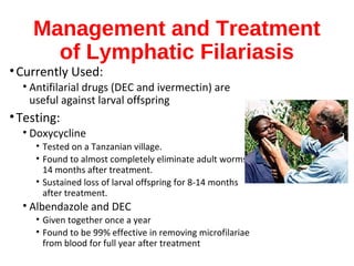
•By decreasing contact between humans
and vectors or by decreasing the
amount of infection the vector can
acquire
•Population: through reducing the
numbers of mosquito vectors
•2-drug treatment regimens (selecting
among albendazole and either
ivermectin or diethylcarbamazine [DEC])
How to prevent the Filarial
infection
•Individuals: personal
repellents, bednets or
insecticide-impregnated
materials.
•Prophylactic regimen of
DEC (6 mg/kg per day x 2
How can I prevent infection?
•How can I prevent infection?
•Avoiding mosquito bites is the best form of
prevention. The mosquitoes that carry the
microscopic worms usually bite between the hours
of dusk and dawn. If you live in or travel to an area
with lymphatic filariasis:
•Sleep under a mosquito net.
•Wear long sleeves and trousers.
•Use mosquito repellent on exposed skin between
dusk and dawn.
Treating the individual.
•Both albendazole and DEC have
been shown to be effective in killing
the adult-stage filarial parasites. It is
clear that this antiparasite treatment
can result in improvement of patients'
elephantiasis and hydrocele (especially
in the early stages of disease)
WHO's Strategy to EliminateWHO's Strategy to Eliminate
Lymphatic FilariasisLymphatic Filariasis
•The strategy of the World Health
Organization (WHO) of the Global
Programme to Eliminate Lymphatic
Filariasis has 2 aims: a) to stop the
spread of infection (interrupt
transmission), and secondly b) to
alleviate the suffering of affected
individuals.
Mass Treatments for
Prevention
•To interrupt transmission, districts in which
lymphatic filariasis is endemic must be
identified, and then community-wide ("mass
treatment") programs implemented to treat the
entire at risk population. In most countries, the
program will be based on once-yearly
administration of single doses of 2 drugs given
together: albendazole plus either
diethylcarbamazine (DEC) or ivermectin, the
latter in areas where either onchocerciasis,
loiasis or another may also be endemic.
Community Treatments
•To alleviate the suffering caused by the
disease, it will be necessary to implement
community education programmes to
raise awareness in affected patients. This
would promote the benefits of intensive
local hygiene and the possible
improvement, both in the damage that has
already occurred, and in preventing the
debilitating and painful, acute episodes of
inflammation.
International communities help for
elimination of Disease
•The pledge in 1998 by GlaxcoSmithKline
to collaborate with the WHO in its
elimination efforts included the donation
of numerous resources, but especially
albendazole free of charge, for as long as
necessary. This donation, coupled with
the recent decision by Merck to expand
its wellknown Mectizan®
(iverme
Economic and Social Impact.Economic and Social Impact.
•Program Created and Designed by
Dr.T.V.Rao MD for Medical and
Paramedical students on global
Education on Communicable
diseases
•Email
•doctortvrao@gmail.com

Filariasis

  • 1.
  • 2.
    WHAT IS Filariasis •Filariasis(or philariasis) is a parasitic disease caused by an infection with roundworms of the Filarioidea type. These are spread by blood-feeding black flies and mosquitoes. This disease belongs to the group of diseases called helminthiasis. •Eight known filarial nematodes use humans as their definitive hosts.
  • 3.
    Epidemiology- International •120 million in80 countries •1 billion at risk •90% - Wucheraria Bancrofti •Remainder – Brugia Malayi
  • 5.
    Parasites • White, slenderroundworms • Three types: Wuchereria bancrofti, Brugia malayi, Brugia timori • Live for 5-7 years, produce millions of offspring •Block the lymphatic system • Network of channels and lymph nodes that help maintain fluid levels in the body • Blockage leads to edema (collection of fluid in tissues)
  • 6.
    Mosquitos are Vectorsand spread the Infection •A mosquito is the intermediate host and carrier. The most common vectors/carriers are: •in Africa: Anopheles species •in the Americas: Culex quinquefasciatus •in the Pacific and in Asia: Mansonia and Aedes species.
  • 7.
    Millions are Infectedwith filariasis •One hundred and twenty million people in at least 80 nations of the world have lymphatic filariasis One billion people are at risk of getting infected. Ninety percent of these infections are caused by Wuchereria bancrofti, and most of the remainder by Brugia malayi. For W. bancrofti, humans are the exclusive host, and even though certain strains of B. malayi can also infect some felines and monkeys.
  • 8.
    Wucheraria bancrofti • Primarycausative agent of lymphatic filariasis • Overt bancroftian filariasis : 115 million cases worldwide (45.5 million India, 40 million sub-Saharan Africa) • Widespread throughout the subtropics and tropics (for e.g. Central Africa, India, Thailand, Malaysia, Phillipines, Pacific Islands, Haiti, coastal Brazil)
  • 12.
    Microfilaria of Mansonellaozzardi. Apparently, it died out of Campeche, Mexico. Stain: methylene blue.
  • 13.
    Characters of theAdult Parasites •An Adult female Wuchereria bancrofti is about 80–100 mm long and 0.24–0.30 mm in diameter, whereas a male is about 40 mm long and 0.1 mm in diameter.
  • 14.
    How the LarvaAppear •A microfilaria is about 240–300 µm (micrometers) long and 7.5–10 µm thick. It is sheathed and has nocturnal periodicity, except the South Pacific microfilaria which does not have marked periodicity. It has a gently curved body, and a tail that is tapered to a point.
  • 15.
    How the LarvaAppear •The nuclear column (the cells that constitute its body) is loosely packed. The cells can be seen individually under a microscope and do not extend to the tip of the tail.
  • 16.
  • 18.
    Impact of Filariasis •withthe disease can suffer from lymphedema and elephantiasis and in men, swelling of the scrotum, called hydrocele. Lymphatic filariasis is a leading cause of permanent disability worldwide. Communities frequently shun and reject women and men disfigured by the disease. Affected people frequently are unable to work because of their disability, and this harms their families and their communities.
  • 19.
    Life Cycle: •Infective larvaeare transmitted by infected biting mosquitoes during a blood meal. The larvae migrate to lymphatic vessels and lymph nodes, where they develop into microfilariae- producing adults.
  • 20.
    Life Cycle: •The adultsdwell in lymphatic vessels and lymph nodes where they can live for several years. The female worms produce microfilariae which circulate in the blood
  • 21.
    Microfilariae •The microfilariae infectbiting mosquitoes. Inside the mosquito, the microfilariae develop in 1 to 2 weeks into infective filariform (third-stage) larvae. During a subsequent blood meal by the mosquito, the larvae infect the human host. They migrate to the lymphatic vessels and lymph nodes of the human host, where they develop into adults.
  • 22.
    Life cycle ofBrugia that also applies to Wuchereria by CDC
  • 25.
    Multiplication and Life Cycle •Adultfemale worms produce microfilariae. Feeding vector mosquitoes ingest microfilariae from the bloodstream. In the mosquito the microfilariae mature to infective larvae, which migrate to the mosquito's mouth- parts, enter a new host via the vector's puncture wound, migrate to the lymphatics, mature, and mate.
  • 26.
    Why Clinical Manifestations •Diseasemanifestations are due to lymphatic dysfunction resulting from the presence of living and dead worms, lymph thrombi, inflammation, and immune reactions to worms and worm products.
  • 27.
    Pathogenesis and Pathology •Complex interplayof the pathogenic potential of the parasite, the immune response of the host, and external ('complicating') bacterial and fungal infections. •Most recognizable – Genital damage ( Hydroceles ) and Lymphoedema/elephantiasis
  • 28.
    Clinical features. There arechronic, acute and asymptomatic presentations of lymphatic filarial disease, as well as some syndromes associated with these infections. Among chronic manifestations, hydrele, even though found only with W. bancrofti infections not in Brugia infections is the most common clinical manifestation of lymphatic filariasis.
  • 29.
    Disease Manifestations •Although theparasite damages the lymph system, most infected people have no symptoms and will never develop clinical symptoms. These people do not know they have lymphatic filariasis unless tested. A small percentage of persons will develop lymphedema.
  • 30.
    Disease Manifestations •This iscaused by fluid collection because of improper functioning of the lymph system resulting in swelling. This mostly affects the legs, but can also occur in the arms, breasts, and genitalia. Most people develop these symptoms years after being infected.
  • 31.
    What is elephantiasis characterizedby? •Thickening and hardening of the skin •Correct. •+ B) Increased body size due to masses of worms all over the body, especially in the nose •+ C) Eosinophilia, heart failure and breathing difficulty
  • 32.
    What causes elephantiasis? •A) Decreaseof blood flow due to worms inside blood vessels •+ B) Blockage of lymph fluid due to worms inside lymph vessels •+ C) Masses of microfilaria in skin tissue
  • 33.
    Tropical pulmonary eosinophilia (TPE) •Distinct syndrome in some individuals • Paroxysmal cough and wheezing • Weight loss, low grade fever, pronounced blood eosinophilia • Total serum IgE and antifilarial Ab titres raised • Responds well to treatment but in its absence progressive pulmonary damage
  • 34.
    Symptoms Fever Kidney damage Skin abnormalitiesdue to bacterial infection. Elephantiasis Swelling of limbs and genitalia Male: Enlargement of scrotum, penis retracted under skin, spermatic cords thickened Female: Long tumorous mass covered by thickened ulcerated skin develops on the vulva
  • 35.
    Social Impact ofDisease Sexual Disability Communities frequently shun those disfigured. Inability to work Women with visible signs may never marry or spouses and families will reject them.
  • 36.
    Clinical features. There arechronic, acute and asymptomatic presentations of lymphatic filarial disease, as well as some syndromes associated with these infections. Among chronic manifestations, hydrele, even though found only with W. bancrofti infections not in Brugia infections is the most common clinical manifestation of lymphatic filariasis.
  • 39.
    Pathogenesis •Men can develophydrocele or swelling of the scrotum due to infection with one of the parasites that causes LF specifically W. bancrofti. •Filarial infection can also cause tropical pulmonary eosinophilia syndrome, although this syndrome is typically found in persons living with the disease in Asia. Symptoms of tropical pulmonary eosinophilia syndrome include cough, shortness of breath, and wheezing. The eosinophilia is often accompanied by high levels of IgE (Immunoglobulin E) and antifilarial antibodies.
  • 40.
    •Histologically - dilatationand proliferation of lymphatic endothelium & abnormal lymphatic function •'non-inflammatory pathway' •‘inflammatory pathway‘ - adenitis and retrograde lymphangitis •bacterial and fungal superinfections
  • 41.
    Clinical Features •Chronic manifestations:Hydrocoele (most common ), elephantiasis, Chyluria •Acute manifestations: Acute inflammatory episodes 'DLA'(dermatolymphangioadenitis) , 'filarial fever' , tropical pulmonary eosinophilia, acute inflammatory reaction •Asymptomatic Presentations •Other Syndromes: arthritis (typically monoarticular), endomyocardial fibrosis, tenosynovitis, thrombophlebitis, glomerulonephritis, lateral popliteal nerve palsy, and others.
  • 42.
    While lymphedema candevelop in the absence of overt inflammatory reactions and in the early stages be associated with microfilaremia, the development of elephantiasis (either of the limbs or the genitals) is most often associated with a history of recurrent inflammation. The early pitting edema gives rise to a stronger edema with the hardening of the tissues.
  • 44.
    Diagnosis. Until very recently,diagnosing lymphatic filariasis had been extremely difficult, since parasites had to be detected microscopically in the blood, and in most parts of the world, the parasites have a nocturnal periodicity that restricts their appearance in the blood to only the hours around midnight.
  • 45.
    Diagnosis Until recently, verydifficult to diagnose Nocturnal periodicity: The worms can only be detected in the blood of those infected around the hour of midnight. New specific card test: Detects parasites using only finger prick blood tests any time of day. Ultrasound can identify rapidly moving adult worms.
  • 46.
    Diagnosis Until recently, diagnosisdepended on the direct demonstration of the parasite Antigen detection: Circulating filarial antigen (CFA) - 'gold standard' for diagnosing Wuchereria bancrofti infections. Clinical Diagnosis
  • 48.
    Serology •Serologic techniques providean alternative to microscopic detection of microfilariae for the diagnosis of lymphatic filariasis. Patients with active filarial infection typically have elevated levels of ant filarial IgG4 in the blood and these can be detected using routine assays.
  • 49.
    Treatment. Communities where filariasisis endemic. The primary goal of treating the affected community is to eliminate microfilariae from the blood of infected individuals so that transmission of the infection by the mosquito can be interrupted.
  • 50.
    Management •Treating the infection:DEC (6 mg/kg per day) for 12 days in bancroftian filariasis and for 6 days in brugian filariasis, repeated at 1-6 monthly intervals if necessary •Ivermectin •Albendazole •Side effects : headaches, fever, myalgia, lymphadenopathy and occasionally rash, itching
  • 51.
    Treatment and Managementof Elephantiasis Prevention Mosquito nets, insect repellents Voodoo healing techniques Elevate and exercise affected body part Skin treatment Wash area twice daily Antibacterial cream CDP (Complex decongestive physiotherapy) Lymph drainage, massage, compressive bandages
  • 52.
    Management and Treatment ofLymphatic Filariasis •Currently Used: • Antifilarial drugs (DEC and ivermectin) are useful against larval offspring •Testing: • Doxycycline • Tested on a Tanzanian village. • Found to almost completely eliminate adult worms 14 months after treatment. • Sustained loss of larval offspring for 8-14 months after treatment. • Albendazole and DEC • Given together once a year • Found to be 99% effective in removing microfilariae from blood for full year after treatment
  • 53.
    Prevention •By decreasing contactbetween humans and vectors or by decreasing the amount of infection the vector can acquire •Population: through reducing the numbers of mosquito vectors •2-drug treatment regimens (selecting among albendazole and either ivermectin or diethylcarbamazine [DEC])
  • 54.
    How to preventthe Filarial infection •Individuals: personal repellents, bednets or insecticide-impregnated materials. •Prophylactic regimen of DEC (6 mg/kg per day x 2
  • 55.
    How can Iprevent infection? •How can I prevent infection? •Avoiding mosquito bites is the best form of prevention. The mosquitoes that carry the microscopic worms usually bite between the hours of dusk and dawn. If you live in or travel to an area with lymphatic filariasis: •Sleep under a mosquito net. •Wear long sleeves and trousers. •Use mosquito repellent on exposed skin between dusk and dawn.
  • 56.
    Treating the individual. •Bothalbendazole and DEC have been shown to be effective in killing the adult-stage filarial parasites. It is clear that this antiparasite treatment can result in improvement of patients' elephantiasis and hydrocele (especially in the early stages of disease)
  • 57.
    WHO's Strategy toEliminateWHO's Strategy to Eliminate Lymphatic FilariasisLymphatic Filariasis •The strategy of the World Health Organization (WHO) of the Global Programme to Eliminate Lymphatic Filariasis has 2 aims: a) to stop the spread of infection (interrupt transmission), and secondly b) to alleviate the suffering of affected individuals.
  • 58.
    Mass Treatments for Prevention •Tointerrupt transmission, districts in which lymphatic filariasis is endemic must be identified, and then community-wide ("mass treatment") programs implemented to treat the entire at risk population. In most countries, the program will be based on once-yearly administration of single doses of 2 drugs given together: albendazole plus either diethylcarbamazine (DEC) or ivermectin, the latter in areas where either onchocerciasis, loiasis or another may also be endemic.
  • 59.
    Community Treatments •To alleviatethe suffering caused by the disease, it will be necessary to implement community education programmes to raise awareness in affected patients. This would promote the benefits of intensive local hygiene and the possible improvement, both in the damage that has already occurred, and in preventing the debilitating and painful, acute episodes of inflammation.
  • 60.
    International communities helpfor elimination of Disease •The pledge in 1998 by GlaxcoSmithKline to collaborate with the WHO in its elimination efforts included the donation of numerous resources, but especially albendazole free of charge, for as long as necessary. This donation, coupled with the recent decision by Merck to expand its wellknown Mectizan® (iverme
  • 61.
    Economic and SocialImpact.Economic and Social Impact.
  • 62.
    •Program Created andDesigned by Dr.T.V.Rao MD for Medical and Paramedical students on global Education on Communicable diseases •Email •doctortvrao@gmail.com

Editor's Notes

  • #11 Filariasis may be difficult to eradicate just because it occupies vast terrains Central America and Mexico are underestimated for Onchocerca volvulu
  • #12 Microfilaria of Wuchereria bancrofti. Giemsa stain.
  • #29 Uncommon in childhood, it is seen more frequently postpuberty and with a progressive increase in prevalence with age. In many endemic communities 40-60% of all adult males have hydrocele. It often develops in the absence of overt inflammatory reactions. Many patients with hydrocele also have microfilarias in the circulation. In bancroftian filariasis, the scrotal lymphatics are the preferred site for localization of the adult worms, and their presence stimulates the proliferation of lymphatic endothelium. The localization of adult worms in the lymphatics of the spermatic cord leads to a thickening of the cord. The hydroceles can become massive, but still occur without lymphedema or elephantiasis developing in the penis and scrotum, since the lymphatic drainage of these tissues is separate and more superficial.
  • #37 Uncommon in childhood, it is seen more frequently postpuberty and with a progressive increase in prevalence with age. In many endemic communities 40-60% of all adult males have hydrocele. It often develops in the absence of overt inflammatory reactions. Many patients with hydrocele also have microfilarias in the circulation. In bancroftian filariasis, the scrotal lymphatics are the preferred site for localization of the adult worms, and their presence stimulates the proliferation of lymphatic endothelium. The localization of adult worms in the lymphatics of the spermatic cord leads to a thickening of the cord. The hydroceles can become massive, but still occur without lymphedema or elephantiasis developing in the penis and scrotum, since the lymphatic drainage of these tissues is separate and more superficial.
  • #43 Still later, hyperpigmentation and hyperkeratosis develop, often with wartlike protuberances which, on histological section, reveal dilated loops of lymphatic vessels within the nodular lesions. Patients with chronic lymphedema or elephantiasis rarely are microfilaremic. Redundant skin folds, of the skin provide havens for bacteria and fungi to thrive and intermittently penetrate the epidermis to lead to either local or systemic infections. Sometimes the skin over the breaks down, causing the dilated lymphatic within to rupture and discharge its lymph fluid directly into the environment, at the same time serving as a causeway for penetration of bacterial or fungal organisms directly into the lymphatic system. Chyluria, another of the chronic filarial syndromes, is caused by the intermittent discharge of intestinal lymph (chyle) into the renal pelvis and subsequently into the . The clinical course is intermittent, sometimes remitting.
  • #45 The new development of a very sensitive, very specific simple card test to detect circulating parasite antigens without the need for laboratory facilities and using only finger-prick blood droplets taken anytime of the day has completely transformed the approach to diagnosis. With this and other new diagnostic tools, it will now be possible both to improve our understanding of where the infection actually occurs and to monitor more easily the effectiveness of treatment and control programs.
  • #50 Recent studies have shown that single doses of diethylcarbamazine (DEC) have the same long-term (1-year) effect in decreasing microfilaremia as the formerly-recommended 12-day regimens of DEC and, even more importantly, that the use of single doses of 2 drugs administered concurrently (optimally albendazole or ivermectin with DEC) is 99% effective in removing microfilarias from the blood.