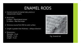
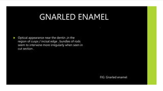
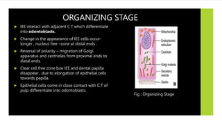
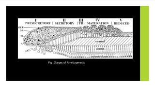
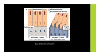
SlideShare a Scribd company logo
ENAMEL
DR. SARGAM R. PARATE , 1ST YEAR PG
DEPARTMENT OF CONSERVATIVE DENTISTRY AND ENDODONTICS.
BHABHA COLLEGE OF DENTAL SCIENCES , BHOPAL.
 INTRODUCTION
 PHYSICAL PROPERTIES
 CHEMICAL PROPERTIES
 STRUCTURE OF ENAMEL
 DEVELOPMENT OF ENAMEL
 AGE CHANGES IN ENAMEL
 CLINICAL IMPLICATIONS
 DEFECTS IN ENAMEL
• EPITHETIAL ENAMEL ORGAN
• AMELOGENESIS
• LIFE CYCLE OF AMELOBLASTS
CONTENTS
INTRODUCTION
 Enamel is the hardest substance in the
human body .
 Highly mineralized structure present as
the outer most covering of the crown.
 Epithelial derived hard tissue ,formed by
ameloblasts which cannot reform itself.
 Origin - Ectodermal
 Non-vital , acellular , avascular.
 Modulus of elasticity – Higher
 Hardness – 296 KHN
 Specific gravity -2.8
 Density – 2.8-3.0 gm/sq.cm
 Non –electrical conductive material
(Insulator at RTM)
 Brittle
 Translucent tissue
 Colour : Yellowish white – grayish white
 Thickness varies
 Semi-permeable membrane
PHYSICAL
PROPERTIES
Enamel
Inorganic
96%
Hydroxyapetite
Crystalline calcium
phosphate
Ca10 (Po4 )6 (OH4)2
Hexagonal crystals
Enamel matrix protein
Proline
Histidine
Glutamine
Leucine
Organic
+ Water
4%
Amelogenins
90%
Hydrophobhic
Non-amelogenins
10%
Enamel matrix protein
Glycine
Aspartic acid
Serine
Eg : Enamelin,Ameloblastin,
Tuftelin
CHEMICAL PROPERTIES
FIG : Structure of Hydroxyapatite crystal
STRUCTURE OF ENAMEL
 Enamel lamellae
 Enamel tufts
 Dentinoenamel Junction
 Enamel spindles.
 Enamel rods
 Incremental lines
 Hunter –Schreger bands
 Gnarled enamel
 Surface structures
 Enamel cuticle
ENAMEL RODS
 Enamel consists of enamel rods, prisms or
interprismatic sheaths.
 No.of rods :
5 million – Mand lateral incisor
12 million – Max first molar
 Torturous course from DEJ to outer surface
 Length is greater than thickness – oblique direction
 Dimensions :
Diameter – 4 um
Breadth – 5 um
Length – 9 um
Fig : Enamel rod
SUBMICROSCOPIC STRUCTURE
Cross
-section
Fish -scales
Light microscope – hexagonal / round /
Confocal laser scanning microscope– Keyhole
/paddle shaped
Fig : KEYHOLE SHAPED STRUCTURE
ULTRASTRUCTURE
Fig : CRYSTAL ORIENTATION SHOWING ENAMEL
BODY AND TAIL
Fig : ULTRASTRICTURE OF ENAMEL ROD
DIRECTION OF ENAMEL RODS
 Directions of rods :
• Right angle to the dentin surface.
• Horizontal – cervical part of deciduous
teeth.
• Oblique –middle third of the crown
• Vertical – cusp tips
Fig : Direction of enamel rods
CLINICAL SIGNIFICANCE
 Unsupported enamel rods should not be left
at cavity margins – soon break and produce
leakage.
 Bacteria lodge in spaces -initiation of
secondary dental caries.
 Uneven dissolution of enamel rods to get
pitted and irregular surface increases
mechanical bonding with enamel in
composites and pit-fissure sealants.
 Beveling- increase surface area – more no. of
enamel rods exposed – more retention. Fig: Unsupported and supported enamel
INCREMENTAL LINES OF RETZIUS
 Ground section : Brownish bands
 Represents incremental deposition of
enamel i.e. successive apposition during
formation of enamel layers.
 Longitudinal section – surrounds tip of the
dentin
 Transverse section – concentric circles.
 Directions :
Cervically – Run obliquely
DEJ-Surface – deviate occlusally Fig : Incremental lines of Retzius
Fig : Longitudinal Fig : Transverse section
Fig: Longitudinal and
Transverse section
CLINICAL SIGNIFICANCE
 Lines become prominent during carious attack.
 Reflect variations in structure and mineralization
during growth.
 Helps in chronological mapping of dental
development.
STRIATIONS
 Dark transverse lines seen crossing the
enamel giving striated appearance to
enamel rods.
 Represents daily increments of growth
during enamel formation.
 Rate of enamel formation : 4 um
Fig: Cross-striations of Enamel rod
HUNTER-SCHREGER BANDS
 Alternating dark and light bands seen in
longitudinal section under oblique reflected
light.
 Optical phenomenon caused due to change in
direction of enamel rods.
 Found in inner 2/3rd of enamel starting from DEJ.
 Prisms when cut :
Longitudinally – Dark bands – Diazones
Transversely - light bands - Parazones
 Angle b/w parazones and diazones – 40 degrees.
Fig : Hunter-schreger bands
GNARLED ENAMEL
 Optical appearance near the dentin ,in the
region of cusps / incisal edge , bundles of rods
seem to interwine more irregularly when seen in
cut section .
FIG: Gnarled enamel
SURFACE STRUCTURES
 PRISMLESS ENAMEL
 PERIKYMATA
 ENAMEL ROD ENDS
 PITS
 CAPS
 ENAMEL BROCHS
 CRACKS
 NEO-NATAL LINE
Fig : Different surface structures of enamel
PRISMLESS ENAMEL
 Structure less layer of enamel .
 30 um thick
 Seen in 70% permanent and all deciduous
teeth.
 Commonly seen – cervical areas
least seen - cusp tips
Fig : A. Prism less enamel , B. Prismatic enamel
PERIKYMATA
 Transverse wave like grooves.
 External manifestation of Striae of Retzius.
 Parallel to each other and to CEJ.
 CEJ- 30 perikymata /mm
occlusal /incisal edge – 10 perikymata /mm
 Absent - occlusal part of primary teeth
present – postnatal cervical areas.
Fig: Perikymata
ENAMEL ROD ENDS
 Circular depressions
 Shallowest – cervical region
Deepest – incisal / occlusal edges
CLINICAL SIGNIFICANCE :
Contribute to adherence of plaque which
result to caries attack
Fig : Enamel rod ends
PITS
 Small depressions on enamel
surface.
 Diameter – 1-1.5 um
 Represent ends of ameloblasts
ENAMEL CAPS
 Small elevations on enamel
surface.
 Diameter – 10-15 um
 Caused due to enamel deposition
on non-mineralizable debris.
ENAMEL BROCHS
 Large enamel elevations .
 Diameter - > 10-15 um
CRACKS
 Narrow fissure-like structures seen on all
surfaces.
 Outer edges of enamel lamellae .
 Disappear on decalcification.
 Right angle to DEJ.
Fig: Cracks on enamel surface
NEONATAL LINE OR RING
 Accentuated incremental line of Retzius.
 Boundary b/w pre-natal and post-natal
enamel.
 Result of abrupt change in the nutrition of
newborn infant.
 Usually seen in deciduous teeth and
permanent first molars.
Fig: Neo-natal line
ENAMEL CUTICLE
 Nasmyth’s membrane / primary enamel cuticle ,
delicate membrane covers entire crown of newly
erupted tooth , soon removed by mastication.
 Secreted after epithelial enamel organ retracts from
cervical region during tooth formation.
 Function: protects the enamel surface from
resorptive activity of adjacent vascular tissue.
Fig : Enamel cuticle
ENAMEL LAMALLAE
 Thin leaf-like hypocalcified structures.
 Extend from enamel surface – DEJ
 Develops in planes of tension during enamel
maturation .
 Best visualized – Transverse section
 CLINICAL SIGNIFICANCE –
Can act as pathways for caries producing
bacteria.
Fig: B. Enamel lamellae
TYPES OF LAMALLAE Based on contents
of lamellae
TYPE A
Only enamel
Poorly calcified rod
segments
TYPE B
May reach dentin
Degenerated cells
TYPE C
May reach dentin
Organic matter
from saliva
More common
ENAMEL TUFTS
 Arise at DEJ – enamel to about 1/5th -1/3rd
of its thickness.
 Consists of hypocalcified enamel rods and
interprismatic substances.
 Consists of highest enamel protein
concentration.
 According to TENCATES , Develop due to
abrupt change in the direction of group of
rods that arise from DEJ.
 Best visualized – Transverse section
Fig : Enamel tufts
DENTINOENAMEL JUNCTION
 DEJ is scalloped with convexity directed
towards dentin.
 Also k/as Amelo-dentinal junction.
Fig : Dentinoenamel junction
CLINICAL SIGNIFICANCE
 Scalloping increases the surface area of enamel.
 Lateral spread of dental caries.
 Stress distribution and resist enamel crack propagation.
 In some pathological condition DEJ becomes flat and enamel get chipped off
easily.
1. Dentinogenesis imperfecta
2. Ehler-Danlos Syndrome
Fig : Dentinoenamel junction
ENAMEL SPINDLES
 Thickened odontoblastic processes passing
across DEJ to enamel.
 Appears dark in transmitted light when seen in
ground section.
 Commonly seen – cusp region (crowding of
odontoblast occur)
 Best visualized – longitudinal section
 CLINICAL SIGNIFICANCE :
Sudden sensitivity occurs during cavity
preparation as bur reaches DEJ
Fig : Enamel Spindles
DEVELOPMENT
OF ENAMEL
EPITHETIAL ENAMEL ORGAN
LIFE CYCLE OF
AMELOGENESIS
EPITHELIAL ENAMEL ORGAN
 Enamel organ, also known as the dental organ, is a
cellular aggregation seen in a developing tooth.
 Function :
 Formation of enamel
 Initiation of dentin formation,
 Establishment of the shape of a tooth's crown
 Establishment of the DEJ.
 Originate from stratified epithelium of primitive oral
cavity.
Fig : Enamel organ
EPITHELIAL ENAMEL ORGAN
CONSISTS OF FOUR LAYERS :
 Outer enamel epithelium
 Stellate reticulum
 Stratum intermedium
 Inner enamel epithelium
Fig : Epithelial Enamel Organ
 In Early stages of development of enamel
organ ,OEE has single layer of cuboidal
cells , separated from dental sac by delicate
basement membrane.
 Prior to formation of hard tissue , regular
arrangement of OEE is maintained only at
cervical parts of enamel organ.
 At highest convexity of organ , OEE cells are
irregular in shape
 During enamel formation ,OEE cells develop
villi, cytoplasmic vesicles and mitochondria
for active transport of materials.
 Forms the middle part of enamel organ.
 Star shaped, with long processes
reaching all directions from central
body.
 Connected to each other and to stratum
intermedium by desmosomes.
 Function : act as buffer against physical
forces that might distort developing
DEJ.
 Reduced in thickness after first layer of
dentin is laid.
OUTER ENAMEL EPITHETIUM (OEE) STELLATE RETICULUM
INNER ENAMEL EPITHELIUM (IEE)
STRATUM INTERMEDIUM
 Situated between stellate reticulum and
inner enamel epithelium.
 Flat to cuboid in shape , arranged in 1-3
layers.
 Connected to each other and to
neighbouring cells by desmosomes.
 Function : believed , role in production of
enamel itself through control of fluid
diffusion in and out of ameloblasts
 Show mitotic division even after cells of
IEE cease to divide.
 Derived from basal cell layer of oral
epithelium.
 Columnar shape.
 Differentiate into ameloblasts to produce
enamel matrix.
Fig : showing OEE &IEE
CERVICAL LOOP
 Location on an enamel organ in
a developing tooth where the outer
enamel epithelium and the inner enamel
epithelium join.
 During root formation the inner layers of
epithelium disappear and only the basal
layers are left creating Hertwig's epithelial
root sheath (HERS).
FIG : Cervical Loop
LIFE CYCLE OF
AMELOBLAST
According to Function :
Fig: Life cycle of Ameloblast
MORPHOGENIC STAGE
 Stage determine morphology of the crown and DEJ.
 Cells are short and columnar
 Large oval nuclei -fill the cell body.
 Golgi apparatus and centrioles -proximal end of
cells
mitochondria –dispersed in cytoplasm
 During ameloblast differentiation terminal bars
appear , migrating the mitochondria to basal region
of cell.
 IEE is separated from C.T of dental papilla by
delicate basal lamina.
Fig: Morphogenic Stage
ORGANIZING STAGE
 IEE interact with adjacent C.T which differentiate
into odontoblasts.
 Change in the appearance of IEE cells occur-
longer , nucleus free –zone at distal ends .
 Reversal of polarity – migration of Golgi
apparatus and centrioles from proximal ends to
distal ends.
 Clear cell free zone b/w IEE and dental papilla
disappear , due to elongation of epithelial cells
towards papilla.
 Epithelial cells come in close contact with C.T of
pulp differentiate into odontoblasts.
Fig : Organizing Stage
 Pre-ameloblast secrete proteins- play role
in epithelial mesenchymal interaction.
 Terminal phase – formation of dentin by
odontoblasts begins.
 When dentin forms , original source of
nourishment of ameloblast cuts off – then
supplied by capillaries .
Fig : Organizing Stage
FORMATIVE STAGE
 Ameloblast enter formative stage after first
layer of dentin is formed.
 Presence of dentin necessary for the beginning
of enamel matrix formation.
 During formation of enamel matrix :
Ameloblast –retain same length and
arrangement.
Cytoplasmic inclusion and organelles – change
in organization and number.
 Earliest change –development of blunt cell
processes on ameloblast , which penetrate
basal lamina and enter predentin.
Fig : Formative Stage
MATURATIVE STAGE
 Enamel maturation occurs after most of the
thickness of enamel matrix is formed in occlusal
/incisal area.
 At cervical area the enamel matrix formation is
still progressing.
 Stratum intermedium cells –lose cuboidal shape
and regular arrangement , converts into spindle
shape.
 Ameloblasts – reduced in length and attached
to enamel matrix. They display microvilli at
distal extremities .
 Cytoplasmic vacuoles contains materials
resembling enamel matrix.
 These structures indicate resorptive function of
these cells.
Fig : Maturative Stage
PROTECTIVE STAGE
 When enamel is fully developed , fully
calcified – ameloblast is present as well-
defined layer and cease to get differentiated
from startum intermedium and OEE .
 Cells form stratified epithelial covering of
enamel k/as Reduced enamel epithelium.
 Function : protecting mature enamel by
separating from C.T until tooth erupts.
 If C.T comes in contact with enamel ,
anomalies develop .Enamel may either get
resorbed or may get covered by cementum.
Fig: Reduced enamel epithelium
DESMOLYTIC STAGE
 REE proliferates and induce atrophy of C.T
separating it from OEE , so that fusion of
two epithelia occur.
 Epithelial cells elaborates enzymes –
capable of destroying C.T fibers by
desmolysis.
 Premature degeneration of REE may
prevent eruption of tooth.
Fig : Fusion of OEE & IEE
AMELOGENESIS
Amelogenesi
Organic
formation
Mineralizatio
 Enamel formation.
 Begins at Late bell stage of
tooth development.
 Formation begins as soon as
IEE &OEE differentiate.
Stages of
Amelogenesis
PRESECRETORY STAGE
MORPHOGENIC PHASE
DIFFERENCIATION PHASE
SECRETORY STAGE
MATURATION STAGE
TRANSITIONAL PHASE
MATURATION PROPER
UNDER ELECTRON MICROSCOPY
Fig : Stages of Amelogenesis
PRESECRETORY STAGE
MORPHOGENIC PHASE :
 Shape of crown is determined.
 Low columnar cells with centrally located
nucleus and poorly developed Golgi
bodies, mitochondria are scattered
throughout cell.
 First junctional complex develop (
ameloblasts near St. intermedium)
DIFFERENCIATION PHASE:
 Cells of IEE become tall columnar and
nucleus shifts towards St. intermedium.
 Increase in RER and Golgi bodies shift
distally.
 Second junctional complex develop(b/w
tome’s process towards enamel)
 Development of tome’s processes occur.
FORMATION OF TOME’S PROCESSES
Projection of ameloblast
enamel matrix - Tome’s
processes.
Initially only proximal
is formed.
After initial layer of
deposition , distal portion
formed.
Fig : Formation of Tome’s Processes
SECRETORY STAGE
Formation
secretory
globules
Golgi bodies are
surrounded by
cisternae of RER
mRNA are
translated into
ribosomes of
RNA enters RER
and enamel
proteins are
secreted.
Proteins are
bounded to
membrane
bound secretory
granules.
Secretory
granules are
migrated into
Tome’s
processes.
ENAMEL FORMATION
Ameloblasts deposit
secretory globules via
Tome’s process
Interdigitation with
dentin. Enamel without
rods are formed.
Distal portion of Tomes
formed by elongation
of ameloblasts
Proximal part starts to
secrete enamel proteins
for interrod formation.
Distal part lays down
enamel.
Enamel and interrod
made of same material
but differ in direction
of deposition.
As ameloblasts lay
down enamel at the
end they lose their
distal part and there is
no more interrod.
Sandwiched prismatic
enamel between non
prismatic enamel
formed.
Fig : Enamel Formation
MATURATION STAGE
 Before tooth erupts in the oral
cavity, enamel hardens by the
process of maturation.
 Takes place at the expense of
enamel fluid and matrix proteins.
 During this stage ameloblats are
called pre-secretory cells (secrete
min amount of ameloblastin and
amelogenin)
TRANSITIONAL
Transformation of
ameloblast into post
secretory cells.
Decrease in cell
organelle , cell
size,apoptosis.
MATURATION
Bulk removal of water
and organic material.
Introduction of ruffled
border and smooth
ended ameloblasts for
incorporation of
inorganic content.
SMOOTH BORDER
• Causes balancing
of pH.
• Distal junctions
tight and leaking
proximal
• No membrane
calcium ATP ,
contains only
proteins.
• Constitute 20% of
cell life
RUFFLED BORDER
• Prepares acidic
environment for
enamel
• Calcium binding
proteins,
lysosomes,
adenosine
triphosphates
present.
• Constitute 80% of
cell life.
MINERALIZATION
2
STAGE
MINERALIZATIO
70%
Maturation
30%
Immediate partial
Immediate partial
mineralization
Occurs in matrix segments and
interprismatic enamel
Apatite crystals of dentin –
nucleation – enamel is laid.
Initial mineral – octacalcium
phosphate (unstable)
Hydroxyapatite
1 unit HA -> 2 unit HA Fig : Immediate partial mineralization
Maturation
(gradual completion of
mineralization)
Process starts – height
of crown – cervically
Begins before enamel
matrix has reached full
thickness.
Apatite crystals increases in
thickness
Size – 1.5-25 microns
Tuftelin –nucleation of
enamel crystals.
Other proteins – inhibit
enamel deposition.
Rate – 4 um / day
1 layer of enamel – 1mm
thickness – 240 days
Permanent teeth > deciduous
teeth
Crystal size increases
after tooth eruption –
ionic exchange with
saliva
After formation of
enamel
Ameloblast – apoptosis
Enamel formation
ceases.
FIG: Mineralization in molar from
occlusal to cervical region
Fig : Maturation stage of mineralization
AGE CHANGES
IN ENAMEL
ATTRITION
• Wear facets seen in older people
• Loss of vertical dimension of
crown and flattening of proximal
contour.
DISCOLOURATION
• Teeth darken with age.
• Addition of organic
material / deepening of
dentin colour.
PERMEABILITY
• Teeth become less
permeable.
• With age pores diminishes
as crystals acquire more
ions.
MODIFICATION OF
SURFACE
• Generalized loss of
enamel rods.
• Slower flattening of
perikymata later
disappear completely.
CLINICAL IMPLICATIONS
DENTAL FLOURIDATIO
ACID ETCHING
TECHNIQUE
BLEACHING
DENTAL CARIES
 Destruction of enamel surface with acid, lead
to the dissolution of enamel matrix, following
carious attack.
 Caries preferentially attack cores of rods and
more permeable striae of Retzius – promote
lateral spread of caries and undermining
adjacent enamel.
Fig : Dental caries
FLOURIDATION
 Incorporation of fluoride ions make
hydroxyapatite crystals more resistant to
carious attack.
 Decreases rate of demineralization.
 Increases rate of remineralisation.
Fig : Fluoride Application
ACID ETCHING TECHNIQUE
 Removes plaque , debris and thin layer of
enamel.
 By dissolving minerals in enamel, etchants
can remove outer 10 um of enamel
surface, and makes a porous layer of 5-50
um deep into enamel.
 Increases porosity through dissolution of
crystals.
 Helps in mechanical bonding of
composites to enamel surface.
Fig : Acid etching technique
BLEACHING
 Lightening of colour of tooth through the
application of chemical agent helps in
oxidizing the organic pigmentation of
enamel.
Fig : Bleaching of teeth
DEFECTS IN ENAMEL
GENETIC
 Amelogenesis Imperfecta
1. Hypoplastic (Type 1)
2. Hypomaturation (Type 2)
3. Hypocalcified (Type 3)
NON –GENETIC
 Dental caries
 Attrition
 Abrasion
 Abfraction
 Erosion
 Localised non-hereditary enamel
hypoplasia
 Localised non-hereditary enamel hypo
calcification.
 Fluorosis
SR.
NO
DISEASE DEFECT CLINICAL FEATURE PICTURE
1. AMELOGENESIS IMPERFECTA Defect in gene encoding enamel
matrix protein.
Type 1- Hypo plastic AI • Defect in formation of
matrix protein
• Ameloblast fail to lay down
sufficient matrix
• Enamel – pitted, grooved,
thin ,hard translucent
• Small teeth with open
contacts.
Type 2-Hypomaturation AI • Defect in maturation stage. • Enamel softer and chips off
easily.
• Mottled brown -yellow
white
Type 3-Hypocalcified AI • Defect in calcification stage. • Most common
• Enamel – normal thickness,
easily lost by attrition.
• Dull , lustrous, honey
colored , stains easily.
GENETIC DEFECT
SR.NO DISEASE DEFECT PICTURE
1. Dental caries Demineralization of inorganic part and
destruction of organic substance .
2. Attrition Loss of tooth structure from direct
frictional forces.
3. Abrasion Mechanical wear other than mastication
4. Abfraction Caused by forces placed on teeth during
biting, eating, chewing .
NON-GENETIC DEFECT IN ENAMEL
SR.NO DISEASE DEFECT PICTURE
5. Erosion Dissolution of mineralized tooth
structure by chemical process.
6. Localized non hereditary enamel hypoplasia Defect in ameloblasts during
formation stage.
7. Localized non hereditary enamel hypo
calcification
Defect in mineralization stage.
8. Fluorosis • Hypo mineralization of enamel
caused due to excessive ingestion
of fluoride during enamel
formation.
Enamel.pptx

More Related Content

What's hot

Tooth Dentin and dentinogenesis ppt
Tooth Dentin and dentinogenesis pptTooth Dentin and dentinogenesis ppt
Tooth Dentin and dentinogenesis ppt
madhusudhan reddy
 
Dentin
DentinDentin
Pulp dentin complex
Pulp dentin complexPulp dentin complex
Pulp dentin complex
Upama Sishan
 
Enamel done
Enamel   doneEnamel   done
Enamel done
kusumvyas41164
 
Dentin -- Structural aspect
Dentin -- Structural aspectDentin -- Structural aspect
Dentin -- Structural aspect
saloni7pathak
 
Enamel
EnamelEnamel
Histology of Pulp
Histology of PulpHistology of Pulp
Histology of Pulp
Aurelian Jovita Alexander
 
Enamel
EnamelEnamel
1 cementum
1  cementum1  cementum
1 cementum
sherifsayed65
 
1st BDS Lecture on tooth Enamel
1st BDS Lecture on tooth Enamel1st BDS Lecture on tooth Enamel
1st BDS Lecture on tooth Enamel
varun surya
 
Amelogenesis by dr.suriya visithiriyan
Amelogenesis by dr.suriya visithiriyanAmelogenesis by dr.suriya visithiriyan
Amelogenesis by dr.suriya visithiriyan
drSuriya2
 
Enamel
EnamelEnamel
Histology of dentin
Histology of dentinHistology of dentin
Histology of dentin
Amin Abusallamah
 
Enamel
EnamelEnamel
Enamelmtbj
 
Enamel
EnamelEnamel
Amelogenesis
Amelogenesis Amelogenesis
Amelogenesis
varun surya
 
Dental Anatomy: Enamel
Dental Anatomy: Enamel Dental Anatomy: Enamel
Dental Anatomy: Enamel
Ozident
 

What's hot (20)

Tooth Dentin and dentinogenesis ppt
Tooth Dentin and dentinogenesis pptTooth Dentin and dentinogenesis ppt
Tooth Dentin and dentinogenesis ppt
 
Dentin
DentinDentin
Dentin
 
Pulp dentin complex
Pulp dentin complexPulp dentin complex
Pulp dentin complex
 
7 dentinogenesis
7 dentinogenesis7 dentinogenesis
7 dentinogenesis
 
Enamel done
Enamel   doneEnamel   done
Enamel done
 
Dentin -- Structural aspect
Dentin -- Structural aspectDentin -- Structural aspect
Dentin -- Structural aspect
 
Enamel
EnamelEnamel
Enamel
 
Histology of Pulp
Histology of PulpHistology of Pulp
Histology of Pulp
 
Enamel
EnamelEnamel
Enamel
 
1 cementum
1  cementum1  cementum
1 cementum
 
1st BDS Lecture on tooth Enamel
1st BDS Lecture on tooth Enamel1st BDS Lecture on tooth Enamel
1st BDS Lecture on tooth Enamel
 
Amelogenesis by dr.suriya visithiriyan
Amelogenesis by dr.suriya visithiriyanAmelogenesis by dr.suriya visithiriyan
Amelogenesis by dr.suriya visithiriyan
 
Enamel
EnamelEnamel
Enamel
 
Histology of dentin
Histology of dentinHistology of dentin
Histology of dentin
 
Dentin
DentinDentin
Dentin
 
Enamel
EnamelEnamel
Enamel
 
Enamel
EnamelEnamel
Enamel
 
Amelogenesis
Amelogenesis Amelogenesis
Amelogenesis
 
Dental Anatomy: Enamel
Dental Anatomy: Enamel Dental Anatomy: Enamel
Dental Anatomy: Enamel
 
DENTIN
DENTINDENTIN
DENTIN
 

Similar to Enamel.pptx

ENAMEL
 ENAMEL ENAMEL
ENAMEL
CmenonMenon
 
Enamel.pptx
Enamel.pptxEnamel.pptx
Enamel.pptx
AjuAnto1
 
TOOTH ENAMEL - HISTOPATHOLOGY FOR STUDENTS
TOOTH ENAMEL - HISTOPATHOLOGY FOR STUDENTSTOOTH ENAMEL - HISTOPATHOLOGY FOR STUDENTS
TOOTH ENAMEL - HISTOPATHOLOGY FOR STUDENTS
drdhaval3
 
Enamel
EnamelEnamel
Enamel
Ruchi Sharma
 
enamel.ppt
enamel.pptenamel.ppt
enamel.ppt
ASHELROSE1
 
Enamel presentation. prepared by mohammed yahia
Enamel presentation. prepared by mohammed yahiaEnamel presentation. prepared by mohammed yahia
Enamel presentation. prepared by mohammed yahiaMaher Aziz
 
Enamel
EnamelEnamel
Enamel
ddert
 
Enamel
EnamelEnamel
Enamel
hattan2
 
Enamel - DCPS
Enamel - DCPSEnamel - DCPS
Enamel - DCPS
Muznah Sultan
 
Dento enamel junction
Dento enamel junction Dento enamel junction
Dento enamel junction
Indian dental academy
 
Enamel
EnamelEnamel
Enamel
EnamelEnamel
Enamel
EnamelEnamel
Enamel seminar.pptx
Enamel seminar.pptxEnamel seminar.pptx
Enamel seminar.pptx
Vivarnaah
 
Enamel
EnamelEnamel
Enamel significance in operative dentistry /certified fixed orthodontic cour...
Enamel significance in operative dentistry  /certified fixed orthodontic cour...Enamel significance in operative dentistry  /certified fixed orthodontic cour...
Enamel significance in operative dentistry /certified fixed orthodontic cour...
Indian dental academy
 
enamel and dentin biology.ppt
enamel and dentin biology.pptenamel and dentin biology.ppt
enamel and dentin biology.ppt
AkshaFati
 
Development of periodontium
Development of periodontiumDevelopment of periodontium
Development of periodontium
Jignesh Tate
 
Development of Teeth by Dr.Smitha Rao
Development of Teeth by Dr.Smitha RaoDevelopment of Teeth by Dr.Smitha Rao
Development of Teeth by Dr.Smitha Rao
smithanaik1980
 

Similar to Enamel.pptx (20)

ENAMEL
 ENAMEL ENAMEL
ENAMEL
 
Enamel.pptx
Enamel.pptxEnamel.pptx
Enamel.pptx
 
TOOTH ENAMEL - HISTOPATHOLOGY FOR STUDENTS
TOOTH ENAMEL - HISTOPATHOLOGY FOR STUDENTSTOOTH ENAMEL - HISTOPATHOLOGY FOR STUDENTS
TOOTH ENAMEL - HISTOPATHOLOGY FOR STUDENTS
 
Enamel
EnamelEnamel
Enamel
 
enamel.ppt
enamel.pptenamel.ppt
enamel.ppt
 
Enamel presentation. prepared by mohammed yahia
Enamel presentation. prepared by mohammed yahiaEnamel presentation. prepared by mohammed yahia
Enamel presentation. prepared by mohammed yahia
 
Enamel
EnamelEnamel
Enamel
 
Enamel
EnamelEnamel
Enamel
 
Enamel
EnamelEnamel
Enamel
 
Enamel - DCPS
Enamel - DCPSEnamel - DCPS
Enamel - DCPS
 
Dento enamel junction
Dento enamel junction Dento enamel junction
Dento enamel junction
 
Enamel
EnamelEnamel
Enamel
 
Enamel
EnamelEnamel
Enamel
 
Enamel
EnamelEnamel
Enamel
 
Enamel seminar.pptx
Enamel seminar.pptxEnamel seminar.pptx
Enamel seminar.pptx
 
Enamel
EnamelEnamel
Enamel
 
Enamel significance in operative dentistry /certified fixed orthodontic cour...
Enamel significance in operative dentistry  /certified fixed orthodontic cour...Enamel significance in operative dentistry  /certified fixed orthodontic cour...
Enamel significance in operative dentistry /certified fixed orthodontic cour...
 
enamel and dentin biology.ppt
enamel and dentin biology.pptenamel and dentin biology.ppt
enamel and dentin biology.ppt
 
Development of periodontium
Development of periodontiumDevelopment of periodontium
Development of periodontium
 
Development of Teeth by Dr.Smitha Rao
Development of Teeth by Dr.Smitha RaoDevelopment of Teeth by Dr.Smitha Rao
Development of Teeth by Dr.Smitha Rao
 

Recently uploaded

Novas diretrizes da OMS para os cuidados perinatais de mais qualidade
Novas diretrizes da OMS para os cuidados perinatais de mais qualidadeNovas diretrizes da OMS para os cuidados perinatais de mais qualidade
Novas diretrizes da OMS para os cuidados perinatais de mais qualidade
Prof. Marcus Renato de Carvalho
 
The Normal Electrocardiogram - Part I of II
The Normal Electrocardiogram - Part I of IIThe Normal Electrocardiogram - Part I of II
The Normal Electrocardiogram - Part I of II
MedicoseAcademics
 
Triangles of Neck and Clinical Correlation by Dr. RIG.pptx
Triangles of Neck and Clinical Correlation by Dr. RIG.pptxTriangles of Neck and Clinical Correlation by Dr. RIG.pptx
Triangles of Neck and Clinical Correlation by Dr. RIG.pptx
Dr. Rabia Inam Gandapore
 
Hemodialysis: Chapter 3, Dialysis Water Unit - Dr.Gawad
Hemodialysis: Chapter 3, Dialysis Water Unit - Dr.GawadHemodialysis: Chapter 3, Dialysis Water Unit - Dr.Gawad
Hemodialysis: Chapter 3, Dialysis Water Unit - Dr.Gawad
NephroTube - Dr.Gawad
 
micro teaching on communication m.sc nursing.pdf
micro teaching on communication m.sc nursing.pdfmicro teaching on communication m.sc nursing.pdf
micro teaching on communication m.sc nursing.pdf
Anurag Sharma
 
KDIGO 2024 guidelines for diabetologists
KDIGO 2024 guidelines for diabetologistsKDIGO 2024 guidelines for diabetologists
KDIGO 2024 guidelines for diabetologists
د.محمود نجيب
 
Ozempic: Preoperative Management of Patients on GLP-1 Receptor Agonists
Ozempic: Preoperative Management of Patients on GLP-1 Receptor Agonists  Ozempic: Preoperative Management of Patients on GLP-1 Receptor Agonists
Ozempic: Preoperative Management of Patients on GLP-1 Receptor Agonists
Saeid Safari
 
BENIGN PROSTATIC HYPERPLASIA.BPH. BPHpdf
BENIGN PROSTATIC HYPERPLASIA.BPH. BPHpdfBENIGN PROSTATIC HYPERPLASIA.BPH. BPHpdf
BENIGN PROSTATIC HYPERPLASIA.BPH. BPHpdf
DR SETH JOTHAM
 
Evaluation of antidepressant activity of clitoris ternatea in animals
Evaluation of antidepressant activity of clitoris ternatea in animalsEvaluation of antidepressant activity of clitoris ternatea in animals
Evaluation of antidepressant activity of clitoris ternatea in animals
Shweta
 
NVBDCP.pptx Nation vector borne disease control program
NVBDCP.pptx Nation vector borne disease control programNVBDCP.pptx Nation vector borne disease control program
NVBDCP.pptx Nation vector borne disease control program
Sapna Thakur
 
263778731218 Abortion Clinic /Pills In Harare ,
263778731218 Abortion Clinic /Pills In Harare ,263778731218 Abortion Clinic /Pills In Harare ,
263778731218 Abortion Clinic /Pills In Harare ,
sisternakatoto
 
Report Back from SGO 2024: What’s the Latest in Cervical Cancer?
Report Back from SGO 2024: What’s the Latest in Cervical Cancer?Report Back from SGO 2024: What’s the Latest in Cervical Cancer?
Report Back from SGO 2024: What’s the Latest in Cervical Cancer?
bkling
 
ARTIFICIAL INTELLIGENCE IN HEALTHCARE.pdf
ARTIFICIAL INTELLIGENCE IN  HEALTHCARE.pdfARTIFICIAL INTELLIGENCE IN  HEALTHCARE.pdf
ARTIFICIAL INTELLIGENCE IN HEALTHCARE.pdf
Anujkumaranit
 
BRACHYTHERAPY OVERVIEW AND APPLICATORS
BRACHYTHERAPY OVERVIEW  AND  APPLICATORSBRACHYTHERAPY OVERVIEW  AND  APPLICATORS
BRACHYTHERAPY OVERVIEW AND APPLICATORS
Krishan Murari
 
Charaka Samhita Sutra sthana Chapter 15 Upakalpaniyaadhyaya
Charaka Samhita Sutra sthana Chapter 15 UpakalpaniyaadhyayaCharaka Samhita Sutra sthana Chapter 15 Upakalpaniyaadhyaya
Charaka Samhita Sutra sthana Chapter 15 Upakalpaniyaadhyaya
Dr KHALID B.M
 
TEST BANK for Operations Management, 14th Edition by William J. Stevenson, Ve...
TEST BANK for Operations Management, 14th Edition by William J. Stevenson, Ve...TEST BANK for Operations Management, 14th Edition by William J. Stevenson, Ve...
TEST BANK for Operations Management, 14th Edition by William J. Stevenson, Ve...
kevinkariuki227
 
ACUTE SCROTUM.....pdf. ACUTE SCROTAL CONDITIOND
ACUTE SCROTUM.....pdf. ACUTE SCROTAL CONDITIONDACUTE SCROTUM.....pdf. ACUTE SCROTAL CONDITIOND
ACUTE SCROTUM.....pdf. ACUTE SCROTAL CONDITIOND
DR SETH JOTHAM
 
Maxilla, Mandible & Hyoid Bone & Clinical Correlations by Dr. RIG.pptx
Maxilla, Mandible & Hyoid Bone & Clinical Correlations by Dr. RIG.pptxMaxilla, Mandible & Hyoid Bone & Clinical Correlations by Dr. RIG.pptx
Maxilla, Mandible & Hyoid Bone & Clinical Correlations by Dr. RIG.pptx
Dr. Rabia Inam Gandapore
 
How STIs Influence the Development of Pelvic Inflammatory Disease.pptx
How STIs Influence the Development of Pelvic Inflammatory Disease.pptxHow STIs Influence the Development of Pelvic Inflammatory Disease.pptx
How STIs Influence the Development of Pelvic Inflammatory Disease.pptx
FFragrant
 
Surat @ℂall @Girls ꧁❤8527049040❤꧂@ℂall @Girls Service Vip Top Model Safe
Surat @ℂall @Girls ꧁❤8527049040❤꧂@ℂall @Girls Service Vip Top Model SafeSurat @ℂall @Girls ꧁❤8527049040❤꧂@ℂall @Girls Service Vip Top Model Safe
Surat @ℂall @Girls ꧁❤8527049040❤꧂@ℂall @Girls Service Vip Top Model Safe
Savita Shen $i11
 

Recently uploaded (20)

Novas diretrizes da OMS para os cuidados perinatais de mais qualidade
Novas diretrizes da OMS para os cuidados perinatais de mais qualidadeNovas diretrizes da OMS para os cuidados perinatais de mais qualidade
Novas diretrizes da OMS para os cuidados perinatais de mais qualidade
 
The Normal Electrocardiogram - Part I of II
The Normal Electrocardiogram - Part I of IIThe Normal Electrocardiogram - Part I of II
The Normal Electrocardiogram - Part I of II
 
Triangles of Neck and Clinical Correlation by Dr. RIG.pptx
Triangles of Neck and Clinical Correlation by Dr. RIG.pptxTriangles of Neck and Clinical Correlation by Dr. RIG.pptx
Triangles of Neck and Clinical Correlation by Dr. RIG.pptx
 
Hemodialysis: Chapter 3, Dialysis Water Unit - Dr.Gawad
Hemodialysis: Chapter 3, Dialysis Water Unit - Dr.GawadHemodialysis: Chapter 3, Dialysis Water Unit - Dr.Gawad
Hemodialysis: Chapter 3, Dialysis Water Unit - Dr.Gawad
 
micro teaching on communication m.sc nursing.pdf
micro teaching on communication m.sc nursing.pdfmicro teaching on communication m.sc nursing.pdf
micro teaching on communication m.sc nursing.pdf
 
KDIGO 2024 guidelines for diabetologists
KDIGO 2024 guidelines for diabetologistsKDIGO 2024 guidelines for diabetologists
KDIGO 2024 guidelines for diabetologists
 
Ozempic: Preoperative Management of Patients on GLP-1 Receptor Agonists
Ozempic: Preoperative Management of Patients on GLP-1 Receptor Agonists  Ozempic: Preoperative Management of Patients on GLP-1 Receptor Agonists
Ozempic: Preoperative Management of Patients on GLP-1 Receptor Agonists
 
BENIGN PROSTATIC HYPERPLASIA.BPH. BPHpdf
BENIGN PROSTATIC HYPERPLASIA.BPH. BPHpdfBENIGN PROSTATIC HYPERPLASIA.BPH. BPHpdf
BENIGN PROSTATIC HYPERPLASIA.BPH. BPHpdf
 
Evaluation of antidepressant activity of clitoris ternatea in animals
Evaluation of antidepressant activity of clitoris ternatea in animalsEvaluation of antidepressant activity of clitoris ternatea in animals
Evaluation of antidepressant activity of clitoris ternatea in animals
 
NVBDCP.pptx Nation vector borne disease control program
NVBDCP.pptx Nation vector borne disease control programNVBDCP.pptx Nation vector borne disease control program
NVBDCP.pptx Nation vector borne disease control program
 
263778731218 Abortion Clinic /Pills In Harare ,
263778731218 Abortion Clinic /Pills In Harare ,263778731218 Abortion Clinic /Pills In Harare ,
263778731218 Abortion Clinic /Pills In Harare ,
 
Report Back from SGO 2024: What’s the Latest in Cervical Cancer?
Report Back from SGO 2024: What’s the Latest in Cervical Cancer?Report Back from SGO 2024: What’s the Latest in Cervical Cancer?
Report Back from SGO 2024: What’s the Latest in Cervical Cancer?
 
ARTIFICIAL INTELLIGENCE IN HEALTHCARE.pdf
ARTIFICIAL INTELLIGENCE IN  HEALTHCARE.pdfARTIFICIAL INTELLIGENCE IN  HEALTHCARE.pdf
ARTIFICIAL INTELLIGENCE IN HEALTHCARE.pdf
 
BRACHYTHERAPY OVERVIEW AND APPLICATORS
BRACHYTHERAPY OVERVIEW  AND  APPLICATORSBRACHYTHERAPY OVERVIEW  AND  APPLICATORS
BRACHYTHERAPY OVERVIEW AND APPLICATORS
 
Charaka Samhita Sutra sthana Chapter 15 Upakalpaniyaadhyaya
Charaka Samhita Sutra sthana Chapter 15 UpakalpaniyaadhyayaCharaka Samhita Sutra sthana Chapter 15 Upakalpaniyaadhyaya
Charaka Samhita Sutra sthana Chapter 15 Upakalpaniyaadhyaya
 
TEST BANK for Operations Management, 14th Edition by William J. Stevenson, Ve...
TEST BANK for Operations Management, 14th Edition by William J. Stevenson, Ve...TEST BANK for Operations Management, 14th Edition by William J. Stevenson, Ve...
TEST BANK for Operations Management, 14th Edition by William J. Stevenson, Ve...
 
ACUTE SCROTUM.....pdf. ACUTE SCROTAL CONDITIOND
ACUTE SCROTUM.....pdf. ACUTE SCROTAL CONDITIONDACUTE SCROTUM.....pdf. ACUTE SCROTAL CONDITIOND
ACUTE SCROTUM.....pdf. ACUTE SCROTAL CONDITIOND
 
Maxilla, Mandible & Hyoid Bone & Clinical Correlations by Dr. RIG.pptx
Maxilla, Mandible & Hyoid Bone & Clinical Correlations by Dr. RIG.pptxMaxilla, Mandible & Hyoid Bone & Clinical Correlations by Dr. RIG.pptx
Maxilla, Mandible & Hyoid Bone & Clinical Correlations by Dr. RIG.pptx
 
How STIs Influence the Development of Pelvic Inflammatory Disease.pptx
How STIs Influence the Development of Pelvic Inflammatory Disease.pptxHow STIs Influence the Development of Pelvic Inflammatory Disease.pptx
How STIs Influence the Development of Pelvic Inflammatory Disease.pptx
 
Surat @ℂall @Girls ꧁❤8527049040❤꧂@ℂall @Girls Service Vip Top Model Safe
Surat @ℂall @Girls ꧁❤8527049040❤꧂@ℂall @Girls Service Vip Top Model SafeSurat @ℂall @Girls ꧁❤8527049040❤꧂@ℂall @Girls Service Vip Top Model Safe
Surat @ℂall @Girls ꧁❤8527049040❤꧂@ℂall @Girls Service Vip Top Model Safe
 

Enamel.pptx

  • 1. ENAMEL DR. SARGAM R. PARATE , 1ST YEAR PG DEPARTMENT OF CONSERVATIVE DENTISTRY AND ENDODONTICS. BHABHA COLLEGE OF DENTAL SCIENCES , BHOPAL.
  • 2.  INTRODUCTION  PHYSICAL PROPERTIES  CHEMICAL PROPERTIES  STRUCTURE OF ENAMEL  DEVELOPMENT OF ENAMEL  AGE CHANGES IN ENAMEL  CLINICAL IMPLICATIONS  DEFECTS IN ENAMEL • EPITHETIAL ENAMEL ORGAN • AMELOGENESIS • LIFE CYCLE OF AMELOBLASTS CONTENTS
  • 3. INTRODUCTION  Enamel is the hardest substance in the human body .  Highly mineralized structure present as the outer most covering of the crown.  Epithelial derived hard tissue ,formed by ameloblasts which cannot reform itself.  Origin - Ectodermal  Non-vital , acellular , avascular.
  • 4.  Modulus of elasticity – Higher  Hardness – 296 KHN  Specific gravity -2.8  Density – 2.8-3.0 gm/sq.cm  Non –electrical conductive material (Insulator at RTM)  Brittle  Translucent tissue  Colour : Yellowish white – grayish white  Thickness varies  Semi-permeable membrane PHYSICAL PROPERTIES
  • 5. Enamel Inorganic 96% Hydroxyapetite Crystalline calcium phosphate Ca10 (Po4 )6 (OH4)2 Hexagonal crystals Enamel matrix protein Proline Histidine Glutamine Leucine Organic + Water 4% Amelogenins 90% Hydrophobhic Non-amelogenins 10% Enamel matrix protein Glycine Aspartic acid Serine Eg : Enamelin,Ameloblastin, Tuftelin CHEMICAL PROPERTIES
  • 6. FIG : Structure of Hydroxyapatite crystal
  • 7. STRUCTURE OF ENAMEL  Enamel lamellae  Enamel tufts  Dentinoenamel Junction  Enamel spindles.  Enamel rods  Incremental lines  Hunter –Schreger bands  Gnarled enamel  Surface structures  Enamel cuticle
  • 8. ENAMEL RODS  Enamel consists of enamel rods, prisms or interprismatic sheaths.  No.of rods : 5 million – Mand lateral incisor 12 million – Max first molar  Torturous course from DEJ to outer surface  Length is greater than thickness – oblique direction  Dimensions : Diameter – 4 um Breadth – 5 um Length – 9 um Fig : Enamel rod
  • 9. SUBMICROSCOPIC STRUCTURE Cross -section Fish -scales Light microscope – hexagonal / round / Confocal laser scanning microscope– Keyhole /paddle shaped Fig : KEYHOLE SHAPED STRUCTURE
  • 10. ULTRASTRUCTURE Fig : CRYSTAL ORIENTATION SHOWING ENAMEL BODY AND TAIL Fig : ULTRASTRICTURE OF ENAMEL ROD
  • 11. DIRECTION OF ENAMEL RODS  Directions of rods : • Right angle to the dentin surface. • Horizontal – cervical part of deciduous teeth. • Oblique –middle third of the crown • Vertical – cusp tips Fig : Direction of enamel rods
  • 12. CLINICAL SIGNIFICANCE  Unsupported enamel rods should not be left at cavity margins – soon break and produce leakage.  Bacteria lodge in spaces -initiation of secondary dental caries.  Uneven dissolution of enamel rods to get pitted and irregular surface increases mechanical bonding with enamel in composites and pit-fissure sealants.  Beveling- increase surface area – more no. of enamel rods exposed – more retention. Fig: Unsupported and supported enamel
  • 13. INCREMENTAL LINES OF RETZIUS  Ground section : Brownish bands  Represents incremental deposition of enamel i.e. successive apposition during formation of enamel layers.  Longitudinal section – surrounds tip of the dentin  Transverse section – concentric circles.  Directions : Cervically – Run obliquely DEJ-Surface – deviate occlusally Fig : Incremental lines of Retzius
  • 14. Fig : Longitudinal Fig : Transverse section Fig: Longitudinal and Transverse section
  • 15. CLINICAL SIGNIFICANCE  Lines become prominent during carious attack.  Reflect variations in structure and mineralization during growth.  Helps in chronological mapping of dental development.
  • 16. STRIATIONS  Dark transverse lines seen crossing the enamel giving striated appearance to enamel rods.  Represents daily increments of growth during enamel formation.  Rate of enamel formation : 4 um Fig: Cross-striations of Enamel rod
  • 17. HUNTER-SCHREGER BANDS  Alternating dark and light bands seen in longitudinal section under oblique reflected light.  Optical phenomenon caused due to change in direction of enamel rods.  Found in inner 2/3rd of enamel starting from DEJ.  Prisms when cut : Longitudinally – Dark bands – Diazones Transversely - light bands - Parazones  Angle b/w parazones and diazones – 40 degrees. Fig : Hunter-schreger bands
  • 18. GNARLED ENAMEL  Optical appearance near the dentin ,in the region of cusps / incisal edge , bundles of rods seem to interwine more irregularly when seen in cut section . FIG: Gnarled enamel
  • 19. SURFACE STRUCTURES  PRISMLESS ENAMEL  PERIKYMATA  ENAMEL ROD ENDS  PITS  CAPS  ENAMEL BROCHS  CRACKS  NEO-NATAL LINE Fig : Different surface structures of enamel
  • 20. PRISMLESS ENAMEL  Structure less layer of enamel .  30 um thick  Seen in 70% permanent and all deciduous teeth.  Commonly seen – cervical areas least seen - cusp tips Fig : A. Prism less enamel , B. Prismatic enamel
  • 21. PERIKYMATA  Transverse wave like grooves.  External manifestation of Striae of Retzius.  Parallel to each other and to CEJ.  CEJ- 30 perikymata /mm occlusal /incisal edge – 10 perikymata /mm  Absent - occlusal part of primary teeth present – postnatal cervical areas. Fig: Perikymata
  • 22. ENAMEL ROD ENDS  Circular depressions  Shallowest – cervical region Deepest – incisal / occlusal edges CLINICAL SIGNIFICANCE : Contribute to adherence of plaque which result to caries attack Fig : Enamel rod ends
  • 23. PITS  Small depressions on enamel surface.  Diameter – 1-1.5 um  Represent ends of ameloblasts ENAMEL CAPS  Small elevations on enamel surface.  Diameter – 10-15 um  Caused due to enamel deposition on non-mineralizable debris. ENAMEL BROCHS  Large enamel elevations .  Diameter - > 10-15 um
  • 24. CRACKS  Narrow fissure-like structures seen on all surfaces.  Outer edges of enamel lamellae .  Disappear on decalcification.  Right angle to DEJ. Fig: Cracks on enamel surface
  • 25. NEONATAL LINE OR RING  Accentuated incremental line of Retzius.  Boundary b/w pre-natal and post-natal enamel.  Result of abrupt change in the nutrition of newborn infant.  Usually seen in deciduous teeth and permanent first molars. Fig: Neo-natal line
  • 26. ENAMEL CUTICLE  Nasmyth’s membrane / primary enamel cuticle , delicate membrane covers entire crown of newly erupted tooth , soon removed by mastication.  Secreted after epithelial enamel organ retracts from cervical region during tooth formation.  Function: protects the enamel surface from resorptive activity of adjacent vascular tissue. Fig : Enamel cuticle
  • 27. ENAMEL LAMALLAE  Thin leaf-like hypocalcified structures.  Extend from enamel surface – DEJ  Develops in planes of tension during enamel maturation .  Best visualized – Transverse section  CLINICAL SIGNIFICANCE – Can act as pathways for caries producing bacteria. Fig: B. Enamel lamellae
  • 28. TYPES OF LAMALLAE Based on contents of lamellae TYPE A Only enamel Poorly calcified rod segments TYPE B May reach dentin Degenerated cells TYPE C May reach dentin Organic matter from saliva More common
  • 29. ENAMEL TUFTS  Arise at DEJ – enamel to about 1/5th -1/3rd of its thickness.  Consists of hypocalcified enamel rods and interprismatic substances.  Consists of highest enamel protein concentration.  According to TENCATES , Develop due to abrupt change in the direction of group of rods that arise from DEJ.  Best visualized – Transverse section Fig : Enamel tufts
  • 30. DENTINOENAMEL JUNCTION  DEJ is scalloped with convexity directed towards dentin.  Also k/as Amelo-dentinal junction. Fig : Dentinoenamel junction
  • 31. CLINICAL SIGNIFICANCE  Scalloping increases the surface area of enamel.  Lateral spread of dental caries.  Stress distribution and resist enamel crack propagation.  In some pathological condition DEJ becomes flat and enamel get chipped off easily. 1. Dentinogenesis imperfecta 2. Ehler-Danlos Syndrome Fig : Dentinoenamel junction
  • 32. ENAMEL SPINDLES  Thickened odontoblastic processes passing across DEJ to enamel.  Appears dark in transmitted light when seen in ground section.  Commonly seen – cusp region (crowding of odontoblast occur)  Best visualized – longitudinal section  CLINICAL SIGNIFICANCE : Sudden sensitivity occurs during cavity preparation as bur reaches DEJ Fig : Enamel Spindles
  • 33. DEVELOPMENT OF ENAMEL EPITHETIAL ENAMEL ORGAN LIFE CYCLE OF AMELOGENESIS
  • 34. EPITHELIAL ENAMEL ORGAN  Enamel organ, also known as the dental organ, is a cellular aggregation seen in a developing tooth.  Function :  Formation of enamel  Initiation of dentin formation,  Establishment of the shape of a tooth's crown  Establishment of the DEJ.  Originate from stratified epithelium of primitive oral cavity. Fig : Enamel organ
  • 35. EPITHELIAL ENAMEL ORGAN CONSISTS OF FOUR LAYERS :  Outer enamel epithelium  Stellate reticulum  Stratum intermedium  Inner enamel epithelium Fig : Epithelial Enamel Organ
  • 36.  In Early stages of development of enamel organ ,OEE has single layer of cuboidal cells , separated from dental sac by delicate basement membrane.  Prior to formation of hard tissue , regular arrangement of OEE is maintained only at cervical parts of enamel organ.  At highest convexity of organ , OEE cells are irregular in shape  During enamel formation ,OEE cells develop villi, cytoplasmic vesicles and mitochondria for active transport of materials.  Forms the middle part of enamel organ.  Star shaped, with long processes reaching all directions from central body.  Connected to each other and to stratum intermedium by desmosomes.  Function : act as buffer against physical forces that might distort developing DEJ.  Reduced in thickness after first layer of dentin is laid. OUTER ENAMEL EPITHETIUM (OEE) STELLATE RETICULUM
  • 37. INNER ENAMEL EPITHELIUM (IEE) STRATUM INTERMEDIUM  Situated between stellate reticulum and inner enamel epithelium.  Flat to cuboid in shape , arranged in 1-3 layers.  Connected to each other and to neighbouring cells by desmosomes.  Function : believed , role in production of enamel itself through control of fluid diffusion in and out of ameloblasts  Show mitotic division even after cells of IEE cease to divide.  Derived from basal cell layer of oral epithelium.  Columnar shape.  Differentiate into ameloblasts to produce enamel matrix. Fig : showing OEE &IEE
  • 38. CERVICAL LOOP  Location on an enamel organ in a developing tooth where the outer enamel epithelium and the inner enamel epithelium join.  During root formation the inner layers of epithelium disappear and only the basal layers are left creating Hertwig's epithelial root sheath (HERS). FIG : Cervical Loop
  • 40. Fig: Life cycle of Ameloblast
  • 41. MORPHOGENIC STAGE  Stage determine morphology of the crown and DEJ.  Cells are short and columnar  Large oval nuclei -fill the cell body.  Golgi apparatus and centrioles -proximal end of cells mitochondria –dispersed in cytoplasm  During ameloblast differentiation terminal bars appear , migrating the mitochondria to basal region of cell.  IEE is separated from C.T of dental papilla by delicate basal lamina. Fig: Morphogenic Stage
  • 42. ORGANIZING STAGE  IEE interact with adjacent C.T which differentiate into odontoblasts.  Change in the appearance of IEE cells occur- longer , nucleus free –zone at distal ends .  Reversal of polarity – migration of Golgi apparatus and centrioles from proximal ends to distal ends.  Clear cell free zone b/w IEE and dental papilla disappear , due to elongation of epithelial cells towards papilla.  Epithelial cells come in close contact with C.T of pulp differentiate into odontoblasts. Fig : Organizing Stage
  • 43.  Pre-ameloblast secrete proteins- play role in epithelial mesenchymal interaction.  Terminal phase – formation of dentin by odontoblasts begins.  When dentin forms , original source of nourishment of ameloblast cuts off – then supplied by capillaries . Fig : Organizing Stage
  • 44. FORMATIVE STAGE  Ameloblast enter formative stage after first layer of dentin is formed.  Presence of dentin necessary for the beginning of enamel matrix formation.  During formation of enamel matrix : Ameloblast –retain same length and arrangement. Cytoplasmic inclusion and organelles – change in organization and number.  Earliest change –development of blunt cell processes on ameloblast , which penetrate basal lamina and enter predentin. Fig : Formative Stage
  • 45. MATURATIVE STAGE  Enamel maturation occurs after most of the thickness of enamel matrix is formed in occlusal /incisal area.  At cervical area the enamel matrix formation is still progressing.  Stratum intermedium cells –lose cuboidal shape and regular arrangement , converts into spindle shape.  Ameloblasts – reduced in length and attached to enamel matrix. They display microvilli at distal extremities .  Cytoplasmic vacuoles contains materials resembling enamel matrix.  These structures indicate resorptive function of these cells. Fig : Maturative Stage
  • 46. PROTECTIVE STAGE  When enamel is fully developed , fully calcified – ameloblast is present as well- defined layer and cease to get differentiated from startum intermedium and OEE .  Cells form stratified epithelial covering of enamel k/as Reduced enamel epithelium.  Function : protecting mature enamel by separating from C.T until tooth erupts.  If C.T comes in contact with enamel , anomalies develop .Enamel may either get resorbed or may get covered by cementum. Fig: Reduced enamel epithelium
  • 47. DESMOLYTIC STAGE  REE proliferates and induce atrophy of C.T separating it from OEE , so that fusion of two epithelia occur.  Epithelial cells elaborates enzymes – capable of destroying C.T fibers by desmolysis.  Premature degeneration of REE may prevent eruption of tooth. Fig : Fusion of OEE & IEE
  • 48. AMELOGENESIS Amelogenesi Organic formation Mineralizatio  Enamel formation.  Begins at Late bell stage of tooth development.  Formation begins as soon as IEE &OEE differentiate.
  • 49. Stages of Amelogenesis PRESECRETORY STAGE MORPHOGENIC PHASE DIFFERENCIATION PHASE SECRETORY STAGE MATURATION STAGE TRANSITIONAL PHASE MATURATION PROPER UNDER ELECTRON MICROSCOPY
  • 50. Fig : Stages of Amelogenesis
  • 51. PRESECRETORY STAGE MORPHOGENIC PHASE :  Shape of crown is determined.  Low columnar cells with centrally located nucleus and poorly developed Golgi bodies, mitochondria are scattered throughout cell.  First junctional complex develop ( ameloblasts near St. intermedium) DIFFERENCIATION PHASE:  Cells of IEE become tall columnar and nucleus shifts towards St. intermedium.  Increase in RER and Golgi bodies shift distally.  Second junctional complex develop(b/w tome’s process towards enamel)  Development of tome’s processes occur.
  • 52. FORMATION OF TOME’S PROCESSES Projection of ameloblast enamel matrix - Tome’s processes. Initially only proximal is formed. After initial layer of deposition , distal portion formed. Fig : Formation of Tome’s Processes
  • 53. SECRETORY STAGE Formation secretory globules Golgi bodies are surrounded by cisternae of RER mRNA are translated into ribosomes of RNA enters RER and enamel proteins are secreted. Proteins are bounded to membrane bound secretory granules. Secretory granules are migrated into Tome’s processes.
  • 54. ENAMEL FORMATION Ameloblasts deposit secretory globules via Tome’s process Interdigitation with dentin. Enamel without rods are formed. Distal portion of Tomes formed by elongation of ameloblasts Proximal part starts to secrete enamel proteins for interrod formation. Distal part lays down enamel. Enamel and interrod made of same material but differ in direction of deposition. As ameloblasts lay down enamel at the end they lose their distal part and there is no more interrod. Sandwiched prismatic enamel between non prismatic enamel formed.
  • 55. Fig : Enamel Formation
  • 56. MATURATION STAGE  Before tooth erupts in the oral cavity, enamel hardens by the process of maturation.  Takes place at the expense of enamel fluid and matrix proteins.  During this stage ameloblats are called pre-secretory cells (secrete min amount of ameloblastin and amelogenin) TRANSITIONAL Transformation of ameloblast into post secretory cells. Decrease in cell organelle , cell size,apoptosis. MATURATION Bulk removal of water and organic material. Introduction of ruffled border and smooth ended ameloblasts for incorporation of inorganic content.
  • 57. SMOOTH BORDER • Causes balancing of pH. • Distal junctions tight and leaking proximal • No membrane calcium ATP , contains only proteins. • Constitute 20% of cell life RUFFLED BORDER • Prepares acidic environment for enamel • Calcium binding proteins, lysosomes, adenosine triphosphates present. • Constitute 80% of cell life.
  • 59. Immediate partial mineralization Occurs in matrix segments and interprismatic enamel Apatite crystals of dentin – nucleation – enamel is laid. Initial mineral – octacalcium phosphate (unstable) Hydroxyapatite 1 unit HA -> 2 unit HA Fig : Immediate partial mineralization
  • 60. Maturation (gradual completion of mineralization) Process starts – height of crown – cervically Begins before enamel matrix has reached full thickness. Apatite crystals increases in thickness Size – 1.5-25 microns Tuftelin –nucleation of enamel crystals. Other proteins – inhibit enamel deposition. Rate – 4 um / day 1 layer of enamel – 1mm thickness – 240 days Permanent teeth > deciduous teeth Crystal size increases after tooth eruption – ionic exchange with saliva After formation of enamel Ameloblast – apoptosis Enamel formation ceases.
  • 61. FIG: Mineralization in molar from occlusal to cervical region Fig : Maturation stage of mineralization
  • 62. AGE CHANGES IN ENAMEL ATTRITION • Wear facets seen in older people • Loss of vertical dimension of crown and flattening of proximal contour. DISCOLOURATION • Teeth darken with age. • Addition of organic material / deepening of dentin colour. PERMEABILITY • Teeth become less permeable. • With age pores diminishes as crystals acquire more ions. MODIFICATION OF SURFACE • Generalized loss of enamel rods. • Slower flattening of perikymata later disappear completely.
  • 63. CLINICAL IMPLICATIONS DENTAL FLOURIDATIO ACID ETCHING TECHNIQUE BLEACHING
  • 64. DENTAL CARIES  Destruction of enamel surface with acid, lead to the dissolution of enamel matrix, following carious attack.  Caries preferentially attack cores of rods and more permeable striae of Retzius – promote lateral spread of caries and undermining adjacent enamel. Fig : Dental caries
  • 65. FLOURIDATION  Incorporation of fluoride ions make hydroxyapatite crystals more resistant to carious attack.  Decreases rate of demineralization.  Increases rate of remineralisation. Fig : Fluoride Application
  • 66. ACID ETCHING TECHNIQUE  Removes plaque , debris and thin layer of enamel.  By dissolving minerals in enamel, etchants can remove outer 10 um of enamel surface, and makes a porous layer of 5-50 um deep into enamel.  Increases porosity through dissolution of crystals.  Helps in mechanical bonding of composites to enamel surface. Fig : Acid etching technique
  • 67. BLEACHING  Lightening of colour of tooth through the application of chemical agent helps in oxidizing the organic pigmentation of enamel. Fig : Bleaching of teeth
  • 68. DEFECTS IN ENAMEL GENETIC  Amelogenesis Imperfecta 1. Hypoplastic (Type 1) 2. Hypomaturation (Type 2) 3. Hypocalcified (Type 3) NON –GENETIC  Dental caries  Attrition  Abrasion  Abfraction  Erosion  Localised non-hereditary enamel hypoplasia  Localised non-hereditary enamel hypo calcification.  Fluorosis
  • 69. SR. NO DISEASE DEFECT CLINICAL FEATURE PICTURE 1. AMELOGENESIS IMPERFECTA Defect in gene encoding enamel matrix protein. Type 1- Hypo plastic AI • Defect in formation of matrix protein • Ameloblast fail to lay down sufficient matrix • Enamel – pitted, grooved, thin ,hard translucent • Small teeth with open contacts. Type 2-Hypomaturation AI • Defect in maturation stage. • Enamel softer and chips off easily. • Mottled brown -yellow white Type 3-Hypocalcified AI • Defect in calcification stage. • Most common • Enamel – normal thickness, easily lost by attrition. • Dull , lustrous, honey colored , stains easily. GENETIC DEFECT
  • 70. SR.NO DISEASE DEFECT PICTURE 1. Dental caries Demineralization of inorganic part and destruction of organic substance . 2. Attrition Loss of tooth structure from direct frictional forces. 3. Abrasion Mechanical wear other than mastication 4. Abfraction Caused by forces placed on teeth during biting, eating, chewing . NON-GENETIC DEFECT IN ENAMEL
  • 71. SR.NO DISEASE DEFECT PICTURE 5. Erosion Dissolution of mineralized tooth structure by chemical process. 6. Localized non hereditary enamel hypoplasia Defect in ameloblasts during formation stage. 7. Localized non hereditary enamel hypo calcification Defect in mineralization stage. 8. Fluorosis • Hypo mineralization of enamel caused due to excessive ingestion of fluoride during enamel formation.