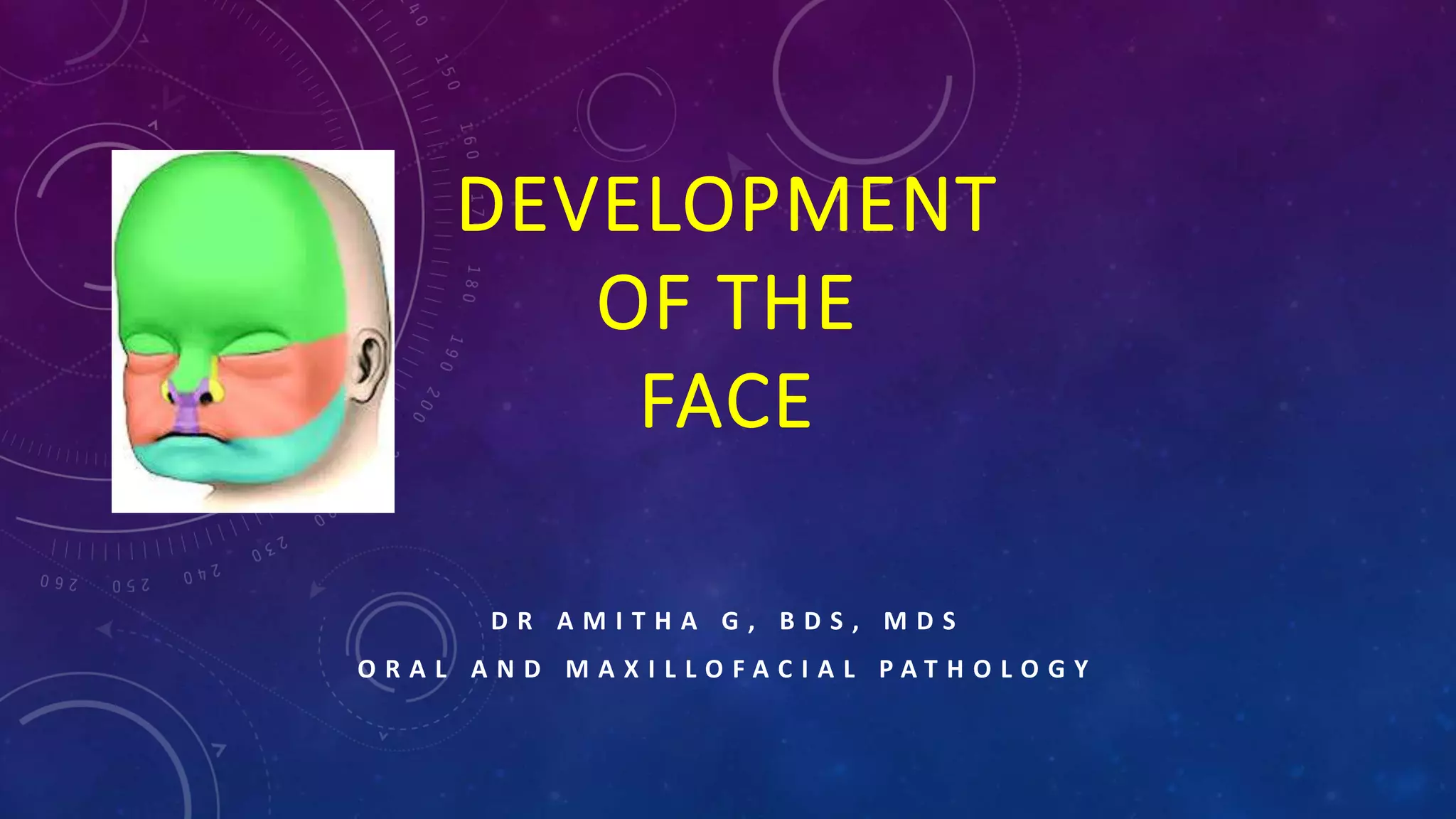
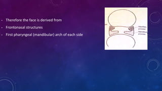
DEVELOPMENT
OF THE
FACE
D R A M I T H A G , B D S , M D S
O R A L A N D M A X I L L O F A C I A L P A T H O L O G Y
Face develops in humans between 4th – 10th
week of intrauterine life.
PRENATAL GROWTH OF THE MAXILLA
4th week of intrauterine life
- Formation of the head fold
- Following which the developing brain and the pericardium form 2 prominent bulges on
the ventral aspect of the embryo.
- The 2 bulges are separated from each other by  a shallow depression called
stomatoedum (corresponding to the primitive mouth).
- Floor of the stomatodeum is formed by the Buccopharyngeal membrane, which
separates the stomatodeum from the foregut.
- Soon, mesoderm covering the developing
forebrain proliferates, and forms a
downward projection that overlaps the
upper part of the stomatodeum – this
downward projection is called frontonasal
process.
- During the same time (4th week of intrauterine life) five
branchial arches form in the region of the future head and
neck region
- The first branchial arch is called – mandibular arch and play
an important role in the development of the nasomaxillary
region.
- The mandibular arch forms the lateral wall of the
stomatodeum.
- This arch gives off a bud from its dorsal end – this is called
maxillary process (4th week).
- It grows ventromedially cranial to the main part of the arch
- Main part of the arch is now called mandibular process.
- Therefore the face is derived from
- Frontonasal structures
- First pharyngeal (mandibular) arch of each side
5th week of intrauterine life
- The ectoderm overlying the frontonasal process soon shows bilateral
localized thickenings that are situated a little above (caudal or
raustral to) the stomatodeum. These are the nasal placodes or
olfactory placodes.
- The formation of these placodes is induced by the underlying
forebrain.
- The placodes soon sink below the surface to form the nasal pits.
- The nasal pits are continuous with the stomatodeum below.
- The edges of each pit are raised above the surface
- Medial raised edge is called – medial nasal process.
- Lateral raised edge – called lateral nasal process
- Now, all the primordial for the formation of lip and primary
palate are present
- Medial nasal process
- Lateral nasal formed
- Maxillary process
DEVELOPMENT OF UPPER LIP
- Maxillary process enlarges
- Maxillary process now grows medially and approaches the
lateral and medial nasal processes but remains separated
from them by distinct grooves (naso-optic groove and
bucco nasal groove)
- From each furrow or groove  a solid ectodermal rod of
cells sink below the surface and canalizes to form
nasolacrimal duct.
- Medial growth of the maxillary process further fuses
first with the lateral nasal process and then medial
nasal process.
- The medial and lateral nasal processes also fuse with
each other.
- This way the nasal pits  now called external nares
are cut off from the stomatodeum
- The maxillary process undergoes considerable growth
 therefore, pushes the medial nasal process towards
the midline, where it merges with its anatomic
counterpart from the opposite side.
- At the same time the frontonasal process becomes
much narrower from side to side, with the result that
the 2 external nares come close to each other.
6th week of intrauterine life
- 2 theories have been proposed for the continued development of the upper lip beyond the
6th week.
1st theory
- Mesenchyme of the maxillary process entirely overgrows the mesenchyme of the medial
nasal process to meet in the midline and thus contribute all the tissue for the upper lip.
- This is based on the fact
 Maxillary process  supplied by maxillary nerve
 Fronto nasal process  by opthalmic nerve
 Fully formed upper lip  supplied by infraorbital branch of the maxillary nerve)
2nd theory
- Maxillary process merely meets the medial nasal process without any overgrowth or
mesenchymal invasion.
- Therefore the middle third of the upper lip is derived from the merged medial nasal process
of frontonasal process.
- Histological evidence favours this
Mouth
- Stomatodeum is now bounded above by the Upper lip which is derived as follows
o Mesoderm of the lateral part of the lip is formed from the maxillary process.
o The overlying skin is derived from ectoderm covering this process
o Mesoderm of the median part of the lips (philtrum) is formed from the frontonasal
process.
o Ectoderm of the maxillary process overgrows this mesoderm to meet that of the opposite
maxillary process in the midline
o As a result skin of the entire upper lip is innervated by the maxillary nerves.
o Muscles of the face (including the lips)  are derived from the mesoderm of the second
branchial arch and are therefore supplied by facial nerve.
Development of lower lip
- At 6th week of embryo
- The mandibular process of the 2 sides grow towards each
other and fuse in the midline
- They form the lower margin of the stomatodeum.
- The fused mandibular process gives rise to lower lip and
the lower jaw
Development of nose
- Nose receives contributions from
 Frontonasal process
 Medial nasal process
 Lateral nasal process
- External nares are formed when the nasal pits are cut off from the stomatodeum by the fusion
of the maxillary process with medial nasal process.
- The external nares gradually approach each other as the maxillary process grows  pushing
the frontonasal process towards each other.
- Deeper part of the frontonasal process  forms nasal septum
- Mesoderm becomes heaped up in the median plane to form the prominence of the nose.
- Simultaneously a groove appear between the region of the nose and the bulging forebrain
(now called forehead)
- As the nose becomes prominent  the external nares comers to open downeards instead
of forwards.
- The external form of the nose is thus formed.
DEVELOPMENT OF CHEEKS
- After formation of the upper and lower lips, the stomatodeum (now called mouth) is very
broad.
- In its lateral part it is bounded
 Above by the maxillary process
 Below by the mandibular process
- These process undergo progressive fusion with each other to form cheeks
- The maxillary process fuses with the lateral nasal process  in the region of the lip + extends
from the stomatodeum to medial amgle of the developing eye this line of fusion is marked
by a groove called the naso optic furrow or nasolacrimal sulcus
- A strip of ectoderm becomes buried along this furrow and gives rise to the nasolacrimal duct.
POST NATAL GROWTH OF THE MAXILLA
- Growth of the nasomaxillary complex is produced by the following mechanisms
o Displacement
o Growth of the sutures
o Surface modeling
Displacement
- 2 types of displacement
 Primary displacement (bone is displaced by its own enlargement)
 Primary displacement of the maxilla  in a forwards direction
 This occurs by growth of the maxillary tuberosity in a posterior direction
 This results in the whole maxilla being carried anteriorly
 The amount of forwards displacement = amount of posterior lengthening
 Secondary or passive displacement (bone is not displaced by its own enlargement, rather a
passive displacement by the growth of the cranial base)
 Maxilla is attached to the cranial base by means of a number of sutures
 Therefore when the cranial base grows  produces a direct effect on the nasomaxillary
complex
 Cranial base grows  Middle cranial fossa grows anteriorly  therefore the passive
pressure produced by the cranial base  pushes the nasomaxillary complex  downwards
and forwards.
Growth at the sutures
- Maxilla is connected to the cranium and cranial base by a number of sutures
 Fronto nasal suture
 Fronto maxillary suture
 Zygomaticotemporal suture
 Zygomatico maxillary suture
 Pterygo palatine suture
- These sutures are oblique and almost parallel to each other
- This allows the downward and forward repositioning of the maxilla as growth occurs at
these sutures.
- Tension related bone formation occurs at sutures.
- Growth of the surrounding soft tissues  carries the maxilla downeards and forward 
leading to opening up of space at the sutural attachements  new bone is now formed on
either side of the suture  increasing the overall size of the bones on either side.
Surface remodeling
- Remodeling of bone by bone deposition and resorption occurs to bring about
 Increase in size
 Change in shape of the bone
 Change in functional relationship.
- Bone remodeling changes taking place in nasomaxillary complex are
 At the orbital rim
 Lateral surface  resorption
To compensate
 Medial surface of the rim  deposition
 External surface of the lateral rim  deposition
 At the Orbital floor
 Orbital floor faces superiorly, laterally and anteriorly
 Surface deposition occurs here and results in  growth in a superior, lateral and anterior direction
 At the maxillary tuberosity
 Bone deposition occurs along the posterior margin  causing the lengthening of the dental arch  and
enlargement of the antero posterior dimension of the entire maxillary body  helps accommodating the
erupting molars.
 At the nose
 Laterall wall of nose  resorption  leading to increase in size of the nasal cavity
 Floor of the nasal cavity
 Floor of the nasal cavity  resorption
To compensate
 Palatal aspect  deposition  therefore net downward shift occurs  leading to increase in maxillary height
 At the zygomatic bone
 Anterior surface  resorption
 Posterior surface  deposition
 The zygomatic bone  moves in a posterior direction.
 At zygomatic arch
 Lateral surface  deposition
 Medial surface  resorption
 Anterior nasal spine  bone deposition  becomes prominent
 Alveolar margins  As teeth erupts  deposition  increasing the maxillary height and
depth of the palate.
 Maxillary sinus  entire wall of the sinus except the mesial wall  undergoes resorption 
results in increase in size of maxillary antrum.
CLINICAL CONSIDERATIONS
- Since the formation of various parts of the face involves fusion of diverse components.
- Occasionally this fusion can be incomplete  give rise to various anomalies
HARE LIP
- Upper lip of the hare normally has a cleft
- Hence the term hare lip is used for defects of the lips.
- When 1 or both maxillary processes do not fuse with the
medial nasal process  this gives rise to defects in upper lip.
- This may vary in degree
- Unilateral or bilateral
b) Defective development of lowermost part of the
frontonasal process  give rise to a midline defect of ht
upper lip
c) When 2 mandibular process  do not fuse  with each
other  lower lip shows a defect in midline  the defect
usually extends to the jaw.
OBLIQUE FACIAL CLEFT
- Non fusion of the maxillary an dlateral nasal processes gives rise to a cleft running from the
medial angle of the eye to mouth.
- Nasolacrimal duct is also not formed.
MACROSTOMIA
- Inadequate fusion of mandibular and maxillary processes with each other  leads to
abnormally wide mouth  macrostomia
LATERAL FACIAL CLEFT
- Unilateral lack of fusion of mandibular and maxillary process  forms LATERAL FACIAL
CLEFT.
MICROSTOMIA
- Too much fusion of mandibular andmaxillary processes  may result  in small mouth
MANDIBULOFACIAL DYSOSTOSIS OR FIRST ARCH SYNDROME
- Entire first arch may remain underdeveloped on one or both sides, affecting
 Lower eyelid
 Maxilla
 Mandible
 External ear.
- Prominence of the cheek is absent
- Ear is displaced ventrally and caudally
HEMIFACIAL HYPERTROPHY
- One half of the face is overdeveloped
HEMIFACIAL ATROPHY
- One half of the face is under developed
RETROGNATHIA
- Mandible may be small compared to the rest of the face resulting in a receding chin.
HYPERTELORISM
- Eyes may be widely separated
PARAMEDIAN OR COMMISURAL PITS
- Lips showing congenital pits or fistula
DOUBLE LIP
- Lips are double
CONGENITAL TUMORS
- May be present in relation to the face.
ANOMALIES OF NOSE
Nose may be bifid
One half of nose is absent
Proboscis – nose forms a cylindrical projection jutting out from just below the forehead.
This anomaly may sometimes affect only one half of the nose and usually associated with
fusion of the eyes  Cyclops
THANK YOU

Development of Face

  • 1.
    DEVELOPMENT OF THE FACE D RA M I T H A G , B D S , M D S O R A L A N D M A X I L L O F A C I A L P A T H O L O G Y
  • 2.
    Face develops inhumans between 4th – 10th week of intrauterine life.
  • 3.
    PRENATAL GROWTH OFTHE MAXILLA 4th week of intrauterine life - Formation of the head fold - Following which the developing brain and the pericardium form 2 prominent bulges on the ventral aspect of the embryo. - The 2 bulges are separated from each other by  a shallow depression called stomatoedum (corresponding to the primitive mouth). - Floor of the stomatodeum is formed by the Buccopharyngeal membrane, which separates the stomatodeum from the foregut.
  • 4.
    - Soon, mesodermcovering the developing forebrain proliferates, and forms a downward projection that overlaps the upper part of the stomatodeum – this downward projection is called frontonasal process.
  • 5.
    - During thesame time (4th week of intrauterine life) five branchial arches form in the region of the future head and neck region - The first branchial arch is called – mandibular arch and play an important role in the development of the nasomaxillary region. - The mandibular arch forms the lateral wall of the stomatodeum. - This arch gives off a bud from its dorsal end – this is called maxillary process (4th week). - It grows ventromedially cranial to the main part of the arch - Main part of the arch is now called mandibular process.
  • 6.
    - Therefore theface is derived from - Frontonasal structures - First pharyngeal (mandibular) arch of each side
  • 7.
    5th week ofintrauterine life - The ectoderm overlying the frontonasal process soon shows bilateral localized thickenings that are situated a little above (caudal or raustral to) the stomatodeum. These are the nasal placodes or olfactory placodes. - The formation of these placodes is induced by the underlying forebrain. - The placodes soon sink below the surface to form the nasal pits. - The nasal pits are continuous with the stomatodeum below. - The edges of each pit are raised above the surface - Medial raised edge is called – medial nasal process. - Lateral raised edge – called lateral nasal process
  • 8.
    - Now, allthe primordial for the formation of lip and primary palate are present - Medial nasal process - Lateral nasal formed - Maxillary process
  • 9.
    DEVELOPMENT OF UPPERLIP - Maxillary process enlarges - Maxillary process now grows medially and approaches the lateral and medial nasal processes but remains separated from them by distinct grooves (naso-optic groove and bucco nasal groove) - From each furrow or groove  a solid ectodermal rod of cells sink below the surface and canalizes to form nasolacrimal duct.
  • 10.
    - Medial growthof the maxillary process further fuses first with the lateral nasal process and then medial nasal process. - The medial and lateral nasal processes also fuse with each other. - This way the nasal pits  now called external nares are cut off from the stomatodeum
  • 11.
    - The maxillaryprocess undergoes considerable growth  therefore, pushes the medial nasal process towards the midline, where it merges with its anatomic counterpart from the opposite side. - At the same time the frontonasal process becomes much narrower from side to side, with the result that the 2 external nares come close to each other.
  • 12.
    6th week ofintrauterine life - 2 theories have been proposed for the continued development of the upper lip beyond the 6th week. 1st theory - Mesenchyme of the maxillary process entirely overgrows the mesenchyme of the medial nasal process to meet in the midline and thus contribute all the tissue for the upper lip. - This is based on the fact  Maxillary process  supplied by maxillary nerve  Fronto nasal process  by opthalmic nerve  Fully formed upper lip  supplied by infraorbital branch of the maxillary nerve)
  • 13.
    2nd theory - Maxillaryprocess merely meets the medial nasal process without any overgrowth or mesenchymal invasion. - Therefore the middle third of the upper lip is derived from the merged medial nasal process of frontonasal process. - Histological evidence favours this
  • 14.
    Mouth - Stomatodeum isnow bounded above by the Upper lip which is derived as follows o Mesoderm of the lateral part of the lip is formed from the maxillary process. o The overlying skin is derived from ectoderm covering this process o Mesoderm of the median part of the lips (philtrum) is formed from the frontonasal process. o Ectoderm of the maxillary process overgrows this mesoderm to meet that of the opposite maxillary process in the midline o As a result skin of the entire upper lip is innervated by the maxillary nerves. o Muscles of the face (including the lips)  are derived from the mesoderm of the second branchial arch and are therefore supplied by facial nerve.
  • 16.
    Development of lowerlip - At 6th week of embryo - The mandibular process of the 2 sides grow towards each other and fuse in the midline - They form the lower margin of the stomatodeum. - The fused mandibular process gives rise to lower lip and the lower jaw
  • 17.
    Development of nose -Nose receives contributions from  Frontonasal process  Medial nasal process  Lateral nasal process - External nares are formed when the nasal pits are cut off from the stomatodeum by the fusion of the maxillary process with medial nasal process. - The external nares gradually approach each other as the maxillary process grows  pushing the frontonasal process towards each other.
  • 18.
    - Deeper partof the frontonasal process  forms nasal septum - Mesoderm becomes heaped up in the median plane to form the prominence of the nose. - Simultaneously a groove appear between the region of the nose and the bulging forebrain (now called forehead) - As the nose becomes prominent  the external nares comers to open downeards instead of forwards. - The external form of the nose is thus formed.
  • 19.
    DEVELOPMENT OF CHEEKS -After formation of the upper and lower lips, the stomatodeum (now called mouth) is very broad. - In its lateral part it is bounded  Above by the maxillary process  Below by the mandibular process - These process undergo progressive fusion with each other to form cheeks - The maxillary process fuses with the lateral nasal process  in the region of the lip + extends from the stomatodeum to medial amgle of the developing eye this line of fusion is marked by a groove called the naso optic furrow or nasolacrimal sulcus - A strip of ectoderm becomes buried along this furrow and gives rise to the nasolacrimal duct.
  • 20.
    POST NATAL GROWTHOF THE MAXILLA - Growth of the nasomaxillary complex is produced by the following mechanisms o Displacement o Growth of the sutures o Surface modeling Displacement - 2 types of displacement  Primary displacement (bone is displaced by its own enlargement)  Primary displacement of the maxilla  in a forwards direction  This occurs by growth of the maxillary tuberosity in a posterior direction  This results in the whole maxilla being carried anteriorly  The amount of forwards displacement = amount of posterior lengthening
  • 21.
     Secondary orpassive displacement (bone is not displaced by its own enlargement, rather a passive displacement by the growth of the cranial base)  Maxilla is attached to the cranial base by means of a number of sutures  Therefore when the cranial base grows  produces a direct effect on the nasomaxillary complex  Cranial base grows  Middle cranial fossa grows anteriorly  therefore the passive pressure produced by the cranial base  pushes the nasomaxillary complex  downwards and forwards.
  • 22.
    Growth at thesutures - Maxilla is connected to the cranium and cranial base by a number of sutures  Fronto nasal suture  Fronto maxillary suture  Zygomaticotemporal suture  Zygomatico maxillary suture  Pterygo palatine suture
  • 23.
    - These suturesare oblique and almost parallel to each other - This allows the downward and forward repositioning of the maxilla as growth occurs at these sutures. - Tension related bone formation occurs at sutures. - Growth of the surrounding soft tissues  carries the maxilla downeards and forward  leading to opening up of space at the sutural attachements  new bone is now formed on either side of the suture  increasing the overall size of the bones on either side.
  • 24.
    Surface remodeling - Remodelingof bone by bone deposition and resorption occurs to bring about  Increase in size  Change in shape of the bone  Change in functional relationship. - Bone remodeling changes taking place in nasomaxillary complex are  At the orbital rim  Lateral surface  resorption To compensate  Medial surface of the rim  deposition  External surface of the lateral rim  deposition
  • 25.
     At theOrbital floor  Orbital floor faces superiorly, laterally and anteriorly  Surface deposition occurs here and results in  growth in a superior, lateral and anterior direction  At the maxillary tuberosity  Bone deposition occurs along the posterior margin  causing the lengthening of the dental arch  and enlargement of the antero posterior dimension of the entire maxillary body  helps accommodating the erupting molars.  At the nose  Laterall wall of nose  resorption  leading to increase in size of the nasal cavity  Floor of the nasal cavity  Floor of the nasal cavity  resorption To compensate  Palatal aspect  deposition  therefore net downward shift occurs  leading to increase in maxillary height
  • 26.
     At thezygomatic bone  Anterior surface  resorption  Posterior surface  deposition  The zygomatic bone  moves in a posterior direction.  At zygomatic arch  Lateral surface  deposition  Medial surface  resorption  Anterior nasal spine  bone deposition  becomes prominent  Alveolar margins  As teeth erupts  deposition  increasing the maxillary height and depth of the palate.  Maxillary sinus  entire wall of the sinus except the mesial wall  undergoes resorption  results in increase in size of maxillary antrum.
  • 27.
    CLINICAL CONSIDERATIONS - Sincethe formation of various parts of the face involves fusion of diverse components. - Occasionally this fusion can be incomplete  give rise to various anomalies
  • 28.
    HARE LIP - Upperlip of the hare normally has a cleft - Hence the term hare lip is used for defects of the lips. - When 1 or both maxillary processes do not fuse with the medial nasal process  this gives rise to defects in upper lip. - This may vary in degree - Unilateral or bilateral
  • 29.
    b) Defective developmentof lowermost part of the frontonasal process  give rise to a midline defect of ht upper lip c) When 2 mandibular process  do not fuse  with each other  lower lip shows a defect in midline  the defect usually extends to the jaw.
  • 30.
    OBLIQUE FACIAL CLEFT -Non fusion of the maxillary an dlateral nasal processes gives rise to a cleft running from the medial angle of the eye to mouth. - Nasolacrimal duct is also not formed.
  • 31.
    MACROSTOMIA - Inadequate fusionof mandibular and maxillary processes with each other  leads to abnormally wide mouth  macrostomia LATERAL FACIAL CLEFT - Unilateral lack of fusion of mandibular and maxillary process  forms LATERAL FACIAL CLEFT. MICROSTOMIA - Too much fusion of mandibular andmaxillary processes  may result  in small mouth
  • 32.
    MANDIBULOFACIAL DYSOSTOSIS ORFIRST ARCH SYNDROME - Entire first arch may remain underdeveloped on one or both sides, affecting  Lower eyelid  Maxilla  Mandible  External ear. - Prominence of the cheek is absent - Ear is displaced ventrally and caudally
  • 33.
    HEMIFACIAL HYPERTROPHY - Onehalf of the face is overdeveloped HEMIFACIAL ATROPHY - One half of the face is under developed RETROGNATHIA - Mandible may be small compared to the rest of the face resulting in a receding chin. HYPERTELORISM - Eyes may be widely separated PARAMEDIAN OR COMMISURAL PITS - Lips showing congenital pits or fistula DOUBLE LIP - Lips are double
  • 34.
    CONGENITAL TUMORS - Maybe present in relation to the face. ANOMALIES OF NOSE Nose may be bifid One half of nose is absent Proboscis – nose forms a cylindrical projection jutting out from just below the forehead. This anomaly may sometimes affect only one half of the nose and usually associated with fusion of the eyes  Cyclops
  • 35.