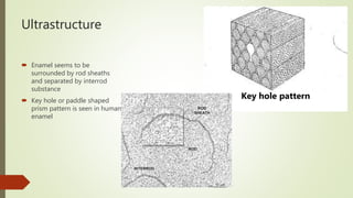
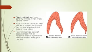
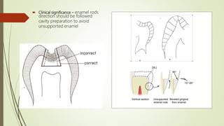
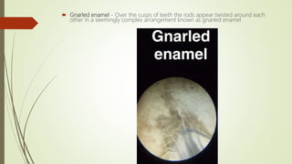
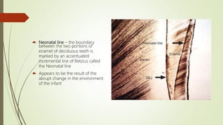
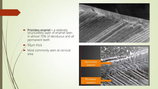
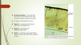
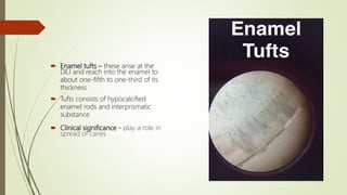
This document provides information on the physical, chemical, structural, and ultrastructural properties of enamel. It discusses how enamel forms a protective covering on the tooth and consists primarily of calcium hydroxyapatite. Enamel's structure includes enamel rods that run from the dentin-enamel junction to the outer surface. It also describes features like Hunter-Schreger bands, incremental lines of Retzius, and surface structures such as enamel tufts. The document outlines the life cycle of ameloblasts and process of amelogenesis, and discusses implications for clinical applications like fluoridation and acid etching.
2. Physical properties
Forms a protective covering over
the tooth structure
Variable thickness seen over entire
surface of crown
Thins down to almost knife edge at
cervical area of crown
Yellowish white to grayish white in
colour
Semi permeable in nature i.e. allows
passage of certain molecules
3. Specific gravity 2.8
Compressive strength 384Mpa
Modulus of elasticity 84Gpa
Knoop hardness number 350-430
KHN
Tensile strength 10Mpa
Co-efficient of thermal expansion
11.4
4. Chemical properties
Inorganic material 96% - Calcium
Hydroxyapatite Ca10(PO4)6(OH4)2
Organic material 4% - Proteins (
Amelogenins, Non Amelogenins, Enamelin,
Ameloblastin, Tuftelin) lipids
Water
5. Structure
Basic structure consists of ENAMEL
RODS or PRISMS
Diameter of rods averages 4µm
Prisms are cylindrical in longitudinal
section
Number of enamel rods ranges
from 5 million to 12 million
6. Enamel rods move from DEJ to
the outer surface of tooth
Enamel rods are obliquely
placed and move in a wavy
course
This leads to increased length
of rods as compared to
thickness of enamel
Enamel rods resemble fish
scale appearance
7. Ultrastructure
Enamel seems to be
surrounded by rod sheaths
and separated by interrod
substance
Key hole or paddle shaped
prism pattern is seen in human
enamel
Key hole pattern
8. Direction of Rods – rods are
generally oriented at right angles
dentinal surface
In deciduous and permanent teeth
rods are in oblique direction until
they become almost vertical at
tips and incisal edges
However in cervical region of
deciduous teeth rods are
horizontal, whereas in permanent
teeth the shift to a more apical
direction
9. Clinical significance – enamel rods
direction should be followed
cavity preparation to avoid
unsupported enamel
10. Gnarled enamel - Over the cusps of teeth the rods appear twisted around each
other in a seemingly complex arrangement known as gnarled enamel
11. Clinical significance - This enamel is not subject to cleavage as regular enamel
This enamel does not yield readily to pressure of hand cutting instruments
12. Hunter-Schreger bands – these are
alternating bands of dark and light
strips of varying widths
Change in the direction of rods is
responsible for the appearance of
Hunter-Schreger bands
Seen in longitudinal ground section
under oblique reflected light
Dark bands are called Diazones and
light bands are called Parazones
Clinical significance - Distribute and
dissipate impact forces
13. Incremental Lines Of Retzius –
appear as brownish lines in ground
section of enamel
Illustrate the incremental
apposition of layers of enamel
during crown formation
Appear as concentric circles in
transverse section
14. Neonatal line – the boundary
between the two portions of
enamel of deciduous teeth is
marked by an accentuated
incremental line of Retzius called
the Neonatal line
Appears to be the result of the
abrupt change in the environment
of the infant
16. Prismless enamel – a relatively
structureless layer of enamel seen
in almost 70% of deciduous and all
permanent teeth
30µm thick
Most commonly seen at cervical
area
17. Enamel cuticle – a delicate membrane called Nasmyth’s Membrane or the
Primary Enamel Cuticle covers the crown of a newly erupted tooth
It is almost as a basal lamina found below most epithelium
Protects the surface of enamel from the resorptive activity of adjacent vascular
tissue prior to eruption of teeth
18. Enamel lamellae – thin leaf like
structures that extend from the
enamel surface towards the DEJ
3 types of lamellae are seen
Type A – composed of poorly
calcified rod segments
Type B – consisting of
degenerated cells
Type C – arising in erupted teeth
where cracks are filled with organic
matter
19. Enamel tufts – these arise at the
DEJ and reach into the enamel to
about one-fifth to one-third of its
thickness
Tufts consists of hypocalcified
enamel rods and interprismatic
substance
Clinical significance - play a role in
spread of caries
20. Dentinoenamel
Junction
The surface of dentin at the
dentinoenamel junction is pitted.
into the shallow depressions of the
dentin fit rounded projections of
the enamel
Scalloped appearance
More pronounced in occlusal area
21. Life Cycle of
Ameloblasts
Life span of ameloblast cells can be
divided into six stages
1. Morphogenic stage
2. Organizing stage
3. Formative
4. Maturative stage
5. Protective stage
6. Desmolytic stage
22. Amelogenesis
Amelogenesis involves 2 processes
Enamel matrix formation
Mineralization
It occurs during the advanced bell stage
Reciprocal epithelial-mesenchymal interactions lead to differentiation of
odontoblasts and ameloblasts
23. Age changes in
enamel
Attrition
Wear of occlusal and proximal contacts
Decreased permeability of enamel
Discoloration
24. Clinical Implications
Fluoridation - fluoride ion incorporated into or adsorbed on the hydroxyapatite
crystal becomes more resistant to acid dissolution
It explains the role of fluoride in caries prevention
Presence of fluoride enhances chemical reactions that lead to the precipitation of
calcium phosphate
Acid Etching - Acid etching of the enamel surface, or enamel conditioning is an
important technique in clinical practice
Fissure sealants, bonding of restorative materials to enamel, and cementing of
orthodontic brackets to tooth surfaces involve acid etching