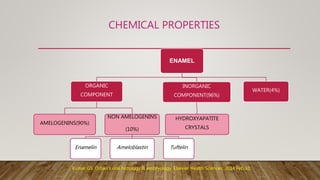
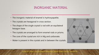
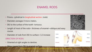
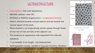
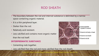
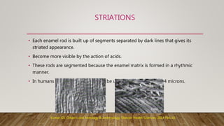
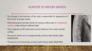
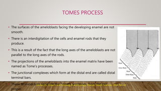
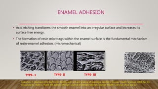
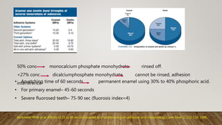
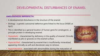
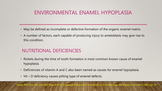
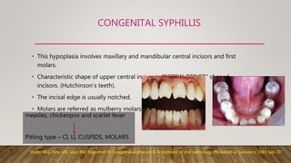
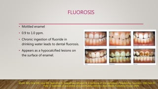
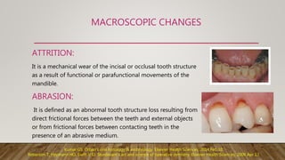
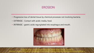
SlideShare a Scribd company logo
ENAMEL
Presented by,
Swapnika.G.
(1 MDS)
Conservative Dentistry and Endodontics
• Introduction
• Functions of enamel
• Physical and chemical properties
• Surface structures of enamel
• Life cycle of ameloblasts and Amelogenesis
• Enamel adhesion
• Effect of bleaching on enamel
• Developmental disturbances of enamel
• Age changes and clinical considerations
• References
CONTENTS
INTRODUCTION
• Enamel is a protective covering of variable thickness over the entire surface of the
crown.
• Enamel is the hardest calcified tissue in the human body.
• The shape and contour of the cusps receive their final modeling in the enamel.
• The cells that are responsible for the formation of enamel (Ameloblasts) are lost as
the tooth erupts into the oral cavity, and hence can’t renew itself.
• To compensate this enamel undergoes complex structural organization and high
degree of mineralization.
FUNCTIONS OF ENAMEL
• Transfers occlusal load to the biomechanical unit.
• It prevents premature wearing of dentine.
• Prevents hypersensitivity of dentine.
• Protects pulp dentine complex from thermal, chemical and electrical substances.
• Provides aesthetics.
• Helps in maintain contacts, contours and vertical dimension.
PHYSICAL PROPERTIES
• Hardest calcified tissue- high content of mineral salts and crystalline arrangement.
• Modulus of elasticity and hardness- more at the surface than at the DEJ.
• Maximum thickness: 2-2.5mm
• Semi-permeable membrane.
• Specific gravity – 2.8
• Translucency- degree of calcification and homogenesity.
• Color : ranges from light yellow to bluish gray.
• Incisal areas may have a bluish tinge where the thin edge consists of only a double layer of
enamel and cervical areas show yellowish tinge because of thinness of the enamel.
Kumar GS. Orban's oral histology & embryology. Elsevier Health Sciences; 2014 Feb 10.
CHEMICAL PROPERTIES
ENAMEL
ORGANIC
COMPONENT
AMELOGENINS(90%)
NON AMELOGENINS
(10%)
Enamelin Ameloblastin Tuftelin
INORGANIC
COMPONENT(96%)
HYDROXYAPATITE
CRYSTALS
WATER(4%)
Kumar GS. Orban's oral histology & embryology. Elsevier Health Sciences; 2014 Feb 10.
INORGANIC MATERIAL
• The inorganic material of enamel is hydroxyapatite.
• The crystals are hexagonal in cross section.
• The shape of the single crystal is rod with an equilateral
hexagon base.
• The crystals are arranged to form enamel rods or prisms.
• The core of the crystal are rich in Mg and carbonate.
• Water is present in the crystals and in between the crystals.
Kumar GS. Orban's oral histology & embryology. Elsevier Health Sciences; 2014 Feb 10.
ENAMEL RODS
• Prisms- cylindrical in longitudinal section. (rods)
• Diameter averages 4 micro meters.
• DEJ to the surface of the tooth- tortuous.
• Length of most of the rods> thickness of enamel—oblique and wavy
course.
• Diameter of rods from DEJ to surface= 1:2 increase
DIRECTION OF RODS:
• Oriented at right angles to dentine.
• Wavy course from DEJ to enamel surface.
Kumar GS. Orban's oral histology & embryology. Elsevier Health Sciences; 2014 Feb 10.
• Cross section—fish scale appearance.
• ARCADE outlines—near DEJ.
• KEYHOLE or PADDLE shaped prism— Longitudinal Sections
• Head is directed towards occlusal aspects and tail towards the
cervical aspect of the tooth.
• When enamel is cut longitudinally section passes through heads
of one row of rods and tails of the adjacent row.
• This produces an appearance rods separated from interrod
substance.
• 5 um breadth, 9 um length– rod measurement.
• Hydroxyapatite crystals– parallel to long axis of prisms, deviation-
40 degrees.
ULTRASTRUCTURE
Kumar GS. Orban's oral histology & embryology. Elsevier Health Sciences; 2014 Feb 10.
ROD SHEATH
• The boundary between the rod and interrod substance is delimited by a narrow
space containing organic material.
• It is a thin peripheral layer.
• Darker than the rod.
• Relatively acid resistant.
• Less calcified and contains more organic matter
than the rod itself.
INTERPRISMATIC SUBSTANCE:
• Cementing rods together.
• Less calcified than the rod and more calcified than the rod sheath.
Kumar GS. Orban's oral histology & embryology. Elsevier Health Sciences; 2014 Feb 10.
STRIATIONS
• Each enamel rod is built up of segments separated by dark lines that gives its
striated appearance.
• Become more visible by the action of acids.
• These rods are segmented because the enamel matrix is formed in a rhythmic
manner.
• In humans these segments seem to be uniform length of about 4 microns.
Kumar GS. Orban's oral histology & embryology. Elsevier Health Sciences; 2014 Feb 10.
HUNTER SCHREGER BANDS
• The change in the direction of the rods is responsible for appearance of
the Hunter Schreger bands.
• Alternating dark and light bands of varying widths seen in longitudinal
sections under oblique reflected light.
• They originate at DEJ and ends at some distance from outer enamel
surface.
• The prisms which are cut longitudinally produce dark bands called
PARAZONES.
• The prisms cut transversely produce light bands called DIAZONES.
• The angle between these two is 40 degrees.
Kumar GS. Orban's oral histology & embryology. Elsevier Health Sciences; 2014 Feb 10.
INCREMENTAL LINES OF RETZIUS AND
PERIKYMATA
• Appears as brownish bands in ground sections.
• They illustrate the incremental pattern of the enamel, that is, the
successive apposition of layers of enamel during formation of the
crown.
• In longitudinal sections– surround tip of dentin.
• In transverse sections— concentric circles.
• Perikymata are the transverse wave like grooves, believed to be the
external manifestation of the striae of Retzius.
• They are continuous around a tooth and usually lie parallel to each
other and to CEJ.
Kumar GS. Orban's oral histology & embryology. Elsevier Health Sciences; 2014 Feb 10.
PRISMLESS ENAMEL OTHER SURFACE
STRUCTURES
• Structureless enamel– 30um
• All deciduous and 70% permanent
teeth.
• Cervical areas of teeth.
• Hydroxyapetite crystals are parelllel
to one another and perpendicular
to striae of Retzius.
• More mineralized than enamel
beneath.
• Pits- 1-1.5um- ends of ameloblasts.
• Enamel caps-small elevations-10-
15um- enamel mineralization on non-
mineralizable debris.
• Enamel brochs-Larger elevations.
Kumar GS. Orban's oral histology & embryology. Elsevier Health Sciences; 2014 Feb 10.
ENAMEL CUTICLE NEONATAL LINE
• A delicate membrane called Nasmyth’s
membrane.
• Primary enamel cuticle.
• Covers entire crown of newly erupted tooth.
• Secreted by ameloblasts when enamel
formation is completed.
• Basal lamina found beneath epithelia.
• Soon disappears due to masticatory forces.
• Erupted enamel- pellicle- precipitate of
salivary proteins.
• Accentuated incremental
line of Retzius.
Kumar GS. Orban's oral histology & embryology. Elsevier Health Sciences; 2014 Feb 10.
ENAMEL LAMELLAE ENAMEL TUFTS
• Thin leaf like structures that extend from enamel
surface towards DEJ.
• Sometimes penetrates dentine.
• More organic less mineral content.
• Seen in transverse ground sections.
Kumar GS. Orban's oral histology & embryology. Elsevier Health Sciences; 2014 Feb 10.
• Arise at DEJ and reach into enamel to
about 1/5 to 1/3 of its thickness.
• Ground sections– grass like.
• Seen in transverse sections.
ENAMEL SPINDLES
• Odontoblastic processes that pass across the DEJ into
enamel.
• Since many are thickened at their end they have been
termed as spindles.
• Direction of odontoblastic process and spindles
corresponds to the original direction of ameloblasts.
• Before hard substances were formed.
• Direction- at right angles to dentin.
• Ground sections– dark in transmitted light.
Kumar GS. Orban's oral histology & embryology. Elsevier Health Sciences; 2014 Feb 10.
GNARLED ENAMEL DEJ
• Over the cusps of teeth the rods
appear twisted around each other in a
seemingly complex arrangement and
this optical appearance known as
gnarled enamel.
GNARLED ENAMEL
Kumar GS. Orban's oral histology & embryology. Elsevier Health Sciences; 2014 Feb 10.
LIFE CYCLE OF AMELOBLASTS
MORPHOGENIC STAGE
ORGANISING STAGE
FORMATIVE STAGE
MATURATIVE STAGE
PROTECTIVE STAGE
DESMOLYTIC STAGE
POST
SECRETORY
PRE
SECRETORY
SSECRETORY
Kumar GS. Orban's oral histology & embryology. Elsevier Health Sciences; 2014 Feb 10.
MORPHOGENIC STAGE
• Before ameloblasts are fully differentiated and produce
enamel, they interact with adjacent mesenchymal cells,
determining the shape of the DEJ.
• Cells are short and columnar, with large oval nuclei that
almost fill the cell body.
• During ameloblasts differentiation, terminal bars appear
concomitantly with migration of mitochondria to the
basal region of the cell.
Kumar GS. Orban's oral histology & embryology. Elsevier Health Sciences; 2014 Feb 10.
ORGANISING STAGE
• Characterized by change in the appearance of the cells of IEE.
• They become longer and the nucleus free zones at the distal ends of
the cells become almost as long as the proximal parts containing the
nuclei.
• Reversal of functional polarity takes place.
• Clear cell free zone between the IEE and dental papilla disappears.
• Thus epithelial cells come into close contact with the connective tissue
cells of the pulp., which differentiate into odontoblasts.
• During terminal phase of organizing stage dentine formation begins.
• First appearance of the dentine is the critical phase of the life cycle of
IEE.
Kumar GS. Orban's oral histology & embryology. Elsevier Health Sciences; 2014 Feb 10.
FORMATIVE STAGE
• Ameloblasts enter into formative stage after the first layer of
dentine is formed.
• Cells retain the same length and arrangement.
• Changes in the organization and number of cytoplasmic
organelles are related to initiation of SECRETION of enamel
matrix.
• Earliest change is development of blunt cell processes on
the ameloblast surface and penetrate the basal lamina and
enter predentine.
Kumar GS. Orban's oral histology & embryology. Elsevier Health Sciences; 2014 Feb 10.
MATURATIVE STAGE
• Full mineralization of enamel forms after most if the
thickness of the enamel matrix has been formed in the
occlusal parts of the crown and incisal areas.
• Ameloblasts are slightly reduced in length and closely attach
to enamel matrix.
• Ameloblasts display microvilli at their distal proximities and
cytoplasmic vacuoles containing material resembling
enamel matrix are present.
• These structures indicate absorptive function of these cells.
Kumar GS. Orban's oral histology & embryology. Elsevier Health Sciences; 2014 Feb 10.
PROTECTIVE STAGE
• The enamel has completely developed, and has fully
calcified, the Ameloblasts cease to be arranged in a well-
defined layer.
• Cells then form a stratified epithelial covering of enamel,
the so-called reduced enamel epithelium.
• The function of REE is that of protecting the mature
enamel by separating it from the connective tissue until
tooth erupts.
• If connective tissue comes in contact with the enamel,
anomalies may develop.
Kumar GS. Orban's oral histology & embryology. Elsevier Health Sciences; 2014 Feb 10.
DESMOLYTIC STAGE
• The reduced enamel epithelium proliferates and
seems to induce atrophy of the connective tissues
separating it from oral epithelium.
• Epithelial cells elaborate enzymes that are able to
destroy connective tissue fibers by desmolysis.
• Premature degeneration of the reduced enamel
epithelium may prevent the eruption of a tooth.
Kumar GS. Orban's oral histology & embryology. Elsevier Health Sciences; 2014 Feb 10.
AMELOGENESIS
Amelogenesis or enamel formation is a two step process.
Organic matrix formation
Mineralization
• Amelogenin a component of enamel matrix proteins, undergo extracellular
degradation by proteolytic enzymes and break into smaller low molecular weight
fragments.
• These are suggested to have specific function in regulating crystal growth.
Kumar GS. Orban's oral histology & embryology. Elsevier Health Sciences; 2014 Feb 10.
TOMES PROCESS
• The surfaces of the ameloblasts facing the developing enamel are not
smooth.
• There is an interdigitation of the cells and enamel rods that they
produce.
• This is a result of the fact that the long axes of the ameloblasts are not
parallel to the long axes of the rods.
• The projections of the ameloblasts into the enamel matrix have been
named as Tome’s processes.
• The junctional complexes which form at the distal end are called distal
terminal bars.
• These terminal bars separate the Tome’s processes from the cell
proper.
Kumar GS. Orban's oral histology & embryology. Elsevier Health Sciences; 2014 Feb 10.
ENAMEL ADHESION
• Acid etching transforms the smooth enamel into an irregular surface and increases its
surface free energy.
• The formation of resin microtags within the enamel surface is the fundamental mechanism
of resin-enamel adhesion. (micromechanical)
TYPE- 1 TYPE- II TYPE- III
Roberson T, Heymann HO, Swift Jr EJ. Sturdevant's art and science of operative dentistry. Elsevier Health Sciences; 2006 Apr 13.
Anusavice KJ, Shen C, Rawls HR, editors. Phillips' science of dental materials. Elsevier Health Sciences; 2012 Sep 27.
50% conc monocalcium phosphate monohydrate rinsed off.
<27% conc dicalciumphosphate monohydrate cannot be rinsed, adhesion
interference
Barkmeier WW, et al: Effects of 15 vs 60 second enamel acid conditioning on adhesion and morphology, Oper Dent 11:111-116, 1986.
• An etching time of 60 seconds permanent enamel using 30% to 40% phosphoric acid.
• For primary enamel– 45-60 seconds
• Severe fluorosed teeth– 75-90 sec (fluorosis index=4)
RETENTIVE FEATURES PLACED IN ENAMEL
• Partial bevel- for cast metal restorations
• Short bevel- for cast metal restorations
• Skirt preperations: Skirts are thin extensions
of the facial or lingual proximal margins of
the cast metal onlay that extend from the
primary flare to a termination just past the
transitional line angle of the tooth.
• Bevelling the composite restorations.
Roberson T, Heymann HO, Swift Jr EJ. Sturdevant's art and science of operative dentistry. Elsevier Health Sciences; 2006 Apr 13.
Marzouk MA, Simonton AL, Gross RD. Operative dentistry. Modern theory and practice, 1st ed. St Louise-Tokyo, Ishiyahu EuroAmerica Inc.
1985.
EFFECT OF BLEACHING ON ENAMEL
• In office extracoronal bleaching- gel with 25% to 38% H2O2.
• The light source can be a laser (e.g., argon, CO2), halogen, plasma
arc, or light-emitting diodes (LED).
• For professional at home bleaching- 3% to 7.5% H2O2 or 10 to 22%
carbamide peroxide, especially those with 10% carbamide peroxide.
• Several investigators reported alteration of enamel surfaces
associated with bleaching treatments, including shallow depression,
increased porosity, and slight erosion.
• Associated with products using acidic pre rinse or gels of low pH.
Ingle JI, Bakland LK, Baumgartner JC, editors. Ingle's endodontics 6. Pmph usa; 2008.
75 volunteers were distributed according to the bleaching technique (n=25):
(a) at-home: 10%HP (Opalescence GO) for 15 days of continuous use (1 hour per day);
(b) in-office: 40%HP (Opalescence Boost) in three clinical sessions (40 minutes each session);
(c) combined: one initial session with 40%HP, and the rest with 10%HP for 15 days of
continuous use.
Clinical evaluations and Ca and P concentration collections were obtained before, during
bleaching treatment, and 15 days after conclusion of treatment.
RESULTS:
• The in-office technique presented the lowest tooth sensitivity.
• All techniques resulted in lower Ca and P concentrations in enamel at each time point,
compared with the baseline concentrations.
• Calcium concentrations did not differ significantly among the treatments (P= 0.9360).
• Phosphorus concentration at the 8th day was higher for the in-office technique group (P<
0.05).
Dourado AP, Carlos NR, Amaral FL, França FM, Turssi CP, Basting RT. At-home, in-office and combined dental bleaching techniques
using hydrogen peroxide: Randomized clinical trial evaluation of effectiveness, clinical parameters and enamel mineral content.
American journal of dentistry. 2019 Jun;32(3):124-32.
TOOTH WEAR INDEX
DEVELOPMENTAL DISTURBANCES OF ENAMEL
AMELOGENESIS IMPERFECTA:
• A developmental disturbance in the structure of the enamel.
• Etiology : a genetic autosomal defective gene linked to the locus DXS85 at
Xp22.
• This is identified as a general location of human gene for amelogenin., a
principle protein in developing enamel.
• Hypoplastic : characterized by deficiency in the quality of enamel. Clinically
manifested as pits or grooves on the enamel surface.
• Hypocalcified : characterized by enamel that is insufficiently mineralized,
appearing clinically as soft and discolored, easy to remove.
• Hypomaturative : associated with abnormalities during the maturation of
enamel formation, giving the enamel an opaque and chalky appearance.Shafer WG, Hine MK, Levy BM, Rajendran R, Sivapathasundharam B. A textbook of oral pathology. Philadelphia: Saunders; 1983 Sep 20.
ENVIRONMENTAL ENAMEL HYPOPLASIA
• May be defined as incomplete or defective formation of the organic enamel matrix.
• A number of factors, each capable of producing injury to ameloblasts may give rise to
this condition.
• Rickets during the time of tooth formation is most common known cause of enamel
hypoplasia.
• Deficiencies of vitamin A and C also been named as causes for enamel hypoplasia.
• Vit – D deficiency causes pitting type of enamel defects.
NUTRITIONAL DEFICIENCIES
Shafer WG, Hine MK, Levy BM, Rajendran R, Sivapathasundharam B. A textbook of oral pathology. Philadelphia: Saunders; 1983 Sep 20.
CONGENITAL SYPHILLIS
• This hypoplasia involves maxillary and mandibular central incisors and first
molars.
• Characteristic shape of upper central incisor is “SCREW-DRIVER” shaped
incisors. (Hutchinson’s teeth).
• The incisal edge is usually notched.
• Molars are referred as mulberry molars.
Shafer WG, Hine MK, Levy BM, Rajendran R, Sivapathasundharam B. A textbook of oral pathology. Philadelphia: Saunders; 1983 Sep 20.
measles, chickenpox and scarlet fever
Pitting type – CI, LI, CUSPIDS, MOLARS
FLUOROSIS
• Mottled enamel
• 0.9 to 1.0 ppm.
• Chronic ingestion of fluoride in
drinking water leads to dental fluorosis.
• Appears as a hypocalcified lesions on
the surface of enamel.
Shafer WG, Hine MK, Levy BM, Rajendran R, Sivapathasundharam B. A textbook of oral pathology. Philadelphia: Saunders; 1983 Sep 20.
Peter S. Essentials of preventive and community dentistry. Arya (Medi) Publishing House; 2009.
AGE CHANGES AND CLINICAL CONSIDERATIONS
MICROSCOPIC CHANGES:
• Loss of Perikymata - generalized loss of rod ends
- more on buccal and lingual
- height of contour
- anterior teeth than posterior
• Localized increase in elements such as nitrogen and fluorine – darker color.
• Increase in the size of the crystal reduces permeability.
• Increase in the organic content of enamel.
Kumar GS. Orban's oral histology & embryology. Elsevier Health Sciences; 2014 Feb 10.
MACROSCOPIC CHANGES
ATTRITION:
It is a mechanical wear of the incisal or occlusal tooth structure
as a result of functional or parafunctional movements of the
mandible.
ABRASION:
It is defined as an abnormal tooth structure loss resulting from
direct frictional forces between the teeth and external objects
or from frictional forces between contacting teeth in the
presence of an abrasive medium.
Kumar GS. Orban's oral histology & embryology. Elsevier Health Sciences; 2014 Feb 10.
Roberson T, Heymann HO, Swift Jr EJ. Sturdevant's art and science of operative dentistry. Elsevier Health Sciences; 2006 Apr 13.
EROSION
• Progressive loss of dental tissue by chemical processes not involving bacteria.
• EXTRINSIC : Contact with acidic media, food.
• INTRINSIC : gastric acids regurgitated into esophagus and mouth.
Kumar GS. Orban's oral histology & embryology. Elsevier Health Sciences; 2014 Feb 10.
Roberson T, Heymann HO, Swift Jr EJ. Sturdevant's art and science of operative dentistry. Elsevier Health Sciences; 2006 Apr 13.
REFERENCES
• Kumar GS. Orban's oral histology & embryology. Elsevier Health Sciences; 2014 Feb 10.
• Ten Cate AR, Nanci A. Ten Cate's oral histology: development, structure, and function.
Elsevier; 2013.
• Shafer WG, Hine MK, Levy BM, Rajendran R, Sivapathasundharam B. A textbook of oral
pathology. Philadelphia: Saunders; 1983 Sep 20.
• Roberson T, Heymann HO, Swift Jr EJ. Sturdevant's art and science of operative
dentistry. Elsevier Health Sciences; 2006 Apr 13.
• Anusavice KJ, Shen C, Rawls HR, editors. Phillips' science of dental materials. Elsevier
Health Sciences; 2012 Sep 27.
• Phillips RW. Skinner's science of dental materials. Philadelphia: Saunders; 1973.
• Barkmeier WW, et al: Effects of 15 vs 60 second enamel acid conditioning on adhesion
and morphology, Oper Dent 11:111-116, 1986.
• Marzouk MA, Simonton AL, Gross RD. Operative dentistry. Modern theory and
practice, 1st ed. St Louise-Tokyo, Ishiyahu EuroAmerica Inc. 1985.
• Ingle JI, Bakland LK, Baumgartner JC, editors. Ingle's endodontics 6. Pmph usa; 2008.
• Peter S. Essentials of preventive and community dentistry. Arya (Medi) Publishing
House; 2009.
Enamel CONSERVATIVE DENTISTRY AND ENDODONTICS

More Related Content

What's hot

Enamel
EnamelEnamel
Dentinogenesis & histology of dentin
Dentinogenesis & histology of dentinDentinogenesis & histology of dentin
Dentinogenesis & histology of dentin
Hesham Dameer
 
Differences between primary and permanent dentition
Differences between primary and permanent dentitionDifferences between primary and permanent dentition
Differences between primary and permanent dentition
AkshMinhas
 
Dental Pulp
Dental PulpDental Pulp
Dental Pulp
drmadhubilla
 
Enamel
EnamelEnamel
cementum by dr. ishu singla.pptx
cementum by dr. ishu singla.pptxcementum by dr. ishu singla.pptx
cementum by dr. ishu singla.pptx
Dr. Ishu SINGLA
 
Pulp dentin complex
Pulp dentin complexPulp dentin complex
Pulp dentin complex
Upama Sishan
 
Enamel &amp; amelogenesis part 1
Enamel &amp; amelogenesis part 1Enamel &amp; amelogenesis part 1
Enamel &amp; amelogenesis part 1
dentyqueen
 
1st BDS Lecture on tooth Enamel
1st BDS Lecture on tooth Enamel1st BDS Lecture on tooth Enamel
1st BDS Lecture on tooth Enamel
varun surya
 
Dentin
DentinDentin
Enamel
EnamelEnamel
Enamel
liwen28
 
Cementum
Cementum Cementum
Cementum
Dr Gauri Kapila
 
Oral Mucosa
Oral MucosaOral Mucosa
Oral Mucosa
Claiddin Bangalisan
 
ENAMEL
 ENAMEL ENAMEL
ENAMEL
CmenonMenon
 
Cementum
CementumCementum
Cementum
benita regi
 
Anatomical structures of teeth
Anatomical structures of teethAnatomical structures of teeth
Anatomical structures of teeth
Jamil Kifayatullah
 
Histology of dentin
Histology of dentinHistology of dentin
Histology of dentin
Amin Abusallamah
 
Gingiva
GingivaGingiva
Gingiva
DrAtulKoundel
 
cementum
cementumcementum
cementum
Parth Thakkar
 
Tooth Dentin and dentinogenesis ppt
Tooth Dentin and dentinogenesis pptTooth Dentin and dentinogenesis ppt
Tooth Dentin and dentinogenesis ppt
madhusudhan reddy
 

What's hot (20)

Enamel
EnamelEnamel
Enamel
 
Dentinogenesis & histology of dentin
Dentinogenesis & histology of dentinDentinogenesis & histology of dentin
Dentinogenesis & histology of dentin
 
Differences between primary and permanent dentition
Differences between primary and permanent dentitionDifferences between primary and permanent dentition
Differences between primary and permanent dentition
 
Dental Pulp
Dental PulpDental Pulp
Dental Pulp
 
Enamel
EnamelEnamel
Enamel
 
cementum by dr. ishu singla.pptx
cementum by dr. ishu singla.pptxcementum by dr. ishu singla.pptx
cementum by dr. ishu singla.pptx
 
Pulp dentin complex
Pulp dentin complexPulp dentin complex
Pulp dentin complex
 
Enamel &amp; amelogenesis part 1
Enamel &amp; amelogenesis part 1Enamel &amp; amelogenesis part 1
Enamel &amp; amelogenesis part 1
 
1st BDS Lecture on tooth Enamel
1st BDS Lecture on tooth Enamel1st BDS Lecture on tooth Enamel
1st BDS Lecture on tooth Enamel
 
Dentin
DentinDentin
Dentin
 
Enamel
EnamelEnamel
Enamel
 
Cementum
Cementum Cementum
Cementum
 
Oral Mucosa
Oral MucosaOral Mucosa
Oral Mucosa
 
ENAMEL
 ENAMEL ENAMEL
ENAMEL
 
Cementum
CementumCementum
Cementum
 
Anatomical structures of teeth
Anatomical structures of teethAnatomical structures of teeth
Anatomical structures of teeth
 
Histology of dentin
Histology of dentinHistology of dentin
Histology of dentin
 
Gingiva
GingivaGingiva
Gingiva
 
cementum
cementumcementum
cementum
 
Tooth Dentin and dentinogenesis ppt
Tooth Dentin and dentinogenesis pptTooth Dentin and dentinogenesis ppt
Tooth Dentin and dentinogenesis ppt
 

Similar to Enamel CONSERVATIVE DENTISTRY AND ENDODONTICS

Enamel the basic!
Enamel the basic!Enamel the basic!
Enamel the basic!
ShreyaSingh450
 
enamel and dentin biology.ppt
enamel and dentin biology.pptenamel and dentin biology.ppt
enamel and dentin biology.ppt
AkshaFati
 
HYPOCALCIFIED STRUTURES OF ENAMEL.pptx
HYPOCALCIFIED STRUTURES OF ENAMEL.pptxHYPOCALCIFIED STRUTURES OF ENAMEL.pptx
HYPOCALCIFIED STRUTURES OF ENAMEL.pptx
neelamsharma1416
 
Enamel - structure and development
Enamel - structure and developmentEnamel - structure and development
Enamel - structure and development
Dr.Lekshmy Jayan
 
Enamel.pptx
Enamel.pptxEnamel.pptx
Enamel.pptx
DR.Faraz Ali
 
Enamel - DCPS
Enamel - DCPSEnamel - DCPS
Enamel - DCPS
Muznah Sultan
 
Periodontium
PeriodontiumPeriodontium
Periodontium
UE
 
Enamel
EnamelEnamel
DR. FARZEEN LEC 1.ppt
DR. FARZEEN LEC 1.pptDR. FARZEEN LEC 1.ppt
DR. FARZEEN LEC 1.ppt
AneshKumar26
 
Enamel significance in operative dentistry /certified fixed orthodontic cour...
Enamel significance in operative dentistry  /certified fixed orthodontic cour...Enamel significance in operative dentistry  /certified fixed orthodontic cour...
Enamel significance in operative dentistry /certified fixed orthodontic cour...
Indian dental academy
 
Enamel &amp; amelogenesis part 2
Enamel &amp; amelogenesis part 2Enamel &amp; amelogenesis part 2
Enamel &amp; amelogenesis part 2
dentyqueen
 
Enamel
EnamelEnamel
Enamel
ddert
 
1 enamel dentin pulp
1 enamel dentin pulp1 enamel dentin pulp
1 enamel dentin pulp
ashish1801
 
3.periodontal ligament Dr.Manisha
3.periodontal ligament Dr.Manisha3.periodontal ligament Dr.Manisha
3.periodontal ligament Dr.Manisha
ManishaDeol1
 
5. SUPPORTING STRUCTURE prof Tilak2024.pdf
5. SUPPORTING STRUCTURE prof Tilak2024.pdf5. SUPPORTING STRUCTURE prof Tilak2024.pdf
5. SUPPORTING STRUCTURE prof Tilak2024.pdf
nafsiyahxyz
 
Enamel
EnamelEnamel
Enamel
ddert
 
Alveolar bone
Alveolar boneAlveolar bone
Alveolar bone
Hesham Dameer
 
Development of teeth
Development of teethDevelopment of teeth
Development of teeth
Dr. Yumna
 
Enamel presentation. prepared by mohammed yahia
Enamel presentation. prepared by mohammed yahiaEnamel presentation. prepared by mohammed yahia
Enamel presentation. prepared by mohammed yahia
Maher Aziz
 
Enamel
EnamelEnamel

Similar to Enamel CONSERVATIVE DENTISTRY AND ENDODONTICS (20)

Enamel the basic!
Enamel the basic!Enamel the basic!
Enamel the basic!
 
enamel and dentin biology.ppt
enamel and dentin biology.pptenamel and dentin biology.ppt
enamel and dentin biology.ppt
 
HYPOCALCIFIED STRUTURES OF ENAMEL.pptx
HYPOCALCIFIED STRUTURES OF ENAMEL.pptxHYPOCALCIFIED STRUTURES OF ENAMEL.pptx
HYPOCALCIFIED STRUTURES OF ENAMEL.pptx
 
Enamel - structure and development
Enamel - structure and developmentEnamel - structure and development
Enamel - structure and development
 
Enamel.pptx
Enamel.pptxEnamel.pptx
Enamel.pptx
 
Enamel - DCPS
Enamel - DCPSEnamel - DCPS
Enamel - DCPS
 
Periodontium
PeriodontiumPeriodontium
Periodontium
 
Enamel
EnamelEnamel
Enamel
 
DR. FARZEEN LEC 1.ppt
DR. FARZEEN LEC 1.pptDR. FARZEEN LEC 1.ppt
DR. FARZEEN LEC 1.ppt
 
Enamel significance in operative dentistry /certified fixed orthodontic cour...
Enamel significance in operative dentistry  /certified fixed orthodontic cour...Enamel significance in operative dentistry  /certified fixed orthodontic cour...
Enamel significance in operative dentistry /certified fixed orthodontic cour...
 
Enamel &amp; amelogenesis part 2
Enamel &amp; amelogenesis part 2Enamel &amp; amelogenesis part 2
Enamel &amp; amelogenesis part 2
 
Enamel
EnamelEnamel
Enamel
 
1 enamel dentin pulp
1 enamel dentin pulp1 enamel dentin pulp
1 enamel dentin pulp
 
3.periodontal ligament Dr.Manisha
3.periodontal ligament Dr.Manisha3.periodontal ligament Dr.Manisha
3.periodontal ligament Dr.Manisha
 
5. SUPPORTING STRUCTURE prof Tilak2024.pdf
5. SUPPORTING STRUCTURE prof Tilak2024.pdf5. SUPPORTING STRUCTURE prof Tilak2024.pdf
5. SUPPORTING STRUCTURE prof Tilak2024.pdf
 
Enamel
EnamelEnamel
Enamel
 
Alveolar bone
Alveolar boneAlveolar bone
Alveolar bone
 
Development of teeth
Development of teethDevelopment of teeth
Development of teeth
 
Enamel presentation. prepared by mohammed yahia
Enamel presentation. prepared by mohammed yahiaEnamel presentation. prepared by mohammed yahia
Enamel presentation. prepared by mohammed yahia
 
Enamel
EnamelEnamel
Enamel
 

More from v c

14. Presentation1.pptx
14. Presentation1.pptx14. Presentation1.pptx
14. Presentation1.pptx
v c
 
15. Presentation15.pptx
15. Presentation15.pptx15. Presentation15.pptx
15. Presentation15.pptx
v c
 
Biocompatibility of Dental materials
 Biocompatibility of Dental materials Biocompatibility of Dental materials
Biocompatibility of Dental materials
v c
 
structure of root apex
structure of root apexstructure of root apex
structure of root apex
v c
 
Blood supply of head and neck
Blood supply of head and neckBlood supply of head and neck
Blood supply of head and neck
v c
 
Diagnosis and treatment planning in rpd
Diagnosis and treatment planning in rpdDiagnosis and treatment planning in rpd
Diagnosis and treatment planning in rpd
v c
 
Splinting of traumatized teeth
Splinting of traumatized teethSplinting of traumatized teeth
Splinting of traumatized teeth
v c
 
Amalgam
AmalgamAmalgam
Amalgam
v c
 
Wound healing prosthodontics
Wound healing prosthodonticsWound healing prosthodontics
Wound healing prosthodontics
v c
 
BONE prosthodontics
BONE  prosthodonticsBONE  prosthodontics
BONE prosthodontics
v c
 

More from v c (10)

14. Presentation1.pptx
14. Presentation1.pptx14. Presentation1.pptx
14. Presentation1.pptx
 
15. Presentation15.pptx
15. Presentation15.pptx15. Presentation15.pptx
15. Presentation15.pptx
 
Biocompatibility of Dental materials
 Biocompatibility of Dental materials Biocompatibility of Dental materials
Biocompatibility of Dental materials
 
structure of root apex
structure of root apexstructure of root apex
structure of root apex
 
Blood supply of head and neck
Blood supply of head and neckBlood supply of head and neck
Blood supply of head and neck
 
Diagnosis and treatment planning in rpd
Diagnosis and treatment planning in rpdDiagnosis and treatment planning in rpd
Diagnosis and treatment planning in rpd
 
Splinting of traumatized teeth
Splinting of traumatized teethSplinting of traumatized teeth
Splinting of traumatized teeth
 
Amalgam
AmalgamAmalgam
Amalgam
 
Wound healing prosthodontics
Wound healing prosthodonticsWound healing prosthodontics
Wound healing prosthodontics
 
BONE prosthodontics
BONE  prosthodonticsBONE  prosthodontics
BONE prosthodontics
 

Recently uploaded

Executive Directors Chat Leveraging AI for Diversity, Equity, and Inclusion
Executive Directors Chat  Leveraging AI for Diversity, Equity, and InclusionExecutive Directors Chat  Leveraging AI for Diversity, Equity, and Inclusion
Executive Directors Chat Leveraging AI for Diversity, Equity, and Inclusion
TechSoup
 
Hindi varnamala | hindi alphabet PPT.pdf
Hindi varnamala | hindi alphabet PPT.pdfHindi varnamala | hindi alphabet PPT.pdf
Hindi varnamala | hindi alphabet PPT.pdf
Dr. Mulla Adam Ali
 
The Diamonds of 2023-2024 in the IGRA collection
The Diamonds of 2023-2024 in the IGRA collectionThe Diamonds of 2023-2024 in the IGRA collection
The Diamonds of 2023-2024 in the IGRA collection
Israel Genealogy Research Association
 
Chapter 4 - Islamic Financial Institutions in Malaysia.pptx
Chapter 4 - Islamic Financial Institutions in Malaysia.pptxChapter 4 - Islamic Financial Institutions in Malaysia.pptx
Chapter 4 - Islamic Financial Institutions in Malaysia.pptx
Mohd Adib Abd Muin, Senior Lecturer at Universiti Utara Malaysia
 
Life upper-Intermediate B2 Workbook for student
Life upper-Intermediate B2 Workbook for studentLife upper-Intermediate B2 Workbook for student
Life upper-Intermediate B2 Workbook for student
NgcHiNguyn25
 
RPMS TEMPLATE FOR SCHOOL YEAR 2023-2024 FOR TEACHER 1 TO TEACHER 3
RPMS TEMPLATE FOR SCHOOL YEAR 2023-2024 FOR TEACHER 1 TO TEACHER 3RPMS TEMPLATE FOR SCHOOL YEAR 2023-2024 FOR TEACHER 1 TO TEACHER 3
RPMS TEMPLATE FOR SCHOOL YEAR 2023-2024 FOR TEACHER 1 TO TEACHER 3
IreneSebastianRueco1
 
How to Build a Module in Odoo 17 Using the Scaffold Method
How to Build a Module in Odoo 17 Using the Scaffold MethodHow to Build a Module in Odoo 17 Using the Scaffold Method
How to Build a Module in Odoo 17 Using the Scaffold Method
Celine George
 
CACJapan - GROUP Presentation 1- Wk 4.pdf
CACJapan - GROUP Presentation 1- Wk 4.pdfCACJapan - GROUP Presentation 1- Wk 4.pdf
CACJapan - GROUP Presentation 1- Wk 4.pdf
camakaiclarkmusic
 
The History of Stoke Newington Street Names
The History of Stoke Newington Street NamesThe History of Stoke Newington Street Names
The History of Stoke Newington Street Names
History of Stoke Newington
 
Top five deadliest dog breeds in America
Top five deadliest dog breeds in AmericaTop five deadliest dog breeds in America
Top five deadliest dog breeds in America
Bisnar Chase Personal Injury Attorneys
 
BÀI TẬP BỔ TRỢ TIẾNG ANH 8 CẢ NĂM - GLOBAL SUCCESS - NĂM HỌC 2023-2024 (CÓ FI...
BÀI TẬP BỔ TRỢ TIẾNG ANH 8 CẢ NĂM - GLOBAL SUCCESS - NĂM HỌC 2023-2024 (CÓ FI...BÀI TẬP BỔ TRỢ TIẾNG ANH 8 CẢ NĂM - GLOBAL SUCCESS - NĂM HỌC 2023-2024 (CÓ FI...
BÀI TẬP BỔ TRỢ TIẾNG ANH 8 CẢ NĂM - GLOBAL SUCCESS - NĂM HỌC 2023-2024 (CÓ FI...
Nguyen Thanh Tu Collection
 
Digital Artefact 1 - Tiny Home Environmental Design
Digital Artefact 1 - Tiny Home Environmental DesignDigital Artefact 1 - Tiny Home Environmental Design
Digital Artefact 1 - Tiny Home Environmental Design
amberjdewit93
 
Assessment and Planning in Educational technology.pptx
Assessment and Planning in Educational technology.pptxAssessment and Planning in Educational technology.pptx
Assessment and Planning in Educational technology.pptx
Kavitha Krishnan
 
MARY JANE WILSON, A “BOA MÃE” .
MARY JANE WILSON, A “BOA MÃE”           .MARY JANE WILSON, A “BOA MÃE”           .
MARY JANE WILSON, A “BOA MÃE” .
Colégio Santa Teresinha
 
The basics of sentences session 5pptx.pptx
The basics of sentences session 5pptx.pptxThe basics of sentences session 5pptx.pptx
The basics of sentences session 5pptx.pptx
heathfieldcps1
 
The basics of sentences session 6pptx.pptx
The basics of sentences session 6pptx.pptxThe basics of sentences session 6pptx.pptx
The basics of sentences session 6pptx.pptx
heathfieldcps1
 
Liberal Approach to the Study of Indian Politics.pdf
Liberal Approach to the Study of Indian Politics.pdfLiberal Approach to the Study of Indian Politics.pdf
Liberal Approach to the Study of Indian Politics.pdf
WaniBasim
 
Azure Interview Questions and Answers PDF By ScholarHat
Azure Interview Questions and Answers PDF By ScholarHatAzure Interview Questions and Answers PDF By ScholarHat
Azure Interview Questions and Answers PDF By ScholarHat
Scholarhat
 
PIMS Job Advertisement 2024.pdf Islamabad
PIMS Job Advertisement 2024.pdf IslamabadPIMS Job Advertisement 2024.pdf Islamabad
PIMS Job Advertisement 2024.pdf Islamabad
AyyanKhan40
 
ANATOMY AND BIOMECHANICS OF HIP JOINT.pdf
ANATOMY AND BIOMECHANICS OF HIP JOINT.pdfANATOMY AND BIOMECHANICS OF HIP JOINT.pdf
ANATOMY AND BIOMECHANICS OF HIP JOINT.pdf
Priyankaranawat4
 

Recently uploaded (20)

Executive Directors Chat Leveraging AI for Diversity, Equity, and Inclusion
Executive Directors Chat  Leveraging AI for Diversity, Equity, and InclusionExecutive Directors Chat  Leveraging AI for Diversity, Equity, and Inclusion
Executive Directors Chat Leveraging AI for Diversity, Equity, and Inclusion
 
Hindi varnamala | hindi alphabet PPT.pdf
Hindi varnamala | hindi alphabet PPT.pdfHindi varnamala | hindi alphabet PPT.pdf
Hindi varnamala | hindi alphabet PPT.pdf
 
The Diamonds of 2023-2024 in the IGRA collection
The Diamonds of 2023-2024 in the IGRA collectionThe Diamonds of 2023-2024 in the IGRA collection
The Diamonds of 2023-2024 in the IGRA collection
 
Chapter 4 - Islamic Financial Institutions in Malaysia.pptx
Chapter 4 - Islamic Financial Institutions in Malaysia.pptxChapter 4 - Islamic Financial Institutions in Malaysia.pptx
Chapter 4 - Islamic Financial Institutions in Malaysia.pptx
 
Life upper-Intermediate B2 Workbook for student
Life upper-Intermediate B2 Workbook for studentLife upper-Intermediate B2 Workbook for student
Life upper-Intermediate B2 Workbook for student
 
RPMS TEMPLATE FOR SCHOOL YEAR 2023-2024 FOR TEACHER 1 TO TEACHER 3
RPMS TEMPLATE FOR SCHOOL YEAR 2023-2024 FOR TEACHER 1 TO TEACHER 3RPMS TEMPLATE FOR SCHOOL YEAR 2023-2024 FOR TEACHER 1 TO TEACHER 3
RPMS TEMPLATE FOR SCHOOL YEAR 2023-2024 FOR TEACHER 1 TO TEACHER 3
 
How to Build a Module in Odoo 17 Using the Scaffold Method
How to Build a Module in Odoo 17 Using the Scaffold MethodHow to Build a Module in Odoo 17 Using the Scaffold Method
How to Build a Module in Odoo 17 Using the Scaffold Method
 
CACJapan - GROUP Presentation 1- Wk 4.pdf
CACJapan - GROUP Presentation 1- Wk 4.pdfCACJapan - GROUP Presentation 1- Wk 4.pdf
CACJapan - GROUP Presentation 1- Wk 4.pdf
 
The History of Stoke Newington Street Names
The History of Stoke Newington Street NamesThe History of Stoke Newington Street Names
The History of Stoke Newington Street Names
 
Top five deadliest dog breeds in America
Top five deadliest dog breeds in AmericaTop five deadliest dog breeds in America
Top five deadliest dog breeds in America
 
BÀI TẬP BỔ TRỢ TIẾNG ANH 8 CẢ NĂM - GLOBAL SUCCESS - NĂM HỌC 2023-2024 (CÓ FI...
BÀI TẬP BỔ TRỢ TIẾNG ANH 8 CẢ NĂM - GLOBAL SUCCESS - NĂM HỌC 2023-2024 (CÓ FI...BÀI TẬP BỔ TRỢ TIẾNG ANH 8 CẢ NĂM - GLOBAL SUCCESS - NĂM HỌC 2023-2024 (CÓ FI...
BÀI TẬP BỔ TRỢ TIẾNG ANH 8 CẢ NĂM - GLOBAL SUCCESS - NĂM HỌC 2023-2024 (CÓ FI...
 
Digital Artefact 1 - Tiny Home Environmental Design
Digital Artefact 1 - Tiny Home Environmental DesignDigital Artefact 1 - Tiny Home Environmental Design
Digital Artefact 1 - Tiny Home Environmental Design
 
Assessment and Planning in Educational technology.pptx
Assessment and Planning in Educational technology.pptxAssessment and Planning in Educational technology.pptx
Assessment and Planning in Educational technology.pptx
 
MARY JANE WILSON, A “BOA MÃE” .
MARY JANE WILSON, A “BOA MÃE”           .MARY JANE WILSON, A “BOA MÃE”           .
MARY JANE WILSON, A “BOA MÃE” .
 
The basics of sentences session 5pptx.pptx
The basics of sentences session 5pptx.pptxThe basics of sentences session 5pptx.pptx
The basics of sentences session 5pptx.pptx
 
The basics of sentences session 6pptx.pptx
The basics of sentences session 6pptx.pptxThe basics of sentences session 6pptx.pptx
The basics of sentences session 6pptx.pptx
 
Liberal Approach to the Study of Indian Politics.pdf
Liberal Approach to the Study of Indian Politics.pdfLiberal Approach to the Study of Indian Politics.pdf
Liberal Approach to the Study of Indian Politics.pdf
 
Azure Interview Questions and Answers PDF By ScholarHat
Azure Interview Questions and Answers PDF By ScholarHatAzure Interview Questions and Answers PDF By ScholarHat
Azure Interview Questions and Answers PDF By ScholarHat
 
PIMS Job Advertisement 2024.pdf Islamabad
PIMS Job Advertisement 2024.pdf IslamabadPIMS Job Advertisement 2024.pdf Islamabad
PIMS Job Advertisement 2024.pdf Islamabad
 
ANATOMY AND BIOMECHANICS OF HIP JOINT.pdf
ANATOMY AND BIOMECHANICS OF HIP JOINT.pdfANATOMY AND BIOMECHANICS OF HIP JOINT.pdf
ANATOMY AND BIOMECHANICS OF HIP JOINT.pdf
 

Enamel CONSERVATIVE DENTISTRY AND ENDODONTICS

  • 2. • Introduction • Functions of enamel • Physical and chemical properties • Surface structures of enamel • Life cycle of ameloblasts and Amelogenesis • Enamel adhesion • Effect of bleaching on enamel • Developmental disturbances of enamel • Age changes and clinical considerations • References CONTENTS
  • 3. INTRODUCTION • Enamel is a protective covering of variable thickness over the entire surface of the crown. • Enamel is the hardest calcified tissue in the human body. • The shape and contour of the cusps receive their final modeling in the enamel. • The cells that are responsible for the formation of enamel (Ameloblasts) are lost as the tooth erupts into the oral cavity, and hence can’t renew itself. • To compensate this enamel undergoes complex structural organization and high degree of mineralization.
  • 4. FUNCTIONS OF ENAMEL • Transfers occlusal load to the biomechanical unit. • It prevents premature wearing of dentine. • Prevents hypersensitivity of dentine. • Protects pulp dentine complex from thermal, chemical and electrical substances. • Provides aesthetics. • Helps in maintain contacts, contours and vertical dimension.
  • 5. PHYSICAL PROPERTIES • Hardest calcified tissue- high content of mineral salts and crystalline arrangement. • Modulus of elasticity and hardness- more at the surface than at the DEJ. • Maximum thickness: 2-2.5mm • Semi-permeable membrane. • Specific gravity – 2.8 • Translucency- degree of calcification and homogenesity. • Color : ranges from light yellow to bluish gray. • Incisal areas may have a bluish tinge where the thin edge consists of only a double layer of enamel and cervical areas show yellowish tinge because of thinness of the enamel. Kumar GS. Orban's oral histology & embryology. Elsevier Health Sciences; 2014 Feb 10.
  • 6. CHEMICAL PROPERTIES ENAMEL ORGANIC COMPONENT AMELOGENINS(90%) NON AMELOGENINS (10%) Enamelin Ameloblastin Tuftelin INORGANIC COMPONENT(96%) HYDROXYAPATITE CRYSTALS WATER(4%) Kumar GS. Orban's oral histology & embryology. Elsevier Health Sciences; 2014 Feb 10.
  • 7. INORGANIC MATERIAL • The inorganic material of enamel is hydroxyapatite. • The crystals are hexagonal in cross section. • The shape of the single crystal is rod with an equilateral hexagon base. • The crystals are arranged to form enamel rods or prisms. • The core of the crystal are rich in Mg and carbonate. • Water is present in the crystals and in between the crystals. Kumar GS. Orban's oral histology & embryology. Elsevier Health Sciences; 2014 Feb 10.
  • 8. ENAMEL RODS • Prisms- cylindrical in longitudinal section. (rods) • Diameter averages 4 micro meters. • DEJ to the surface of the tooth- tortuous. • Length of most of the rods> thickness of enamel—oblique and wavy course. • Diameter of rods from DEJ to surface= 1:2 increase DIRECTION OF RODS: • Oriented at right angles to dentine. • Wavy course from DEJ to enamel surface. Kumar GS. Orban's oral histology & embryology. Elsevier Health Sciences; 2014 Feb 10.
  • 9. • Cross section—fish scale appearance. • ARCADE outlines—near DEJ. • KEYHOLE or PADDLE shaped prism— Longitudinal Sections • Head is directed towards occlusal aspects and tail towards the cervical aspect of the tooth. • When enamel is cut longitudinally section passes through heads of one row of rods and tails of the adjacent row. • This produces an appearance rods separated from interrod substance. • 5 um breadth, 9 um length– rod measurement. • Hydroxyapatite crystals– parallel to long axis of prisms, deviation- 40 degrees. ULTRASTRUCTURE Kumar GS. Orban's oral histology & embryology. Elsevier Health Sciences; 2014 Feb 10.
  • 10. ROD SHEATH • The boundary between the rod and interrod substance is delimited by a narrow space containing organic material. • It is a thin peripheral layer. • Darker than the rod. • Relatively acid resistant. • Less calcified and contains more organic matter than the rod itself. INTERPRISMATIC SUBSTANCE: • Cementing rods together. • Less calcified than the rod and more calcified than the rod sheath. Kumar GS. Orban's oral histology & embryology. Elsevier Health Sciences; 2014 Feb 10.
  • 11. STRIATIONS • Each enamel rod is built up of segments separated by dark lines that gives its striated appearance. • Become more visible by the action of acids. • These rods are segmented because the enamel matrix is formed in a rhythmic manner. • In humans these segments seem to be uniform length of about 4 microns. Kumar GS. Orban's oral histology & embryology. Elsevier Health Sciences; 2014 Feb 10.
  • 12. HUNTER SCHREGER BANDS • The change in the direction of the rods is responsible for appearance of the Hunter Schreger bands. • Alternating dark and light bands of varying widths seen in longitudinal sections under oblique reflected light. • They originate at DEJ and ends at some distance from outer enamel surface. • The prisms which are cut longitudinally produce dark bands called PARAZONES. • The prisms cut transversely produce light bands called DIAZONES. • The angle between these two is 40 degrees. Kumar GS. Orban's oral histology & embryology. Elsevier Health Sciences; 2014 Feb 10.
  • 13. INCREMENTAL LINES OF RETZIUS AND PERIKYMATA • Appears as brownish bands in ground sections. • They illustrate the incremental pattern of the enamel, that is, the successive apposition of layers of enamel during formation of the crown. • In longitudinal sections– surround tip of dentin. • In transverse sections— concentric circles. • Perikymata are the transverse wave like grooves, believed to be the external manifestation of the striae of Retzius. • They are continuous around a tooth and usually lie parallel to each other and to CEJ. Kumar GS. Orban's oral histology & embryology. Elsevier Health Sciences; 2014 Feb 10.
  • 14. PRISMLESS ENAMEL OTHER SURFACE STRUCTURES • Structureless enamel– 30um • All deciduous and 70% permanent teeth. • Cervical areas of teeth. • Hydroxyapetite crystals are parelllel to one another and perpendicular to striae of Retzius. • More mineralized than enamel beneath. • Pits- 1-1.5um- ends of ameloblasts. • Enamel caps-small elevations-10- 15um- enamel mineralization on non- mineralizable debris. • Enamel brochs-Larger elevations. Kumar GS. Orban's oral histology & embryology. Elsevier Health Sciences; 2014 Feb 10.
  • 15. ENAMEL CUTICLE NEONATAL LINE • A delicate membrane called Nasmyth’s membrane. • Primary enamel cuticle. • Covers entire crown of newly erupted tooth. • Secreted by ameloblasts when enamel formation is completed. • Basal lamina found beneath epithelia. • Soon disappears due to masticatory forces. • Erupted enamel- pellicle- precipitate of salivary proteins. • Accentuated incremental line of Retzius. Kumar GS. Orban's oral histology & embryology. Elsevier Health Sciences; 2014 Feb 10.
  • 16. ENAMEL LAMELLAE ENAMEL TUFTS • Thin leaf like structures that extend from enamel surface towards DEJ. • Sometimes penetrates dentine. • More organic less mineral content. • Seen in transverse ground sections. Kumar GS. Orban's oral histology & embryology. Elsevier Health Sciences; 2014 Feb 10. • Arise at DEJ and reach into enamel to about 1/5 to 1/3 of its thickness. • Ground sections– grass like. • Seen in transverse sections.
  • 17. ENAMEL SPINDLES • Odontoblastic processes that pass across the DEJ into enamel. • Since many are thickened at their end they have been termed as spindles. • Direction of odontoblastic process and spindles corresponds to the original direction of ameloblasts. • Before hard substances were formed. • Direction- at right angles to dentin. • Ground sections– dark in transmitted light. Kumar GS. Orban's oral histology & embryology. Elsevier Health Sciences; 2014 Feb 10.
  • 18. GNARLED ENAMEL DEJ • Over the cusps of teeth the rods appear twisted around each other in a seemingly complex arrangement and this optical appearance known as gnarled enamel. GNARLED ENAMEL Kumar GS. Orban's oral histology & embryology. Elsevier Health Sciences; 2014 Feb 10.
  • 19. LIFE CYCLE OF AMELOBLASTS MORPHOGENIC STAGE ORGANISING STAGE FORMATIVE STAGE MATURATIVE STAGE PROTECTIVE STAGE DESMOLYTIC STAGE POST SECRETORY PRE SECRETORY SSECRETORY Kumar GS. Orban's oral histology & embryology. Elsevier Health Sciences; 2014 Feb 10.
  • 20. MORPHOGENIC STAGE • Before ameloblasts are fully differentiated and produce enamel, they interact with adjacent mesenchymal cells, determining the shape of the DEJ. • Cells are short and columnar, with large oval nuclei that almost fill the cell body. • During ameloblasts differentiation, terminal bars appear concomitantly with migration of mitochondria to the basal region of the cell. Kumar GS. Orban's oral histology & embryology. Elsevier Health Sciences; 2014 Feb 10.
  • 21. ORGANISING STAGE • Characterized by change in the appearance of the cells of IEE. • They become longer and the nucleus free zones at the distal ends of the cells become almost as long as the proximal parts containing the nuclei. • Reversal of functional polarity takes place. • Clear cell free zone between the IEE and dental papilla disappears. • Thus epithelial cells come into close contact with the connective tissue cells of the pulp., which differentiate into odontoblasts. • During terminal phase of organizing stage dentine formation begins. • First appearance of the dentine is the critical phase of the life cycle of IEE. Kumar GS. Orban's oral histology & embryology. Elsevier Health Sciences; 2014 Feb 10.
  • 22. FORMATIVE STAGE • Ameloblasts enter into formative stage after the first layer of dentine is formed. • Cells retain the same length and arrangement. • Changes in the organization and number of cytoplasmic organelles are related to initiation of SECRETION of enamel matrix. • Earliest change is development of blunt cell processes on the ameloblast surface and penetrate the basal lamina and enter predentine. Kumar GS. Orban's oral histology & embryology. Elsevier Health Sciences; 2014 Feb 10.
  • 23. MATURATIVE STAGE • Full mineralization of enamel forms after most if the thickness of the enamel matrix has been formed in the occlusal parts of the crown and incisal areas. • Ameloblasts are slightly reduced in length and closely attach to enamel matrix. • Ameloblasts display microvilli at their distal proximities and cytoplasmic vacuoles containing material resembling enamel matrix are present. • These structures indicate absorptive function of these cells. Kumar GS. Orban's oral histology & embryology. Elsevier Health Sciences; 2014 Feb 10.
  • 24. PROTECTIVE STAGE • The enamel has completely developed, and has fully calcified, the Ameloblasts cease to be arranged in a well- defined layer. • Cells then form a stratified epithelial covering of enamel, the so-called reduced enamel epithelium. • The function of REE is that of protecting the mature enamel by separating it from the connective tissue until tooth erupts. • If connective tissue comes in contact with the enamel, anomalies may develop. Kumar GS. Orban's oral histology & embryology. Elsevier Health Sciences; 2014 Feb 10.
  • 25. DESMOLYTIC STAGE • The reduced enamel epithelium proliferates and seems to induce atrophy of the connective tissues separating it from oral epithelium. • Epithelial cells elaborate enzymes that are able to destroy connective tissue fibers by desmolysis. • Premature degeneration of the reduced enamel epithelium may prevent the eruption of a tooth. Kumar GS. Orban's oral histology & embryology. Elsevier Health Sciences; 2014 Feb 10.
  • 26. AMELOGENESIS Amelogenesis or enamel formation is a two step process. Organic matrix formation Mineralization • Amelogenin a component of enamel matrix proteins, undergo extracellular degradation by proteolytic enzymes and break into smaller low molecular weight fragments. • These are suggested to have specific function in regulating crystal growth. Kumar GS. Orban's oral histology & embryology. Elsevier Health Sciences; 2014 Feb 10.
  • 27. TOMES PROCESS • The surfaces of the ameloblasts facing the developing enamel are not smooth. • There is an interdigitation of the cells and enamel rods that they produce. • This is a result of the fact that the long axes of the ameloblasts are not parallel to the long axes of the rods. • The projections of the ameloblasts into the enamel matrix have been named as Tome’s processes. • The junctional complexes which form at the distal end are called distal terminal bars. • These terminal bars separate the Tome’s processes from the cell proper. Kumar GS. Orban's oral histology & embryology. Elsevier Health Sciences; 2014 Feb 10.
  • 28. ENAMEL ADHESION • Acid etching transforms the smooth enamel into an irregular surface and increases its surface free energy. • The formation of resin microtags within the enamel surface is the fundamental mechanism of resin-enamel adhesion. (micromechanical) TYPE- 1 TYPE- II TYPE- III Roberson T, Heymann HO, Swift Jr EJ. Sturdevant's art and science of operative dentistry. Elsevier Health Sciences; 2006 Apr 13. Anusavice KJ, Shen C, Rawls HR, editors. Phillips' science of dental materials. Elsevier Health Sciences; 2012 Sep 27.
  • 29. 50% conc monocalcium phosphate monohydrate rinsed off. <27% conc dicalciumphosphate monohydrate cannot be rinsed, adhesion interference Barkmeier WW, et al: Effects of 15 vs 60 second enamel acid conditioning on adhesion and morphology, Oper Dent 11:111-116, 1986. • An etching time of 60 seconds permanent enamel using 30% to 40% phosphoric acid. • For primary enamel– 45-60 seconds • Severe fluorosed teeth– 75-90 sec (fluorosis index=4)
  • 30. RETENTIVE FEATURES PLACED IN ENAMEL • Partial bevel- for cast metal restorations • Short bevel- for cast metal restorations • Skirt preperations: Skirts are thin extensions of the facial or lingual proximal margins of the cast metal onlay that extend from the primary flare to a termination just past the transitional line angle of the tooth. • Bevelling the composite restorations. Roberson T, Heymann HO, Swift Jr EJ. Sturdevant's art and science of operative dentistry. Elsevier Health Sciences; 2006 Apr 13. Marzouk MA, Simonton AL, Gross RD. Operative dentistry. Modern theory and practice, 1st ed. St Louise-Tokyo, Ishiyahu EuroAmerica Inc. 1985.
  • 31. EFFECT OF BLEACHING ON ENAMEL • In office extracoronal bleaching- gel with 25% to 38% H2O2. • The light source can be a laser (e.g., argon, CO2), halogen, plasma arc, or light-emitting diodes (LED). • For professional at home bleaching- 3% to 7.5% H2O2 or 10 to 22% carbamide peroxide, especially those with 10% carbamide peroxide. • Several investigators reported alteration of enamel surfaces associated with bleaching treatments, including shallow depression, increased porosity, and slight erosion. • Associated with products using acidic pre rinse or gels of low pH. Ingle JI, Bakland LK, Baumgartner JC, editors. Ingle's endodontics 6. Pmph usa; 2008.
  • 32. 75 volunteers were distributed according to the bleaching technique (n=25): (a) at-home: 10%HP (Opalescence GO) for 15 days of continuous use (1 hour per day); (b) in-office: 40%HP (Opalescence Boost) in three clinical sessions (40 minutes each session); (c) combined: one initial session with 40%HP, and the rest with 10%HP for 15 days of continuous use. Clinical evaluations and Ca and P concentration collections were obtained before, during bleaching treatment, and 15 days after conclusion of treatment. RESULTS: • The in-office technique presented the lowest tooth sensitivity. • All techniques resulted in lower Ca and P concentrations in enamel at each time point, compared with the baseline concentrations. • Calcium concentrations did not differ significantly among the treatments (P= 0.9360). • Phosphorus concentration at the 8th day was higher for the in-office technique group (P< 0.05). Dourado AP, Carlos NR, Amaral FL, França FM, Turssi CP, Basting RT. At-home, in-office and combined dental bleaching techniques using hydrogen peroxide: Randomized clinical trial evaluation of effectiveness, clinical parameters and enamel mineral content. American journal of dentistry. 2019 Jun;32(3):124-32.
  • 34. DEVELOPMENTAL DISTURBANCES OF ENAMEL AMELOGENESIS IMPERFECTA: • A developmental disturbance in the structure of the enamel. • Etiology : a genetic autosomal defective gene linked to the locus DXS85 at Xp22. • This is identified as a general location of human gene for amelogenin., a principle protein in developing enamel. • Hypoplastic : characterized by deficiency in the quality of enamel. Clinically manifested as pits or grooves on the enamel surface. • Hypocalcified : characterized by enamel that is insufficiently mineralized, appearing clinically as soft and discolored, easy to remove. • Hypomaturative : associated with abnormalities during the maturation of enamel formation, giving the enamel an opaque and chalky appearance.Shafer WG, Hine MK, Levy BM, Rajendran R, Sivapathasundharam B. A textbook of oral pathology. Philadelphia: Saunders; 1983 Sep 20.
  • 35. ENVIRONMENTAL ENAMEL HYPOPLASIA • May be defined as incomplete or defective formation of the organic enamel matrix. • A number of factors, each capable of producing injury to ameloblasts may give rise to this condition. • Rickets during the time of tooth formation is most common known cause of enamel hypoplasia. • Deficiencies of vitamin A and C also been named as causes for enamel hypoplasia. • Vit – D deficiency causes pitting type of enamel defects. NUTRITIONAL DEFICIENCIES Shafer WG, Hine MK, Levy BM, Rajendran R, Sivapathasundharam B. A textbook of oral pathology. Philadelphia: Saunders; 1983 Sep 20.
  • 36. CONGENITAL SYPHILLIS • This hypoplasia involves maxillary and mandibular central incisors and first molars. • Characteristic shape of upper central incisor is “SCREW-DRIVER” shaped incisors. (Hutchinson’s teeth). • The incisal edge is usually notched. • Molars are referred as mulberry molars. Shafer WG, Hine MK, Levy BM, Rajendran R, Sivapathasundharam B. A textbook of oral pathology. Philadelphia: Saunders; 1983 Sep 20. measles, chickenpox and scarlet fever Pitting type – CI, LI, CUSPIDS, MOLARS
  • 37. FLUOROSIS • Mottled enamel • 0.9 to 1.0 ppm. • Chronic ingestion of fluoride in drinking water leads to dental fluorosis. • Appears as a hypocalcified lesions on the surface of enamel. Shafer WG, Hine MK, Levy BM, Rajendran R, Sivapathasundharam B. A textbook of oral pathology. Philadelphia: Saunders; 1983 Sep 20. Peter S. Essentials of preventive and community dentistry. Arya (Medi) Publishing House; 2009.
  • 38. AGE CHANGES AND CLINICAL CONSIDERATIONS MICROSCOPIC CHANGES: • Loss of Perikymata - generalized loss of rod ends - more on buccal and lingual - height of contour - anterior teeth than posterior • Localized increase in elements such as nitrogen and fluorine – darker color. • Increase in the size of the crystal reduces permeability. • Increase in the organic content of enamel. Kumar GS. Orban's oral histology & embryology. Elsevier Health Sciences; 2014 Feb 10.
  • 39. MACROSCOPIC CHANGES ATTRITION: It is a mechanical wear of the incisal or occlusal tooth structure as a result of functional or parafunctional movements of the mandible. ABRASION: It is defined as an abnormal tooth structure loss resulting from direct frictional forces between the teeth and external objects or from frictional forces between contacting teeth in the presence of an abrasive medium. Kumar GS. Orban's oral histology & embryology. Elsevier Health Sciences; 2014 Feb 10. Roberson T, Heymann HO, Swift Jr EJ. Sturdevant's art and science of operative dentistry. Elsevier Health Sciences; 2006 Apr 13.
  • 40. EROSION • Progressive loss of dental tissue by chemical processes not involving bacteria. • EXTRINSIC : Contact with acidic media, food. • INTRINSIC : gastric acids regurgitated into esophagus and mouth. Kumar GS. Orban's oral histology & embryology. Elsevier Health Sciences; 2014 Feb 10. Roberson T, Heymann HO, Swift Jr EJ. Sturdevant's art and science of operative dentistry. Elsevier Health Sciences; 2006 Apr 13.
  • 41. REFERENCES • Kumar GS. Orban's oral histology & embryology. Elsevier Health Sciences; 2014 Feb 10. • Ten Cate AR, Nanci A. Ten Cate's oral histology: development, structure, and function. Elsevier; 2013. • Shafer WG, Hine MK, Levy BM, Rajendran R, Sivapathasundharam B. A textbook of oral pathology. Philadelphia: Saunders; 1983 Sep 20. • Roberson T, Heymann HO, Swift Jr EJ. Sturdevant's art and science of operative dentistry. Elsevier Health Sciences; 2006 Apr 13. • Anusavice KJ, Shen C, Rawls HR, editors. Phillips' science of dental materials. Elsevier Health Sciences; 2012 Sep 27. • Phillips RW. Skinner's science of dental materials. Philadelphia: Saunders; 1973.
  • 42. • Barkmeier WW, et al: Effects of 15 vs 60 second enamel acid conditioning on adhesion and morphology, Oper Dent 11:111-116, 1986. • Marzouk MA, Simonton AL, Gross RD. Operative dentistry. Modern theory and practice, 1st ed. St Louise-Tokyo, Ishiyahu EuroAmerica Inc. 1985. • Ingle JI, Bakland LK, Baumgartner JC, editors. Ingle's endodontics 6. Pmph usa; 2008. • Peter S. Essentials of preventive and community dentistry. Arya (Medi) Publishing House; 2009.

Editor's Notes

  1. 2- 2.5, knife edge. Density Non- electric condutive
  2. Low and high mol wt
  3. mg and carbonate Parallel to prisms and make prisms Surrounded by organic substance.
  4. 5 million- LI 12 million- molars
  5. Seen in enamel which is insufficiently calcified. Light microscope-Diurnal rhythms in enamel formation
  6. Functional adaptation
  7. Variation in mineralization. 30 perikymata per mm- cej 10 per mm- occlusal or incisal
  8. Surface structures- perikymata, prismless enamel, pits, enamel caps, brochs Hypocalcified structures-rod sheath, in lines of retzius, lamella, tufts, crack, spindles, neonatal line
  9. Function of cuticle- to protect the surface of newly formed enamel by the resorptive activity of the surrounding vascular tissue prior to it eruption.
  10. A- poorly calcified rod segments B- degenerated cells C- erupted teeth cracks filled with organic matter. Functional adaptation, imperfections
  11. MOF- MPD
  12. Picket fence arrangement of tomes process.
  13. Resin tags- 6um in diameter and 10 to 20 um in length.
  14. 30-40 degree marginal metal.