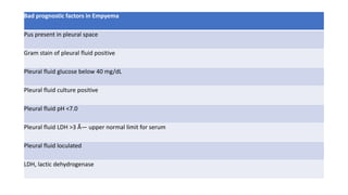
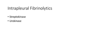
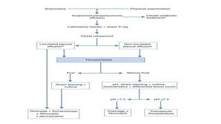
EMPYEMA
DR RAVI ASHOK DOSI
ASSOSCIATE PROFESSOR
SAMC & PGI , INDORE
Parapneumonic Effusions and Empyema
• bacterial pneumonia still results in significant morbidity and mortality
• The annual incidence of bacterial pneumonia is estimated to be 4 million, with approximately 25% of patients requiring
hospitalization .
• 40% of hospitalized patients with bacterial pneumonia have an accompanying pleural effusion , effusions associated with
pneumonia account for a large percentage of pleural effusions (PPE).
• The morbidity and mortality rates in patients with pneumonia and pleural effusions are higher than those in patients with
pneumonia alone.
• In one study of 1,424 patients hospitalized with community-acquired pneumonia, patients with pleural effusions were 2.7 times
more likely to be treatment failures than were those without pleural effusions (3). In another study, the relative risk of mortality in
patients with community-acquired pneumonia was 7.0 times higher for patients with bilateral pleural effusions and 3.4 times
higher for patients with unilateral pleural effusion of moderate or greater size as compared with other patients with community-
acquired pneumonia alone (4).
• In assessing risks of patients with community-acquired pneumonia, the presence of a pleural effusion is given the same weight as
a PO2 less than 60 mm Hg (5).
• Espana et al. (6) recommend that any patient with pneumonia and a loculated effusion or an effusion greater than 2 cm in
thickness on the decubitus be hospitalized.
• Some of the increased morbidity and mortality in patients with parapneumonic effusions are due to mismanagement of the
pleural effusion (7).
• Most pleural effusions associated with pneumonia resolve without
any specific therapy directed toward the pleural fluid
• Approximately 10% of patients require operative intervention.
• Delay in instituting proper therapy for these effusions is responsible
for some of the morbidity associated with parapneumonic effusions.
History
• Hippocrates, the treatment of empyema remained essentially
unchanged until the middle of the nineteenth century.
• At this time Bowditch (10) in the United States and Trousseau (11) in
France popularized the use of thoracentesis and demonstrated that
open drainage was not necessary in many patients.
• The next advance in the management of empyema came in 1876
when Hewitt (12) described a method of closed drainage of the chest
in which a rubber tube was placed into the empyema cavity through a
cannula. He was the first to use the water seal for chest tubes.
Definitions
• Any pleural effusion associated with bacterial pneumonia, lung abscess, or
bronchiectasis is a parapneumonic effusion .
• An empyema, by definition, is pus in the pleural space
• Weese et al. (29) defined an empyema as pleural fluid with a specific gravity
greater than 1.018, a WBC count greater than 500 cells/mm3, or a protein level
greater than 2.5 g/dL.
• Vianna (30) defined an empyema as pleural fluid on which the bacterial cultures
are positive or the WBC is greater than 15,000/mm3 and the protein level is
above 3.0 g/dL.
• Empyema are those pleural effusions with thick, purulent appearing pleural fluid.
• Many complicated parapneumonic effusions are empyemas, but some
parapneumonic effusions with nonpurulent appearing pleural fluid are also
complicated parapneumonic effusions.
Etiology
Cause Prevalence %
Pulmonary infection 55
Surgical procedure 21
Trauma 6
Esophageal perforation 5
Spontaneous pneumothorax 2
Thoracentesis 2
Sub Diaphragmatic Infection 1
Septicemia 1
Pathogenesis Of Empyema – 3 stages
Stage Origin WBC
COUNT
LDH Ph Glucose INTERVE
NTION
FLUID
Exudative INTERSTITIAL SPACE LOW LOW NORMAL NORMAL ANTIBIO
TICS
FREE
FLOW
Fibropurulent bacteria invade the
pleural fluid from
the contiguous
pneumonic process
HIGH
PMN
,BACTERIA
,DEBRIS
HIGH LOW LOW LOCULAT
ED
PL
THICKENI
NG +
the organization
stage
which fibroblasts
grow into the
exudate
Thick HIGH LOW LOW EMPYEM
A
NECESSIT
ANS
the
pleural
peel.
MICROBIOLOGY
Gram-Positive
Organisms
Gram-Negative
Organisms
Anaerobic
Organisms
Staphylococcus
aureus
36 Escherichia coli 30 Bacteroides
species
20
Staphylococcus
epidermidis
3 Klebsiella species 21 Peptostreptococc
us species
20
Streptococcus
pneumoniae
35 Proteus species 7 Fusobacterium
species
14
Enterococcus
faecalis
6 Pseudomonas
species
25 Prevotella species13
Streptococcus
pyogenes
8 Enterobacter
species
3 Streptococcus
species
10
Other
streptococci
12 Hemophilus
influenzae
12 Clostridium
species
7
Clinical Manifestations
Aerobic Bacterial Infections
• The clinical presentation of patients with aerobic bacterial pneumonia
and a pleural effusion is no different from that of patients with
bacterial pneumonia without effusion
• The patients first manifest an acute febrile illness with chest pain,
sputum production, and leukocytosis.
• A complicated parapneumonic effusion is suggested by the presence
of fever for more than 48 hours after antibiotic therapy is instituted.
• A complicated parapneumonic effusion should be suspected in febrile
patients in the intensive care unit who have temperature elevations.
CLINICAL MANIFESTATIONS
Anaerobic Bacterial Infections
• Usually first seen with subacute illnesses.
• Symptoms for more than 7 days before presentation, with a median
symptom duration of 10 days
• Substantial weight loss (mean 29 lb).
• A history of alcoholism
• An episode of unconsciousness
• Factors that predisposes them to aspiration
• Poor oral hygiene.
DIAGNOSIS
• Basic Labs
• Radiology
• Microbiology
• Biochemistry
Chest x ray
Ultrasonography
CT Scan
Procedures
BIOCHEMISTRY
Bad prognostic factors in Empyema
Pus present in pleural space
Gram stain of pleural fluid positive
Pleural fluid glucose below 40 mg/dL
Pleural fluid culture positive
Pleural fluid pH <7.0
Pleural fluid LDH >3 × upper normal limit for serum
Pleural fluid loculated
LDH, lactic dehydrogenase
Antibiotic Selection
• All patients with parapneumonic effusions or empyema should be treated with antibiotics.
• Metronidazole penetrated most easily, followed by penicillin, clindamycin, vancomycin, ceftriaxone, and gentamicin ,the
quinolones and clarithromycin penetrate the infected pleural space well .
• For patients with severe community-acquired pneumonia in whom pseudomonas infection is not an issue, the recommended
agents are β-lactam plus either an advanced macrolide or a respiratory fluoroquinolone . If a pseudomonas infection is suspected,
an anti-pseudomonas antibiotic such as piperacillin, piperacillin-tazobactam, imipenem, meropenem or cefepime should be
included .
• Because anaerobic bacteria cause a sizable percentage of parapneumonic effusions, anaerobic coverage is recommended for all
patients with parapneumonic effusions with either clindamycin or metronidazole (118). In addition, because a large percentage of
the complicated parapneumonic effusions that occur in hospitalized patients are due to MRSA (119), vancomycin should be
administered to such patients until the culture results are available.
• Current standard of practice is to continue antibiotics for several weeks (119).
 Intrapleural Antibiotics
• Intrapleural antibiotics were first used to treat an infected pneumonectomy space by Clagett and Geraci (120) in 1963. Since that
time, there have been several reports (121,122,123,124,125) regarding the use of intrapleural antibiotics in the treatment of
empyema complicating pneumonia. All of these reports have indicated positive results, but in none was there a randomized
control group. Until such controlled studies documenting the efficacy of intrapleural antibiotics are completed, they are not
recommended for patients with parapneumonic effusions.
Intrapleural Fibrinolytics
• Streptokinase
• Urokinase
Treatment Option for Complicated Parapneumonic Effusions
Therapeutic thoracentesis
Tube thoracostomy
Tube thoracostomy with the intrapleural administration of fibrinolytics
Thoracoscopy with the breakdown of adhesions
Thoracotomy with decortication
Thoracotomy
• THANKYOU

Empyema

  • 1.
    EMPYEMA DR RAVI ASHOKDOSI ASSOSCIATE PROFESSOR SAMC & PGI , INDORE
  • 2.
    Parapneumonic Effusions andEmpyema • bacterial pneumonia still results in significant morbidity and mortality • The annual incidence of bacterial pneumonia is estimated to be 4 million, with approximately 25% of patients requiring hospitalization . • 40% of hospitalized patients with bacterial pneumonia have an accompanying pleural effusion , effusions associated with pneumonia account for a large percentage of pleural effusions (PPE). • The morbidity and mortality rates in patients with pneumonia and pleural effusions are higher than those in patients with pneumonia alone. • In one study of 1,424 patients hospitalized with community-acquired pneumonia, patients with pleural effusions were 2.7 times more likely to be treatment failures than were those without pleural effusions (3). In another study, the relative risk of mortality in patients with community-acquired pneumonia was 7.0 times higher for patients with bilateral pleural effusions and 3.4 times higher for patients with unilateral pleural effusion of moderate or greater size as compared with other patients with community- acquired pneumonia alone (4). • In assessing risks of patients with community-acquired pneumonia, the presence of a pleural effusion is given the same weight as a PO2 less than 60 mm Hg (5). • Espana et al. (6) recommend that any patient with pneumonia and a loculated effusion or an effusion greater than 2 cm in thickness on the decubitus be hospitalized. • Some of the increased morbidity and mortality in patients with parapneumonic effusions are due to mismanagement of the pleural effusion (7).
  • 3.
    • Most pleuraleffusions associated with pneumonia resolve without any specific therapy directed toward the pleural fluid • Approximately 10% of patients require operative intervention. • Delay in instituting proper therapy for these effusions is responsible for some of the morbidity associated with parapneumonic effusions.
  • 4.
    History • Hippocrates, thetreatment of empyema remained essentially unchanged until the middle of the nineteenth century. • At this time Bowditch (10) in the United States and Trousseau (11) in France popularized the use of thoracentesis and demonstrated that open drainage was not necessary in many patients. • The next advance in the management of empyema came in 1876 when Hewitt (12) described a method of closed drainage of the chest in which a rubber tube was placed into the empyema cavity through a cannula. He was the first to use the water seal for chest tubes.
  • 5.
    Definitions • Any pleuraleffusion associated with bacterial pneumonia, lung abscess, or bronchiectasis is a parapneumonic effusion . • An empyema, by definition, is pus in the pleural space • Weese et al. (29) defined an empyema as pleural fluid with a specific gravity greater than 1.018, a WBC count greater than 500 cells/mm3, or a protein level greater than 2.5 g/dL. • Vianna (30) defined an empyema as pleural fluid on which the bacterial cultures are positive or the WBC is greater than 15,000/mm3 and the protein level is above 3.0 g/dL. • Empyema are those pleural effusions with thick, purulent appearing pleural fluid. • Many complicated parapneumonic effusions are empyemas, but some parapneumonic effusions with nonpurulent appearing pleural fluid are also complicated parapneumonic effusions.
  • 6.
    Etiology Cause Prevalence % Pulmonaryinfection 55 Surgical procedure 21 Trauma 6 Esophageal perforation 5 Spontaneous pneumothorax 2 Thoracentesis 2 Sub Diaphragmatic Infection 1 Septicemia 1
  • 7.
    Pathogenesis Of Empyema– 3 stages Stage Origin WBC COUNT LDH Ph Glucose INTERVE NTION FLUID Exudative INTERSTITIAL SPACE LOW LOW NORMAL NORMAL ANTIBIO TICS FREE FLOW Fibropurulent bacteria invade the pleural fluid from the contiguous pneumonic process HIGH PMN ,BACTERIA ,DEBRIS HIGH LOW LOW LOCULAT ED PL THICKENI NG + the organization stage which fibroblasts grow into the exudate Thick HIGH LOW LOW EMPYEM A NECESSIT ANS the pleural peel.
  • 8.
    MICROBIOLOGY Gram-Positive Organisms Gram-Negative Organisms Anaerobic Organisms Staphylococcus aureus 36 Escherichia coli30 Bacteroides species 20 Staphylococcus epidermidis 3 Klebsiella species 21 Peptostreptococc us species 20 Streptococcus pneumoniae 35 Proteus species 7 Fusobacterium species 14 Enterococcus faecalis 6 Pseudomonas species 25 Prevotella species13 Streptococcus pyogenes 8 Enterobacter species 3 Streptococcus species 10 Other streptococci 12 Hemophilus influenzae 12 Clostridium species 7
  • 9.
    Clinical Manifestations Aerobic BacterialInfections • The clinical presentation of patients with aerobic bacterial pneumonia and a pleural effusion is no different from that of patients with bacterial pneumonia without effusion • The patients first manifest an acute febrile illness with chest pain, sputum production, and leukocytosis. • A complicated parapneumonic effusion is suggested by the presence of fever for more than 48 hours after antibiotic therapy is instituted. • A complicated parapneumonic effusion should be suspected in febrile patients in the intensive care unit who have temperature elevations.
  • 10.
    CLINICAL MANIFESTATIONS Anaerobic BacterialInfections • Usually first seen with subacute illnesses. • Symptoms for more than 7 days before presentation, with a median symptom duration of 10 days • Substantial weight loss (mean 29 lb). • A history of alcoholism • An episode of unconsciousness • Factors that predisposes them to aspiration • Poor oral hygiene.
  • 11.
    DIAGNOSIS • Basic Labs •Radiology • Microbiology • Biochemistry
  • 12.
  • 13.
  • 14.
  • 15.
  • 16.
  • 17.
    Bad prognostic factorsin Empyema Pus present in pleural space Gram stain of pleural fluid positive Pleural fluid glucose below 40 mg/dL Pleural fluid culture positive Pleural fluid pH <7.0 Pleural fluid LDH >3 × upper normal limit for serum Pleural fluid loculated LDH, lactic dehydrogenase
  • 18.
    Antibiotic Selection • Allpatients with parapneumonic effusions or empyema should be treated with antibiotics. • Metronidazole penetrated most easily, followed by penicillin, clindamycin, vancomycin, ceftriaxone, and gentamicin ,the quinolones and clarithromycin penetrate the infected pleural space well . • For patients with severe community-acquired pneumonia in whom pseudomonas infection is not an issue, the recommended agents are β-lactam plus either an advanced macrolide or a respiratory fluoroquinolone . If a pseudomonas infection is suspected, an anti-pseudomonas antibiotic such as piperacillin, piperacillin-tazobactam, imipenem, meropenem or cefepime should be included . • Because anaerobic bacteria cause a sizable percentage of parapneumonic effusions, anaerobic coverage is recommended for all patients with parapneumonic effusions with either clindamycin or metronidazole (118). In addition, because a large percentage of the complicated parapneumonic effusions that occur in hospitalized patients are due to MRSA (119), vancomycin should be administered to such patients until the culture results are available. • Current standard of practice is to continue antibiotics for several weeks (119).  Intrapleural Antibiotics • Intrapleural antibiotics were first used to treat an infected pneumonectomy space by Clagett and Geraci (120) in 1963. Since that time, there have been several reports (121,122,123,124,125) regarding the use of intrapleural antibiotics in the treatment of empyema complicating pneumonia. All of these reports have indicated positive results, but in none was there a randomized control group. Until such controlled studies documenting the efficacy of intrapleural antibiotics are completed, they are not recommended for patients with parapneumonic effusions.
  • 19.
  • 20.
    Treatment Option forComplicated Parapneumonic Effusions Therapeutic thoracentesis Tube thoracostomy Tube thoracostomy with the intrapleural administration of fibrinolytics Thoracoscopy with the breakdown of adhesions Thoracotomy with decortication
  • 21.
  • 23.