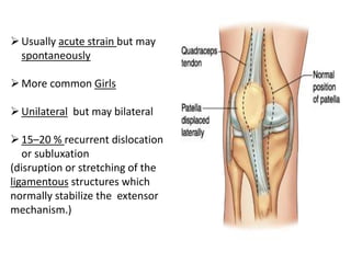
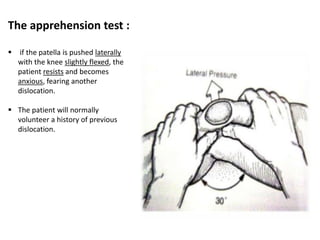
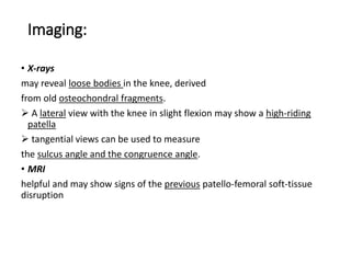
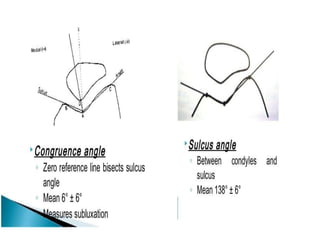
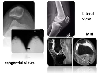
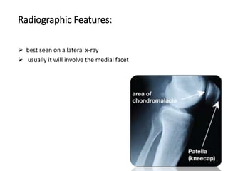
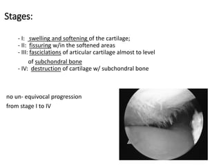
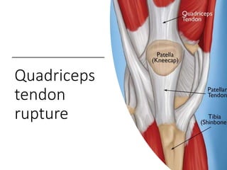
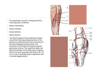
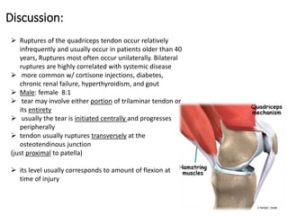
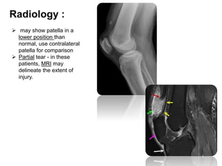
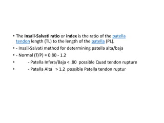
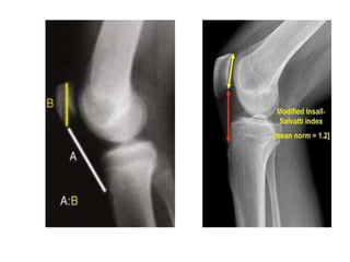
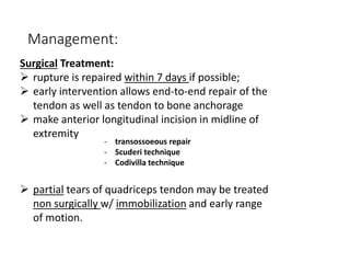
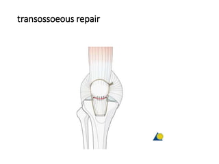
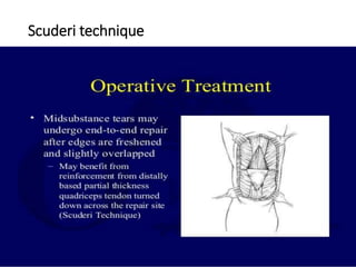
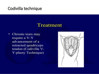
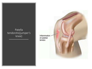
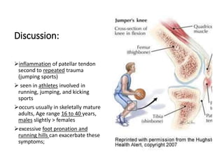
This document summarizes knee disorders including patellar instability, lateral patellar compression syndrome, idiopathic chondromalacia patellae, quadriceps tendon rupture, and patella tendon tendinitis. It describes the causes, clinical presentations, physical exam findings, and treatment options for each condition. Quadriceps tendon rupture most often occurs in patients over 40 years old and is usually caused by an eccentric contraction of the quadriceps muscle with the foot planted and knee partially flexed. Physical exam may reveal swelling, ecchymosis, and a palpable defect in the suprapatellar region.
Dr. Manoj Das' document provides an overview of examining the foot and ankle. It discusses the anatomy of the foot and ankle including bones, joints, ligaments and muscles. The examination involves taking a history, observing gait, posture and deformities, palpating for tenderness, and assessing range of motion, neurovascular status, and performing special tests. The goal is to assess, diagnose and treat conditions of the foot and ankle.
This case report describes a 21-year old female patient with a 10-year history of habitual left patellar dislocation. Examination found lateral subluxation of the patella in flexion and reduced range of motion. X-rays showed patella baja on the left side and lateral dislocation of the patella. The patient underwent a proximal and distal soft tissue realignment procedure involving lateral release, medial reinforcement, and partial medialization of the patellar tendon. Post-operatively, the patella was centrally located with improved range of motion and stability. Habitual patellar dislocation is rare in adults and can be treated with soft tissue realignment surgery to reinforce the medial structures and release tight
This document discusses shoulder instability, including definitions, anatomy, evaluation, and treatment. It defines instability as the inability to maintain the humeral head in the glenoid fossa, ranging from dislocation to laxity. Static stabilizers include the labrum and ligaments, while dynamic stabilizers are the rotator cuff and scapulothoracic muscles. Evaluation involves history, exam, and imaging to classify instability by direction, degree, and etiology. Treatment depends on classification but may include immobilization, rehabilitation, or surgical repair of labral tears or bone defects.
This document provides information about pes planus (flat foot), including its components, classification, examination, and treatment. Pes planus is characterized by a lowered or absent medial longitudinal arch. It can be flexible or rigid depending on joint mobility. Flexible flat foot is more common and usually asymptomatic, especially in children. Treatment focuses on orthotics, exercises, or surgery if conservative measures fail. Surgical options include tendon lengthening, arthrodesis, and osteotomies to realign the foot structure.
Dupuytren's contracture is a condition causing the fingers to bend towards the palm due to fibrosis of the palmar fascia. It typically affects men over 50 years old and has an autosomal dominant inheritance pattern. Clinically, patients present with nodules and cords in the palm leading to finger contractures. Treatment options include observation for slow cases, radiotherapy in early stages, and surgery involving fasciotomy or fasciectomy. Complete fasciectomy has high recurrence rates while partial fasciectomy balances effectiveness and risk of recurrence. Post-operative splinting and exercises are needed to regain finger extension.
Apply gentle pressure proximally
Surgeon: Check distal pulses and capillary refill
If no improvement:
Consider temporary arteriotomy or venous shunt
Delay closure and observe
Flap or graft may be needed
Shoulder impingement syndrome occurs when the tendons of the rotator cuff muscles become irritated and inflamed as they pass through the subacromial space under the coraco-acromial arch. It results in pain, weakness, and loss of movement, especially in an arc between 45-160 degrees of shoulder abduction and elevation. Causes include repeated overhead arm use, trauma, poor posture, and degenerative changes. Clinical features are pain at rest or with movement, and limited range of motion. Diagnosis involves x-rays and MRI, while special tests like Neer's and Hawkins' tests reproduce shoulder pain. Treatment consists of rest, anti-inflammatories, physical therapy including stretching,
The document provides guidance on clinically examining the hip joint. It outlines important points to consider when examining a patient's hip, including examination techniques and order. Key areas that are assessed include inspection, palpation, range of motion, deformities, measurements, special tests like Trendelenburg sign, and making a diagnosis. The examination is thorough and considers multiple factors that could provide clues about a patient's hip condition.
The document provides information on patellar dislocation, including:
- Anatomy of the patella and patellofemoral joint.
- Causes of patellar instability including anatomical abnormalities, trochlear dysplasia, and injury mechanisms.
- Evaluation of patients with patellar instability focusing on the integrity of the medial patellofemoral ligament and examining for patella alta.
- Imaging techniques used to assess patellar instability including x-rays, MRI, and CT which evaluate trochlear morphology, patellar height, and tracking.
This document summarizes stress fractures, which occur through normal bone subjected to repeated stress. Key points:
- They result from repetitive submaximal forces exceeding bone's adaptive ability, common in athletes and military.
- Lower limb weight-bearing bones like tibia are most prone. Specific sites include femoral neck, tibia, navicular.
- Causes include increased activity without rest, muscle fatigue concentrating forces, and nutritional/hormonal imbalances.
- Diagnosis involves history of increased activity and focal bone pain worsened by stress. Imaging includes xray, CT, MRI, bone scan.
- Treatment depends on fracture location and risk but typically involves initial rest, then progressive return to
Meniscus injuries are common in young adults, often caused by twisting or heavy lifting. Symptoms include knee pain, swelling, stiffness, tenderness, pain with squatting, popping or clicking in the knee, and limited motion. Meniscus tears are classified as longitudinal, horizontal, radial, or flap tears. Exams like McMurray's test and Apley's test are used to diagnose tears. Treatment involves medications, surgery if the meniscus cannot be repaired, physiotherapy including exercises and bracing, and rehabilitation protocols after arthroscopic surgery or meniscal repair surgery. Isokinetic training after arthroscopy can help improve knee function and muscle strength recovery.
The document provides information on recurrent patellar dislocation, including:
- Anatomy of the patella and its attachments
- Static and dynamic stabilizers of the patella
- Causes of patellar instability such as trochlear dysplasia, patella alta, increased Q angle
- Mechanisms of injury for acute vs recurrent dislocations
- Evaluation methods like the apprehension test, patellar glide test, and imaging views
This document provides an overview of hallux valgus, including its anatomy, causes, symptoms, diagnosis, and treatment options. Key points include:
- Hallux valgus is a lateral deviation of the great toe and medial deviation of the first metatarsal. It can cause pain over the bunion.
- Risk factors include heredity, footwear, ligament laxity, and pes planus. Diagnosis involves examining range of motion, deformity, and taking x-rays to measure angles.
- Treatment progresses from footwear modifications and stretching to various surgical procedures depending on severity, including distal soft tissue procedures, osteotomies, and joint fusion or replacement in severe cases.
MCL. LCL.ALL injuries
To understand the relevant anatomy of the side ligaments of the knee
To study the mechanism of injury of each ligament and how to diagnose such injury
To highlight the different treatment options in acute or chronic situations
1) The document discusses the process for examining a patient's hip joint, including obtaining history, performing physical examination, and conducting specific tests.
2) The physical examination involves inspecting the hip from various angles, palpating bony landmarks and soft tissues, measuring range of motion, assessing limb length and muscle bulk, and performing stability and special tests.
3) A number of special tests are described that can help identify conditions like labral tears, femoral anteversion, and soft tissue contractures. Taking a thorough history and conducting a complete physical exam are important for accurately diagnosing hip joint pathology.
This document provides an overview of clubfoot (congenital talipes equinovarus), including:
1. The definition, incidence, causes, and typical deformities seen in clubfoot.
2. Evaluation methods like the Pirani scoring system and radiographic assessment.
3. Treatment approaches like the Ponseti method of serial casting and bracing, as well as surgical options like the McKay procedure when non-operative treatment fails.
4. Post-operative casting protocols and complications that can arise from treatment.
The document provides information on ligament injuries of the knee, specifically ACL injury. It describes the anatomy of the ACL and other major ligaments of the knee. It discusses the mechanisms of acute ligament injuries, their clinical features, imaging, and treatment options. For acute injuries, treatment may involve bracing, physiotherapy, or reconstruction depending on the ligaments involved and severity of injury. Chronic ligament instability can lead to abnormal knee motion and giving way, requiring further assessment and possibly reconstruction to restore stability.
This document discusses hallux valgus, a deformity of the great toe. It begins by describing the anatomy and pathophysiology, noting that hallux valgus has no single cause but can be due to conditions like flat feet or footwear. Clinical presentation includes bunion pain that worsens with footwear. Treatment involves modifying footwear, splinting, and surgery if conservative measures fail. Surgical options correct soft tissues like tendons and ligaments as well as bony procedures like osteotomies of the toe bone or metatarsal. Complications of surgery include recurrence of the deformity or issues like nerve damage.
This document provides information on meniscal tears of the knee. It begins with an introduction stating that meniscal tears are common injuries responsible for many arthroscopies annually. It then covers anatomy of the medial and lateral menisci, blood supply, classification of tears, mechanisms of injury, clinical features, investigations like MRI and arthroscopy, and treatment options including non-operative care, meniscectomy, and meniscal repair. The focus is on providing detailed information on meniscal anatomy, tears, and surgical and non-surgical management.
There are three types of patella dislocation: acute, recurrent, and habitual. Acute dislocation occurs suddenly due to quadriceps contraction with the knee flexed and results in the patella dislocating laterally, causing pain, swelling, and inability to straighten the knee. Recurrent dislocations are caused by ligament laxity or anatomical abnormalities and damage bones with repeated dislocations. Habitual dislocations occur every time the knee is flexed and present in early childhood.
The meniscus is a C-shaped fibrocartilage structure in the knee that provides shock absorption, joint stability, and lubrication. It receives minimal blood supply, so tears do not heal well. Bucket handle tears are most common. Symptoms include locking, pain, and swelling. Exams involve McMurray's test and Apley's test to isolate meniscal tears. MRI is very sensitive for diagnosis. Treatment ranges from immobilization and physical therapy for acute tears to arthroscopic surgery to repair or excise chronic tears.
Collapse of medial longitudinal arch, with the entire sole of the foot coming into complete or near-complete contact with the ground.
Books Refered :
Text Book Of ANATOMY - Vishram Singh
Joint Structure And Function – Cynthia Norkin
Therapeutic Exrercise – Carolyn Kisner
Orthopaedic Physical Assessment – Magee
Orthopaedic Medicine – L. Ombregt
Campbell’s Operative Orthopaedics
Recurrent Dislocation of patella -PAWANPawan Yadav
This document discusses recurrent patellar dislocation. It begins by defining recurrent patellar dislocation as the patella shifting laterally with minimal stress on knee flexion. It then discusses the anatomy and Q angle as well as predisposing causes such as increased Q angle, weak medial quads, and tight lateral structures. The document outlines clinical features, tests, x-ray findings, and treatment options including conservative immobilization and surgical procedures like realignment and patellectomy.
This document provides information on various arthrodesis procedures. Arthrodesis is a surgical technique used to fuse a dysfunctional joint to relieve pain. It summarizes techniques for fusing specific joints like the shoulder, elbow, wrist, fingers, hip, and knee. For each joint, it describes common indications, positions, surgical approaches, fixation methods, and post-operative care. Complications are also reviewed. The document is a comprehensive reference for orthopedic surgeons on the principles and techniques of different arthrodesis procedures.
This document discusses flat feet (pes planus), including its anatomy, causes, types, symptoms, physical exam findings, and treatment options. Key points include:
- Pes planus is characterized by a low or absent medial longitudinal arch. It can be flexible or rigid.
- Causes include ligament laxity, obesity, muscle weakness, bony abnormalities, and tarsal coalitions.
- Treatment focuses on orthotics, stretches, braces, and surgery if conservative options fail. Surgical procedures include tendon lengthening, osteotomies, and fusions.
Dr. Ankur Mittal presented on diagnostic tests and imaging for Achilles tendon injuries. [1] Ultrasound is often used to determine tendon thickness and gap size for complete ruptures and is inexpensive and fast. [2] MRI is better for detecting incomplete tears and planning surgery for chronic tears but is more expensive. [3] Imaging is rarely needed for acute cases but can help with chronic cases for diagnosis and surgical planning.
د. عمار الياسري shoulder and pectoral girdle-5 (Muhadharaty).pptxhussainAltaher
The document discusses various disorders of the shoulder and pectoral girdle, including:
- Rotator cuff disorders like impingement syndrome and tears, which can cause pain and weakness. Conservative treatments include physiotherapy, while surgery may be needed for tears.
- Shoulder instability, which can be traumatic from injury, atraumatic from overuse, or muscular in nature. Treatments range from physiotherapy to surgical repairs or plications.
- Other conditions discussed include adhesive capsulitis (frozen shoulder), biceps tendon disorders, calcific tendinitis of the shoulder, and tuberculosis of the glenohumeral joint. A variety of imaging tools and both nonsurgical and surgical treatment approaches
The document provides information on ligament injuries, specifically cranial cruciate ligament rupture in dogs. It discusses the pathophysiology, diagnosis, and treatment of cranial cruciate ligament rupture. Key points include:
- The cranial cruciate ligament limits cranial translation of the tibia and controls internal rotation of the tibia. Rupture can be caused by traumatic injury or degeneration.
- Diagnosis involves physical exam tests like the cranial drawer test and tibial compression test, as well as diagnostic imaging like radiography and arthroscopy.
- Surgical treatment options aim to stabilize the stifle joint and include intracapsular reconstruction, extracapsular reconstruction, imbr
Chondromalacia patellar, or softening of the cartilage under the kneecap, is caused by several factors like malalignment, muscle weakness, or lesions. It begins as softening and progresses to tearing and erosion. Diagnosis involves knee exams and imaging like MRI. Treatment includes rest, braces, exercises to strengthen muscles like quadriceps, and sometimes surgery to realign the patella. Physiotherapy focuses on exercises, taping or bracing to improve tracking, and modalities for pain relief.
Ligamentous Injuries Around Knee by Dr Arun C Raj, Ortho Resident, KIMS HubliArunCRaj1
This document discusses ligamentous injuries around the knee. It begins by describing the relevant anatomy of the knee joint and its ligaments. It then discusses the mechanisms of various knee injuries including injuries to the medial collateral ligament from valgus forces, lateral collateral ligament from varus forces, posterior cruciate ligament from backward tibial forces, and anterior cruciate ligament from twisting forces. It describes the clinical examination and diagnosis of these injuries including various tests. Treatment options discussed include conservative treatment with immobilization for mild injuries and surgical repair or reconstruction for more severe injuries. Complications of untreated injuries like instability and osteoarthritis are also mentioned. The document then discusses meniscal injuries of the knee, their mechanism as twisting injuries
- Ankle fractures are the most common weight-bearing skeletal injury, with the highest incidence in elderly women. The document classifies common ankle fracture patterns and discusses evaluation and treatment.
- Physical exam includes neurovascular assessment and evaluation of deformities, tenderness, and ligament stability. Imaging includes plain films and sometimes CT or MRI to further evaluate fracture patterns and ligament injuries.
- Treatment depends on the fracture classification system used (Lauge-Hansen, Weber, OTA) and whether the injury is stable and can be treated non-operatively or requires operative fixation due to instability, malalignment, or joint incongruity. Surgical techniques are described for common fracture types.
Knee dislocation powerpoint for medical studentsCasualityShift
Knee dislocations are high-energy traumatic injuries that often involve ruptures of three or more major ligaments of the knee. They require emergent treatment due to the risk of neurovascular injury. The most common type is anterior dislocation from hyperextension. A thorough clinical exam is needed to assess ligament stability and neurovascular status before and after reduction. Vascular injury occurs in 20-60% of cases and requires revascularization within 8 hours to prevent limb loss. Immediate surgical exploration is indicated if pulses do not return after reduction.
This document provides an overview of patellofemoral disorders, including the anatomy and biomechanics of the patellofemoral joint. It describes several common patellofemoral conditions such as patellar instability, excessive lateral patellar compression syndrome, chondromalacia patellae, bipartite patella, and patellofemoral arthritis. For each condition, it discusses symptoms, physical exam findings, imaging features, and treatment options both non-surgical and surgical. Surgical procedures discussed include lateral retinacular release, tibial tubercle elevation, drilling/abrasion techniques, and patellectomy.
This document discusses acute knee ligament injuries, including their common causes, mechanisms of injury, classification, and management. The most common injury mechanism is abduction, flexion, and internal rotation of the femur, which typically injures the medial ligaments. Injuries are classified by degree of ligament disruption. Treatment options include nonoperative management, repair, and reconstruction, depending on the specific ligaments injured and degree of instability. Post-operative rehabilitation focuses on regaining range of motion and strengthening.
This document discusses common musculoskeletal problems seen in the community, including knee, shoulder, ankle and foot issues. It provides details on the causes, clinical features, diagnosis and treatment approaches for various conditions like patellofemoral pain syndrome, shoulder instability, adhesive capsulitis, rotator cuff tears, ankle sprains and plantar fasciitis. Special investigations like MRI and treatment methods such as injections, physiotherapy, manipulation and surgery are touched upon.
This document discusses ankle sprains, including the anatomy of the ankle complex and its three joints. It describes the classification, symptoms, physical examination findings, and special tests used to evaluate ankle sprains. Low ankle sprains include lateral and medial sprains, while high ankle sprains involve the syndesmosis. Management involves the PRICE regimen initially, followed by rehabilitation exercises. Surgery may be indicated for persistent pain or instability after conservative treatment or in cases of severe ligament damage or recurrent sprains.
This document discusses ankle sprains, including the anatomy of the ankle complex and its three joints. It describes the classification, symptoms, physical examination findings, and special tests used to evaluate ankle sprains. Low ankle sprains, which include lateral and medial sprains, are more common and involve ligament tears. High ankle sprains involve the syndesmotic ligaments. Management includes the PRICE regimen initially followed by rehabilitation exercises. Surgery may be indicated for severe, recurrent, or unstable sprains.
The knee joint contains bony structures like the patella, femoral condyles, and tibial plateaus. It also contains soft tissues like the medial and lateral menisci and anterior and posterior cruciate ligaments. The menisci function to distribute joint fluid, absorb shock, deepen the joint, stabilize the joint, and bear weight. Meniscal tears are most commonly longitudinal tears of the posterior horn of the medial meniscus caused by rotation of the flexed knee. Diagnosis involves history, physical exam including tests like McMurray's and Thessaly, and imaging like MRI. Treatment involves initial immobilization and rehab followed by possible surgical repair, removal, or replacement of torn meniscal tissue.
This document discusses various injuries to the ankle and foot, including:
- The anatomy of the ankle joint and ligaments that support it.
- Common ankle injuries like sprains, fractures of the medial/lateral malleolus, and fractures of the calcaneum.
- Clinical features, radiological examinations, and treatment approaches for different types of ankle and foot injuries. Conservative treatment involves immobilization, while surgical treatment may be needed for displaced fractures or chronic injuries. Complications can include stiffness, arthritis, and long-term impairment.
Ankle sprains are very common injuries, usually caused by an inversion mechanism, that commonly involve tears of the lateral ligaments. The most commonly injured ligament is the anterior talofibular ligament. Ankle sprains are graded based on the severity of ligament tearing, from grade I (mild stretching) to grade III (complete tear). Physical examination involves assessing for swelling, tenderness, instability and range of motion loss. Special tests like the anterior drawer and talar tilt can help evaluate ligament integrity. X-rays are usually only needed if indicated by the Ottawa ankle rules to check for fractures.
28,000 ankle sprains occur daily in the US (Kaminski 2013)
Ankle is the 2nd most commonly injured body site. (Ferran 2006)
Ankle sprains are the most common type of ankle injury. (Ferran 2006)
A sprained ankle can happen to athletes and non-athletes,
children and adults.
Inversion injury most common mechanism (Ferran 2006)
Only risk factor is previous ankle sprain (Ferran 2006)
Sex , generalized joint laxity or anatomical foot types are
not risk factors. (Beynnon et al. 2002 )
The anterior cruciate ligament (ACL) is commonly ruptured in the knee. It occurs from a twisting force on a bent knee and often accompanies injuries to other knee ligaments and meniscus. The ACL attaches the femur to the tibia and prevents anterior tibial displacement. Diagnosis involves physical exams like the Lachman and pivot shift tests and MRI. Treatment options are conservative rehabilitation or surgical reconstruction, with surgery recommended for athletes or those with instability. Reconstruction uses grafts fixed in the knee with screws or buttons. Post-op rehabilitation is needed to regain strength and function.
This document summarizes common lower limb fractures seen by Dr. Ghassan Al Kefeiri. It describes pelvic fractures which most commonly result from lateral compression and may cause life-threatening hemorrhage. Hip injuries like dislocations and fractures are also discussed in detail. Femoral fractures of the diaphysis and distal regions are outlined along with potential complications. The knee section evaluates various injuries to ligaments, menisci and patella. Common causes, clinical features and treatments are provided for lower limb fractures and orthopedic conditions.
This document summarizes the anatomy of the fetal head, including its bones, sutures, landmarks, and diameters. It describes how the cranial bones are thin and overlapping at birth, allowing molding to the maternal pelvis. The sutures between bones are identified. Landmarks from front to back are listed. Four anteroposterior diameters and two transverse diameters are defined. The average fetal head circumference at term is provided.
This document provides information on burns and sudden death. It begins by defining burns and classifying them based on depth and extent. It then discusses the pathophysiology of burns, including edema, cardiac, renal and immunologic effects. Burn management in the hospital is outlined, including fluid resuscitation, airway management, infection control, pain relief and nutrition. Complications of burns like shock, infections and scarring are also summarized. The document concludes by defining sudden death and noting it is important in forensic analysis of unexpected deaths within 24 hours of burn onset.
This document provides information on empyema, including its definition, etiology, stages, symptoms, investigations, and management. Empyema is defined as infection of the pleural space resulting in pus accumulation. It is usually caused by bacteria spreading from a pneumonia or other infection. It progresses through exudative, fibrinopurulent, and organizing stages. Symptoms include fever, cough, chest pain, and shortness of breath. Diagnosis involves imaging like chest X-ray or CT scan and thoracentesis. Management includes antibiotics, chest tube drainage, fibrinolytics, VATS, and sometimes open drainage or decortication surgery. The goal is to treat infection, drain pus, and re-expand
Calcium channel blockers (CCBs) work by blocking the influx of calcium through L-type calcium channels in cardiac and vascular smooth muscle. This leads to vasodilation, decreased blood pressure, and reduced cardiac contractility. The main classes of CCBs are dihydropyridines like nifedipine, benzothiazepines like diltiazem, and diphenylalkylamines like verapamil. They are used to treat hypertension, angina, and arrhythmias. Common side effects include headache, flushing, edema, and constipation. Cautions include hypotension, heart failure, and avoiding grapefruit juice with some CCBs.
This document provides guidance on assessing geriatric patients in an office setting. It emphasizes the importance of pre-visit preparation, focusing the office environment to meet seniors' needs, and conducting a comprehensive initial assessment covering medical history, functional status, social supports, and finances. Key parts of the assessment include evaluating cognitive status using tests like the Mini-Mental State Exam, reviewing the patient's multiple comorbidities and geriatric syndromes, and understanding their often vague presentations of illness. The goal is to establish rapport, understand priorities for care, and develop a follow-up plan in the first visit.
This document defines ectopic pregnancy and discusses its causes, signs and symptoms, diagnostic tests, and treatment options. Key points:
- Ectopic pregnancy occurs when a fertilized egg implants outside the uterus, most commonly in the fallopian tubes.
- Risk factors include previous ectopic pregnancy, pelvic inflammatory disease, assisted reproduction, and IUD use.
- Patients often experience abdominal pain, amenorrhea, and vaginal bleeding. Serum hCG levels and ultrasound are used for diagnosis.
- Treatment depends on stability and includes observation, methotrexate injection, or surgery like salpingectomy or salpingostomy. The goal is to resolve the ectopic pregnancy safely while preserving fertility when
The document discusses the lens and cataract. It defines the lens as a transparent structure behind the iris that focuses light on the retina. Cataract is defined as any opacity within the lens. The most common cause of cataract is aging, but it can also be caused by trauma, inflammation, diabetes, and other systemic factors. Surgical removal of the cataract involves phacoemulsification, extracapsular extraction, or intracapsular extraction techniques. Post-operative care includes antibiotic and steroid eye drops. Complications include vitreous loss, iris prolapse, and endophthalmitis.
Urinary tract infections (UTIs) can range from asymptomatic bacteriuria to severe kidney infection. Common symptoms include dysuria, urinary frequency and urgency. UTIs are more common in women than men. Types include cystitis, urethritis, prostatitis and pyelonephritis. Pyelonephritis is a kidney infection that can cause loin pain, fever and vomiting. It is generally treated with oral or IV antibiotics depending on severity. Complicated UTIs involve abnormal anatomy or immunity.
Here is the updated list of Top Best Ayurvedic medicine for Gas and Indigestion and those are Gas-O-Go Syp for Dyspepsia | Lavizyme Syrup for Acidity | Yumzyme Hepatoprotective Capsules etc
share - Lions, tigers, AI and health misinformation, oh my!.pptxTina Purnat
• Pitfalls and pivots needed to use AI effectively in public health
• Evidence-based strategies to address health misinformation effectively
• Building trust with communities online and offline
• Equipping health professionals to address questions, concerns and health misinformation
• Assessing risk and mitigating harm from adverse health narratives in communities, health workforce and health system
Clinic ^%[+27633867063*Abortion Pills For Sale In Tembisa Central19various
Clinic ^%[+27633867063*Abortion Pills For Sale In Tembisa Central Clinic ^%[+27633867063*Abortion Pills For Sale In Tembisa CentralClinic ^%[+27633867063*Abortion Pills For Sale In Tembisa CentralClinic ^%[+27633867063*Abortion Pills For Sale In Tembisa CentralClinic ^%[+27633867063*Abortion Pills For Sale In Tembisa Central
Integrating Ayurveda into Parkinson’s Management: A Holistic ApproachAyurveda ForAll
Explore the benefits of combining Ayurveda with conventional Parkinson's treatments. Learn how a holistic approach can manage symptoms, enhance well-being, and balance body energies. Discover the steps to safely integrate Ayurvedic practices into your Parkinson’s care plan, including expert guidance on diet, herbal remedies, and lifestyle modifications.
8 Surprising Reasons To Meditate 40 Minutes A Day That Can Change Your Life.pptxHolistified Wellness
We’re talking about Vedic Meditation, a form of meditation that has been around for at least 5,000 years. Back then, the people who lived in the Indus Valley, now known as India and Pakistan, practised meditation as a fundamental part of daily life. This knowledge that has given us yoga and Ayurveda, was known as Veda, hence the name Vedic. And though there are some written records, the practice has been passed down verbally from generation to generation.
Histololgy of Female Reproductive System.pptxAyeshaZaid1
Dive into an in-depth exploration of the histological structure of female reproductive system with this comprehensive lecture. Presented by Dr. Ayesha Irfan, Assistant Professor of Anatomy, this presentation covers the Gross anatomy and functional histology of the female reproductive organs. Ideal for students, educators, and anyone interested in medical science, this lecture provides clear explanations, detailed diagrams, and valuable insights into female reproductive system. Enhance your knowledge and understanding of this essential aspect of human biology.
Muktapishti is a traditional Ayurvedic preparation made from Shoditha Mukta (Purified Pearl), is believed to help regulate thyroid function and reduce symptoms of hyperthyroidism due to its cooling and balancing properties. Clinical evidence on its efficacy remains limited, necessitating further research to validate its therapeutic benefits.
4. RECURRENT DISLOCATION
Dislocation occurs
unexpectedly when the
quadriceps muscle is
contracted with the
knee in flexion
Dislocation is almost
always towards the
lateral side
medial dislocation is
seen only in rare
iatrogenic cases
5. Usually acute strain but may
spontaneously
More common Girls
Unilateral but may bilateral
15–20 % recurrent dislocation
or subluxation
(disruption or stretching of the
ligamentous structures which
normally stabilize the extensor
mechanism.)
6. predisposing factors :
(1)generalized ligamentous laxity
(2) under -development of the lateral femoral condyle and
flattening of the intercondylar groove
(3) Maldevelopment of the patella, which may be too high or too
Small
(4) valgus deformity of the knee
(5) external tibial torsion
(6) primary muscle defect
7. Clinical presentation :
acute pain
knee is stuck in flexion and the patient may fall to the ground.
Although the patella always dislocates laterally, the patient may think
it has displaced medially because the uncovered medial femoral
condyle stands out prominently.
8. Physical examination :
• The diagnosis is obvious If the knee
is seen while the patella is
dislocated.
1- lump on the lateral side
2-the front of the knee (where the patella ought to be) is flat
medial side are tender
3-may be swollen (aspiration may reveal a blood-stained effusion.)
•More often the patella has reduced
by the time the patient is seen.
1-Tenderness and swelling may still be
2- positive apprehension test
9. The apprehension test :
if the patella is pushed laterally
with the knee slightly flexed, the
patient resists and becomes
anxious, fearing another
dislocation.
The patient will normally
volunteer a history of previous
dislocation.
10. Imaging:
• X-rays
may reveal loose bodies in the knee, derived
from old osteochondral fragments.
A lateral view with the knee in slight flexion may show a high-riding
patella
tangential views can be used to measure
the sulcus angle and the congruence angle.
• MRI
helpful and may show signs of the previous patello-femoral soft-tissue
disruption
13. Management :
If the patella is still dislocated, it is pushed back into
place while the knee is gently extended
The only indications for immediate surgery are :
(1) inability to reduce the patella
(2) the presence of a large, displaced osteochondral fragment.
splint is applied for 2–3 weeks
quadriceps-strengthening exercises (Exercises should be continued for
at least 3 months)
the patient is allowed to walk with the aid of crutches.
15. RECURRENT SUBLUXATION
• Patellar dislocation is sometimes followed by
recurrent subluxation rather than further
episodes of complete displacement.
• This is the borderline between frank instability
and maltracking of the patella
16. Congenital dislocation
-The patella is permanently displaced
-very rare
- Reconstructive procedures
have been tried but the results are unpredictable.
Habitual dislocation
-The patella dislocates every time the knee is bent and reduces each time it is
straightened.
-In longstanding cases the patella may be permanently dislocated.
contracture of the quadriceps
-may be congenital or may result from repeated injections (usually antibiotics) into the
muscle.
-Treatment requires lengthening of the quadriceps.
OTHER TYPES OF NON-TRAUMATIC DISLOCATION
18. • This syndrome is common among active adolescents and young
adults(women).
• It is often (but not invariably) associated with softening and
fibrillation of the articular surface of the patella – chondromalacia
patellae
• The basic disorder is probably mechanical overload of the patello-
femoral joint
• Diagnosed by exclusion
19.
20. Clinical presentation:
• pain over the front of the knee or underneath the knee-cap
• aggravated by activity or climbing stairs, or when standing up after
prolonged sitting
• knee may give way and occasionally swells.
• Often both knees are affected
21. P.E:
• At first sight the knee looks normal but careful
• examination may reveal malalignment or tilting of the patellae.
• Other signs include quadriceps wasting, fluid in the knee, tenderness
under the edge of the patella and crepitus on moving the knee.
22. investigation :
• The most accurate way of showing and measuring patello-femoral
malposition is by CT or MRI with the knees in full extension and varying
degrees of flexion
• arthroscopy is useful in excluding other causes of anterior knee pain
23. Management:
• CONSERVATIVE MANAGEMENT In the vast majority of cases the patient
will be helped by adjustment of stressful activities and physiotherapy
• OPERATIVE TREATMENT Surgery should be considered only if :
(1) there is a demonstrable abnormality that is correctable by operation
(2) conservative treatment has been tried for at least 6 months
• Operation is intended to improve patellar alignment and patello-femoral
congruence and to reduce patello-femoral pressure
25. Discussion:
chondromalacia describes softening & fissuring of articular hyaline
cartilage
chondromalacia may result from an excessive load on patellofemoral
joint, but disuse may be a contributing factor
most common in young women
contributing factors:
- weakness and tightness
of quadriceps muscle
- genu valgum
- increased Q angle
- patella alta
26. Clinical Features and Exam:
pts may report anterior knee pain, esp. while climbing stairs
compression of patella may cause pain
compression of the patella during flexion & extension of knee may elicits
crepitation and discomfort
- patellar tracking
- best seen when examiner
is seated in front of pt
takes knee through full
passive and active ROM.
Note: crepitus may be a normal
finding in young people
28. Stages:
- I: swelling and softening of the cartilage;
- II: fissuring w/in the softened areas
- III: fasciclations of articular cartilage almost to level
of subchondral bone
- IV: destruction of cartilage w/ subchondral bone
no un- equivocal progression
from stage I to IV
29. Management :
• Non Operative Treatment:
- reduced strenuous activities;
- exercises to stretch & strengthen quadriceps muscle are started
- avoid stressing the painful motions
- immobilization is a contributing cause of chondromalacia and its
subsequent symptoms
- Operative:
- debridement-
• Partial lateral patellar facetectomy for treatment of arthritis due to lateral
patellar compression syndrome.
• lateral retinacular release
• distal realignment procedures
• Anteromedialization of the tibial tuberosity in the treatment of
patellofemoral pain and malalignment.
31. • The quadriceps muscle is composed of four
muscle groups, as follows:
• Vastus intermedius
• Vastus medialis
• Vastus lateralis
• Rectus femoris
• the blood supply to the quadriceps tendon
arises from the descending branches of the
lateral circumflex femoral artery, branches of
the descending geniculate artery, and
branches of the medial and lateral superior
geniculate arteries. The superficial layers are
well vascularized. In the deep layer, however,
there is an oval, avascular area that is 30 × 15
mm in size; it probably plays a significant role
in tendon degeneration.
32. Discussion:
Ruptures of the quadriceps tendon occur relatively
infrequently and usually occur in patients older than 40
years, Ruptures most often occur unilaterally. Bilateral
ruptures are highly correlated with systemic disease
more common w/ cortisone injections, diabetes,
chronic renal failure, hyperthyroidism, and gout
Male: female 8:1
tear may involve either portion of trilaminar tendon or
its entirety
usually the tear is initiated centrally and progresses
peripherally
tendon usually ruptures transversely at the
osteotendinous junction
(just proximal to patella)
its level usually corresponds to amount of flexion at
time of injury
33. Pathophysiology
• Various systemic conditions may cause damage to the tendon
vascular supply or may disrupt the tendon structure.
• Diabetes can cause arteriosclerotic changes in tendon vessels.
• Fibrinoid necrosis of tendons is seen with chronic synovitis.
• Hyperparathyroidism causes dystrophic calcifications and
subperiosteal bone resorption at the tendon insertion.
• Obesity causes fatty degenerative changes in tendons and increases
the forces on the tendon.
• Fatty degeneration, fibrinoid degeneration, and decreased collagen
are seen with normal aging.
• 97% of the pathologic changes were degenerative.
34. Etiology
• Quadriceps tendon rupture usually occurs during a rapid, eccentric
contraction of the quadriceps muscle, with the foot planted and the
knee partially flexed.
• This injury commonly occurs during falls. Other mechanisms of injury
include direct blows, lacerations, and iatrogenic causes.
35. Clinical Presentation :
History:
Patients typically present with acute knee pain, swelling, and functional loss after
a stumble, fall, or giving way of the knee. There may be no history of prior knee
pain.
Specifically ask patients about any history of systemic disease, steroid use,
infection, tumors, or prior surgeries. There may be a history of an audible pop at
the time of injury.
36. Physical Examination
• Begin the physical examination by noting any obesity. Patients with recent
ruptures have difficulty ambulating. Usually, obvious suprapatellar swelling,
ecchymosis, and tenderness are present. Carefully evaluate lacerations.
There may be a palpable defect in the suprapatellar area and a low-lying
patella, but swelling initially may obscure this finding.
• Testing for full, active extension against gravity is the most important aspect
of the examination. This may make the defect more apparent. Extension lags
of varying degrees are seen, depending on the amount of retinacular
damage. In incomplete ruptures, the patient may be able to fully extend the
knee from the supine position but not from the flexed position. If only
tendinitis is present, no extension lag should be noted with any test position.
Examine the contralateral knee to rule out bilateral rupture.
• If the patient is not seen in the acute phase, diagnosing the rupture becomes
more difficult, and it can be easily missed.
37. usually associated w/ intense pain
patient is unable to walk
swelling ( large hemarthrosis )
patient unable to extend the knee
freely mobile patella
suprapatellar gap(2nd to hemarthrosis)
Note :
hemarthrosis/swelling may mask defect; aspiration or knee flexion may
widen the gap by shortening the rectus
partial tears
- an extensor lag usually is present
38.
39. Radiology :
may show patella in a
lower position than
normal, use contralateral
patella for comparison
Partial tear - in these
patients, MRI may
delineate the extent of
injury.
40. • The Insall-Salvati ratio or index is the ratio of the patella
tendon length (TL) to the length of the patella (PL).
• - Insall-Salvati method for determining patella alta/baja
• - Normal (T/P) = 0.80 - 1.2
• - Patella Infera/Baja < .80 possible Quad tendon rupture
• - Patella Alta > 1.2 possible Patella tendon ruptur
41.
42. Management:
Surgical Treatment:
rupture is repaired within 7 days if possible;
early intervention allows end-to-end repair of the
tendon as well as tendon to bone anchorage
make anterior longitudinal incision in midline of
extremity
partial tears of quadriceps tendon may be treated
non surgically w/ immobilization and early range
of motion.
- transossoeous repair
- Scuderi technique
- Codivilla technique
47. Discussion:
inflammation of patellar tendon
second to repeated trauma
(jumping sports)
seen in athletes involved in
running, jumping, and kicking
sports
occurs usually in skeletally mature
adults, Age range 16 to 40 years,
males slightly > females
excessive foot pronation and
running hills can exacerbate these
symptoms;
48. Clinical presentation :
• classification:
- phase I: pain only after activity
- phase II: Pain during and after activity, although the patient is
still able to perform satisfactorily in his or her sport
- phase III: Prolonged pain during and after activity, with
increasing difficulty in performing at a satisfactory level
- phase IV: complete tendon disruption
49. Physical Exam:
Perform exam with knee in full extension
- Bassett Sign:
- Tenderness to palpation with knee at full extension and patellar
tendon relaxed
- Non-tender with knee in flexion and patellar tendon taut
Quadriceps atrophy
Quadriceps and hamstring tightness
Knee effusion is rare
Ligaments usually stable
50. Radiology :
X-ray:
- in adolescents, sclerosis,
decalcification and fragmentation
at the inferior pole of the patella is
referred to as Sinding-Larsen-
Johansson disease
MRI or Ultrasound: may confirm
the diagnosis
Bone scan ~ 29% false-negative
rate
51. Management:
Non-Operative Treatment:
- Ice, NSAIDs, Bracing & Strapping, Activity modification(REST)
- Steroid injection – controversial, but not recommended
Surgical Intervention:
- Indications
- High profile athlete
- Failure of 6 months non-operative therapy
- Tendon rupture
- Open Tendon debridement
- Pole excision with reinsertion of tendon
- Realignment
- Arthroscopic Tendon and Fat Pad debridement
53. Discussion :
knee is the commonest of the large joints to be affected by osteoarthritis
Cartilage breakdown usually starts in an area of
excessive loading
predisposing factor:
injury to the articular surface
torn meniscus
ligamentous instability
pre-existing deformity of the hip or knee
male: female distribution is more or less equal ……(black African women)
Osteoarthritis is often bilateral
The natural history of osteoarthritis is one of alternating ‘bad spells’ and ‘good spells’.
Patients may experience long periods of lesser discomfort and only moderate loss of
function, followed by exacerbations of pain and stiffness (perhaps after unaccustomed
activity).
54. Clinical presentation :
Pain is the leading symptom, worse after use, or the patello-femoral
joint is affected) on stairs.
After rest, the joint feels stiff and it hurts to ‘get going’ after sitting for
any length of time.
Swelling is common
giving way or locking may occur.
55. Physical examination :
There may be an obvious deformity(usually Varus)
the scar of a previous operation.
The quadriceps muscle is usually wasted.
Movement is somewhat limited and is often accompanied by patella-femoral
crepitus.
It is useful to test movement applying first a varus and then a valgus force to the
knee; pain indicates which tibio-femoral compartment is involved.
Pressure on the patella may elicit pain.
Except during an exacerbation, there is little fluid
and no warmth; nor is the synovial membrane thickened
56. Radiology:
x-ray
-Anteroposterior the patient standing and bearing weight
-Findings :
- The tibio-femoral joint space is diminished
(often only in one compartment)
- subchondral sclerosis.
- Osteophytes and subchondral cysts
- sometimes there is soft-tissue calcification in the
suprapatellar region or in the joint itself
(chondrocalcinosis).
57.
58. Management:
conservative.
If symptoms are not severe
Joint loading is lessened by using a walking stick.
Quadriceps exercises are important. Analgesics are prescribed
for pain
Intra-articular corticosteroid injections will often relieve pain,
repeated injections may permit (or even predispose to)
progressive cartilage and bone destruction.
oral administration of glucosamine and intra-articular injection
of hyalourans. ( no agreement about the long-term efficacy of
these products)
59. OPERATIVE
usual indications
- Persistent pain
-unresponsive to conservative treatment
-progressive deformity
-instability .
Available surgeries :
• Arthroscopic washouts, with trimming of degenerate meniscal
tissue and osteophytes, may give temporary relief
• Patellectomy is indicated only in those rare cases where
osteoarthritis is strictly confined to the patellofemoral joint
• Realignment osteotomy ideal indication is a ‘young’ patient (under 50
years) with a varus knee and osteoarthritis confined to the
medial compartment
• Replacement arthroplasty is indicated in older patients with
progressive joint destructionArthrodesis is indicated only if there
is a strong contraindication to arthroplasty (e.g. previous
infection)
62. Discussion:
• The usual site is the dome of one of the femoral condyles, but
occasionally the medial tibial condyle is affected
• commonly presents in females over age 60 years of age
• women are affected three times more often than men
• these lesions often show articular degenerative changes, especially
when lesions are large or when there is pre-existing varus deformities;
63. • Two main categories are identified:
(1) osteonecrosis associated with a definite background disorder
e.g. corticosteroid therapy
alcohol abuse
sickle-cell disease
hyperbaric decompression sickness or caisson disease
systemic lupus erythematosus (SLE)
Gaucher’s disease
(2) ‘spontaneous’ osteonecrosis of the knee( known by SONK)
which is due to :
- a small insufficiency fracture of a prominent part of the
osteoarticular surface in osteoporotic bone
- the vascular supply to the free fragment is compromised
A third type, postmeniscectomy osteonecrosis, has been reported; its prevalence
and pathophysiology are still unclear
64. Clinical presentation and P.E:
• pain may or may not be associated w/ acute injury and may be worse at
night;
• Typically sudden, acute pain on the medial side of the joint. Pain at rest
also is common.
• The patient may offer a history of similar symptoms in the hip or the
shoulder(so examined as well)
• small effusion
• The classic feature is tenderness on pressure upon the medial femoral or
tibial condyle rather than along the joint line proper.
65. INVESTIGATION:
• Imaging
• X RAY
- slight flattening of medial femoral condyle on both AP & lateral views
bone scan:
- technetium 99 bone scan may reveal increased uptake in both femoral
condyles and slightly increased in the proximal tibia
- MRI
- evidence of well localized osteonecrosis in lateral condyle & extensive
involvement of the medial condyle
- T1 images show decreased signal
• Special investigations
• Once the diagnosis is confirmed, investigations should be carried out to exclude generalized disorders known to be
associated with osteonecrosis
66.
67.
68. Treatment:
• Treatment is conservative in the first instance (measures to reduce
loading of the joint and analgesics for pain)
• If symptoms or signs increase operative treatment may be considered
• Treatment Options:
- high tibial osteotomy:
- indicated for small (less than 45%) AVN lesions of
the medial femoral condyle in patients with a pre-
existing varus deformity
- total knee arthroplasty:
- indicated for older patients w/ significant collapse and
degenerative changes
Editor's Notes
Sulcus angle and congrounce angle
Vastus medialis muscle
Original Text by Clifford R. Wheeless, III, MD.
Wheeless' Textbook of Orthopaedics
increased Q angle
patella alta:Patella alta (or a high riding patella)
- Associated syndromes: Sinding-Larsen-Johansson Disease: - osteochondrosis of the inferior pole of the patella - presents as anterior knee pain in the active adolescent - usually resolves with skeletal maturation Osgood-Schlatter Disease: - partial avulsion of the tibial tubercle - presents as tenderness at the tibial prominence in active adolescents - usually resolves with rest
Differential diagnosis
Osteonecrosis of the knee should be distinguished
from osteochondritis dissecans, though in truth the
two conditions are closely related; however, the age
group, aetiology, site of the lesion and prognosis are
different and these factors may influence treatment.
Other conditions that have a sudden, painful onset
and tenderness at the joint line are fracture of an
osteoarthritic osteophyte, disruption of a degenerative
meniscus, a stress fracture, pes anserinus bursitis and a
local tendonitis.
Prognosis
Symptoms and signs may stabilize and the patient be
left with no more than slight distortion of the articular
surface; or one of the condyles may collapse, leading
to osteoarthritis of the affected compartment.
The clinical progress depends on the radiographic
size of the lesion, the ratio of size of the lesion to the
size of the condyle (>40 per cent carries a worse
prognosis) and the stage of the lesion (Patel et al.,
1998).