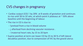
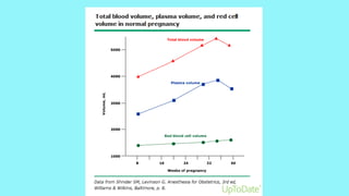
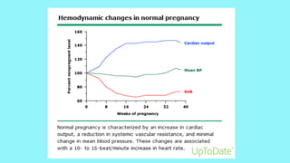
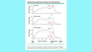
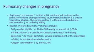
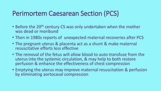
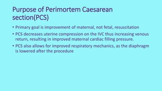
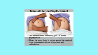
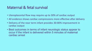
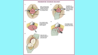
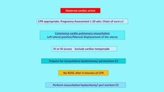
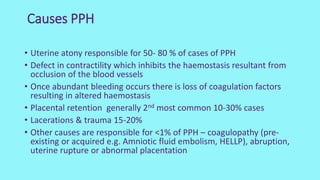
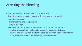
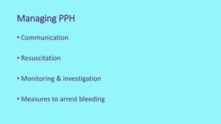
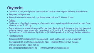
ED Obstetrics
Louise Farrell
CVS changes in pregnancy
• Cardiac output (CO) ↑ by 20% at 8 weeks of gestation and continues
to rise until 30 to 32 wks, at which point it plateaus at ~ 50% above
baseline until the beginning of labour.
• The rise in CO is due to:
- ↑preload from a rise in blood volume
- ↓ afterload from declining vascular resistance .
- ↑maternal heart rate, by 15 to 20 bpm
• Supine position at term can lower CO by 25 to 30 % cf left lateral
decubitus position, due to compression of IVC by the gravid uterus
Pulmonary changes in pregnancy
• Beginning 1st trimester ↑ in tidal vol & respiratory drive (due to the
stimulatory effects of progesterone) cause hyperventilation & a chronic
respiratory alkalosis The compensatory ↓ in the plasma bicarbonate
concentration ↓ its buffering ability.
• Pregnancy-related pulmonary changes include:
- PaO2 may be slightly ↑ at 104 to 108 mmHg as a result of ↑ CO &
minimization of the ventilation-perfusion mismatch in the lung.
- Beginning ~ 20 wks of gestation, upward displacement of the diaphragm
→20% ↓ in functional residual capacity.
- Oxygen consumption ↑ by almost 20%
Perimortem Caesarean Section (PCS)
• Before the 20th century CS was only undertaken when the mother
was dead or moribund
• Then in 1980s reports of unexpected maternal recoveries after PCS
• The pregnant uterus & placenta act as a shunt & make maternal
resuscitative efforts less effective
• The removal of the fetus will allow blood to auto-transfuse from the
uterus into the systemic circulation, & may help to both restore
perfusion & enhance the effectiveness of chest compression
• Emptying the uterus may improve maternal resuscitation & perfusion
by eliminating aortocaval compression
Purpose of Perimortem Caesarean
section(PCS)
• Primary goal is improvement of maternal, not fetal, resuscitation
• PCS decreases uterine compression on the IVC thus increasing venous
return, resulting in improved maternal cardiac filling pressure.
• PCS also allows for improved respiratory mechanics, as the diaphragm
is lowered after the procedure
Special considerations in primary survey
• Airway: There is physiologic narrowing of the upper airways in 3rd trimester
• Use an endotracheal tube 1 size smaller.
• Intubation medications are the same.
• RSI is the preferred method of intubation in 3rd trimester ↑=risk of aspiration.
• Breathing: Pregnant patients are predisposed to rapid ↓ in Pa02 during apnea
• Supplemental O2 all pregnant patient being resuscitated regardless of saturation.
• Circulation: Hypovolemia should be suspected before clinical signs of
hypotension in trauma patients, as the state of hypervolemia & resulting
hemodilution may mask underlying significant blood loss.
• Aggressive volume resuscitation is encouraged regardless of BP.
• Resuscitation of the pregnant patient should include uterine displacement to relieve
compression of the IVC and thus improve cardiac output and restore circulation.
When to perform PCS
• Cardiac arrest when gestational age ≥ 24 wks
• When resuscitative efforts deemed appropriate
• After correction of the causes of cardiac arrest – IV access,
catastrophic haemorrhage controlled, airway patency established &
tension pneumothorax & cardiac tamponade excluded
• Wherever possible PCS if no ROSC following 4 mins active CPR
• There is no need to determine if the fetus is alive
Maternal & fetal survival
• Uteroplacental flow may require up to 30% of cardiac output
• All evidence shows cardiac compressions more effective after delivery
• Delivery of the near term infant provides 30-80% improvement in
cardiac output
• Best outcomes in terms of infant neurologic status appear to
occur if the infant is delivered within 5 minutes of maternal
cardiac arrest
Pregnant trauma patient
Begin Primary survey
Control catastrophic haemorrhage
Airway with C-spine control. Breathing & ventilation
No respiratory effort
Exclude Tension/open pneumothorax
Oxygenate Ventilate
No Palpable Pulse
Maternal cardiac arrest
CPR appropriate: Pregnancy Assessment ≥ 20 wks: Chain of survival
Commence cardio-pulmonary resuscitation
Left lateral position/Manual displacement of the uterus
IV or IO access Exclude cardiac tamponade
Prepare for resuscitative hysterotomy/ perimortem CS
No ROSC after 4 minutes of CPR
Perform resuscitative hysterotomy/ peri mortem CS
Cord prolapse out of hospital
• Cord prolapse may occur with premature rupture of membranes .
Overt cord prolapse - call for help & assume the knee-chest face-
down position or lie on the floor with pillows to elevate the hips
above the heart till ambulance for hospital transfer. During transfer,
the knee-chest position is potentially unsafe for the mother so a left
lateral position, with pillows under the hip. If possible, the presenting
part should be elevated manually or by bladder distension during the
transfer
• Cord prolapse outside of the hospital setting - poor prognosis. In a
review of large series, cord prolapse occurring outside of the hospital
was associated with perinatal mortality rates of 38 to 44 %
Intrauterine resuscitation
• Manually elevate the presenting part (pp) — the most common
intervention for managing cord prolapse - fast, does not require special
equipment, & shown to be effective. While preparations for an emergency
C/S delivery are made, the clinician places his/her hand in the vagina &
gently elevates the fetal head so it is not compressing the cord.
• Place patient in Trendenburg or knee chest position — Attempt to elevate
the fetal head manually to decompress the cord. Placing the patient in
steep Trendelenburg or the knee-chest position may change the
relationship between the fetus & umbilical cord & thereby alleviate cord
compression.
• Retrofill the bladder — with 500 to 700 mls of saline rapidly via a catheter
to distend the bladder→ elevate pp . May be particularly useful as a
temporizing measure when C/S delivery cannot be performed urgently.
• Administer a tocolytic — rapid action to reduce pressure on the cord from
uterine contractions. Terbutaline 0.25mg S/C
Overt cord prolapse
• Minimize manipulating an overtly prolapsed cord & avoid exposing
it to the cold environment, which may exacerbate poor perfusion by
inducing spasm of the umbilical artery. Replace an overtly prolapsed
cord in the vagina & keep it moist with wet gauze.
• Perform emergency delivery by the most rapid & safe route, which
is typically C/S –Assisted vaginal delivery may be considered in select
situations when, in the clinician's judgment, the fetus can be
delivered safely & as quickly, or more quickly, than by C/S..
• If the fetal heart rate is present prior to moving to the operating
room, do not pause to recheck it before surgery as should perform
emergency C/S regardless of the findings & attempt neonatal
resuscitation, if required.
PPH - Definition
• 10 in 1st 24 after delivery
• Traditional PPH loss of ≥ 500mls of blood
• Major PPH ≥ 1000mls
• Severe PPH ≥ 2000mls
• Incidence of PPH varies widely, depending upon criteria used to
define the disorder. A reasonable estimate is 1 to 5% of deliveries
The 4 Ts of PPH
• Tone
• Trauma
• Thrombin
• Tissue
Causes PPH
• Uterine atony responsible for 50- 80 % of cases of PPH
• Defect in contractility which inhibits the haemostasis resultant from
occlusion of the blood vessels
• Once abundant bleeding occurs there is loss of coagulation factors
resulting in altered haemostasis
• Placental retention generally 2nd most common 10-30% cases
• Lacerations & trauma 15-20%
• Other causes are responsible for <1% of PPH – coagulopathy (pre-
existing or acquired e.g. Amniotic fluid embolism, HELLP), abruption,
uterine rupture or abnormal placentation
Arresting the bleeding
• The commonest cause of PPH is uterine atony
• If uterine atony suspected as cause & other causes excluded
- uterine massage
- bimanual uterine compression
- empty bladder
- oxytocics – syntocinon, ergometrine, carboprost, misoprostol
- surgical intervention – balloon tamponade, haemostatic brace
suture, bilateral ligation of uterine arteries, bilateral ligation of internal
iliacs, selective arterial embolization, hysterectomy
Managing PPH
• Communication
• Resuscitation
• Monitoring & investigation
• Measures to arrest bleeding
The uterotonics
• Which type ?
• What dose ?
• Which route of administration?
• What mode of delivery ?
Oxytocics
• Oxytocin is the prophylactic uterotonic of choice after vaginal delivery. Rapid onset
• Requires refrigeration
• Route & dose controversial - probably slow bolus of 5 IU over 1 min
• Others
• Carbetocin - Synthetic analogue of oxytocin with a prolonged duration of action. Dose
also uncertain ? 100 μg
• Ergometrine – somewhat more effective than Syntocinon & longer acting but frequent
adverse events most especially hypertension. Vomiting & nausea is substantially >
Syntocinon. Combination of Syntocinon (5IU) & Ergometrine (0.5mg) better tolerated
• Prostaglandins
Misoprostol (Prostaglandin E1 analogue) – oral, sublingual, rectal or vaginal
Carboprost (15-methyl prostaglandin F2α) – 250mg IMI never IVI. ? given
intramyometrially (but not lic)
Dinoprost (prostaglandin F2α ) – intramyometrial injection only
WOMAN trial
• Tranexamic acid reduces death due to bleeding in women with post-
partum haemorrhage with no adverse effects.
• When used as a treatment for PPH, tranexamic acid should be given
as soon as possible after bleeding onset.
Management of PPH after vaginal delivery
Initial
management
of PPH
≥ 30
min
Call Obstetric & Anaesthetic team
OBSTETRIC TEAM
• Urinary
catheterisation
• Visual inspection of
lower genital tract
• Uterine Massage
•Repeat uterotonics
•Manual removal of
placenta if not yet
delivered
• Manual exploration
of uterus If placenta
already delivered
ANAESTHETIC TEAM
• Monitor
• Assess & maintain
haemodynamics: plasma
expansion by crystalloids
•Provide anaesthesia for
manual exploration of uterus
•Antibiotic therapy
•Prevention of hypothermia,
oxygen therapy
•Haemocue
•Bloods
COMMUNICATION
Failure of initial
management
Failure of initial management
Persistent or
severe PPH
• Carboprost ( beware of
contraindications) 0.25 mg by IMI
repeated at intervals of not < 15 mins to
a max of 8 doses
• Direct intramyometrial injection of
carboprost 0.5 mg with responsibility of
the administering clinician as it is not
recommended for intramyometrial use
•2nd peripheral line
•Bloods – FBP,
Coagulation profile
•Xmatch blood
≥ 30
min
Failure of 2nd line measures
Persistent or
severe PPH
Failure of 2nd line measures
Intra- uterine
balloon
tamponnade
Haemodynamically
unstable &/or
Massive haemorrhage
&/or
Embolisation unavailable
Conservative surgery
•Arterial ligation
•Uterine compression
suture
Failure
Haemodynamically stable
&
Embolisation rapidly
available
EMBOLIZATION
Failure
HYSTERECTOMY +/- rFVIIa (after
checking fibrinogen &
platelets
• Fluid resuscitation
(crystalloids/colloids)
•Transfusion of packed
RBC
•Hypothermia prevention
• Laboratory results
• +/- TXA
• +/-Fresh frozen plasma
• +/- Fibrinogen
• +/- Platelets
• +/- Arterial line
• +/- Central venous
• +/- Noradrenaline
Failure
20 PPH
• Secondary PPH is usually associated with endometritis (with or
without retained products of conception).
• Resuscitation & fluid management
• Antibiotic therapy +/_ uterotonics . In situations of excessive or
continued bleeding surgical intervention is usually required
Thank you
QUESTIONS ?

Emergency Obstetrics

  • 1.
  • 4.
    CVS changes inpregnancy • Cardiac output (CO) ↑ by 20% at 8 weeks of gestation and continues to rise until 30 to 32 wks, at which point it plateaus at ~ 50% above baseline until the beginning of labour. • The rise in CO is due to: - ↑preload from a rise in blood volume - ↓ afterload from declining vascular resistance . - ↑maternal heart rate, by 15 to 20 bpm • Supine position at term can lower CO by 25 to 30 % cf left lateral decubitus position, due to compression of IVC by the gravid uterus
  • 8.
    Pulmonary changes inpregnancy • Beginning 1st trimester ↑ in tidal vol & respiratory drive (due to the stimulatory effects of progesterone) cause hyperventilation & a chronic respiratory alkalosis The compensatory ↓ in the plasma bicarbonate concentration ↓ its buffering ability. • Pregnancy-related pulmonary changes include: - PaO2 may be slightly ↑ at 104 to 108 mmHg as a result of ↑ CO & minimization of the ventilation-perfusion mismatch in the lung. - Beginning ~ 20 wks of gestation, upward displacement of the diaphragm →20% ↓ in functional residual capacity. - Oxygen consumption ↑ by almost 20%
  • 10.
    Perimortem Caesarean Section(PCS) • Before the 20th century CS was only undertaken when the mother was dead or moribund • Then in 1980s reports of unexpected maternal recoveries after PCS • The pregnant uterus & placenta act as a shunt & make maternal resuscitative efforts less effective • The removal of the fetus will allow blood to auto-transfuse from the uterus into the systemic circulation, & may help to both restore perfusion & enhance the effectiveness of chest compression • Emptying the uterus may improve maternal resuscitation & perfusion by eliminating aortocaval compression
  • 11.
    Purpose of PerimortemCaesarean section(PCS) • Primary goal is improvement of maternal, not fetal, resuscitation • PCS decreases uterine compression on the IVC thus increasing venous return, resulting in improved maternal cardiac filling pressure. • PCS also allows for improved respiratory mechanics, as the diaphragm is lowered after the procedure
  • 12.
    Special considerations inprimary survey • Airway: There is physiologic narrowing of the upper airways in 3rd trimester • Use an endotracheal tube 1 size smaller. • Intubation medications are the same. • RSI is the preferred method of intubation in 3rd trimester ↑=risk of aspiration. • Breathing: Pregnant patients are predisposed to rapid ↓ in Pa02 during apnea • Supplemental O2 all pregnant patient being resuscitated regardless of saturation. • Circulation: Hypovolemia should be suspected before clinical signs of hypotension in trauma patients, as the state of hypervolemia & resulting hemodilution may mask underlying significant blood loss. • Aggressive volume resuscitation is encouraged regardless of BP. • Resuscitation of the pregnant patient should include uterine displacement to relieve compression of the IVC and thus improve cardiac output and restore circulation.
  • 14.
    When to performPCS • Cardiac arrest when gestational age ≥ 24 wks • When resuscitative efforts deemed appropriate • After correction of the causes of cardiac arrest – IV access, catastrophic haemorrhage controlled, airway patency established & tension pneumothorax & cardiac tamponade excluded • Wherever possible PCS if no ROSC following 4 mins active CPR • There is no need to determine if the fetus is alive
  • 16.
    Maternal & fetalsurvival • Uteroplacental flow may require up to 30% of cardiac output • All evidence shows cardiac compressions more effective after delivery • Delivery of the near term infant provides 30-80% improvement in cardiac output • Best outcomes in terms of infant neurologic status appear to occur if the infant is delivered within 5 minutes of maternal cardiac arrest
  • 20.
    Pregnant trauma patient BeginPrimary survey Control catastrophic haemorrhage Airway with C-spine control. Breathing & ventilation No respiratory effort Exclude Tension/open pneumothorax Oxygenate Ventilate No Palpable Pulse
  • 21.
    Maternal cardiac arrest CPRappropriate: Pregnancy Assessment ≥ 20 wks: Chain of survival Commence cardio-pulmonary resuscitation Left lateral position/Manual displacement of the uterus IV or IO access Exclude cardiac tamponade Prepare for resuscitative hysterotomy/ perimortem CS No ROSC after 4 minutes of CPR Perform resuscitative hysterotomy/ peri mortem CS
  • 22.
    Cord prolapse outof hospital • Cord prolapse may occur with premature rupture of membranes . Overt cord prolapse - call for help & assume the knee-chest face- down position or lie on the floor with pillows to elevate the hips above the heart till ambulance for hospital transfer. During transfer, the knee-chest position is potentially unsafe for the mother so a left lateral position, with pillows under the hip. If possible, the presenting part should be elevated manually or by bladder distension during the transfer • Cord prolapse outside of the hospital setting - poor prognosis. In a review of large series, cord prolapse occurring outside of the hospital was associated with perinatal mortality rates of 38 to 44 %
  • 23.
    Intrauterine resuscitation • Manuallyelevate the presenting part (pp) — the most common intervention for managing cord prolapse - fast, does not require special equipment, & shown to be effective. While preparations for an emergency C/S delivery are made, the clinician places his/her hand in the vagina & gently elevates the fetal head so it is not compressing the cord. • Place patient in Trendenburg or knee chest position — Attempt to elevate the fetal head manually to decompress the cord. Placing the patient in steep Trendelenburg or the knee-chest position may change the relationship between the fetus & umbilical cord & thereby alleviate cord compression. • Retrofill the bladder — with 500 to 700 mls of saline rapidly via a catheter to distend the bladder→ elevate pp . May be particularly useful as a temporizing measure when C/S delivery cannot be performed urgently. • Administer a tocolytic — rapid action to reduce pressure on the cord from uterine contractions. Terbutaline 0.25mg S/C
  • 24.
    Overt cord prolapse •Minimize manipulating an overtly prolapsed cord & avoid exposing it to the cold environment, which may exacerbate poor perfusion by inducing spasm of the umbilical artery. Replace an overtly prolapsed cord in the vagina & keep it moist with wet gauze. • Perform emergency delivery by the most rapid & safe route, which is typically C/S –Assisted vaginal delivery may be considered in select situations when, in the clinician's judgment, the fetus can be delivered safely & as quickly, or more quickly, than by C/S.. • If the fetal heart rate is present prior to moving to the operating room, do not pause to recheck it before surgery as should perform emergency C/S regardless of the findings & attempt neonatal resuscitation, if required.
  • 25.
    PPH - Definition •10 in 1st 24 after delivery • Traditional PPH loss of ≥ 500mls of blood • Major PPH ≥ 1000mls • Severe PPH ≥ 2000mls • Incidence of PPH varies widely, depending upon criteria used to define the disorder. A reasonable estimate is 1 to 5% of deliveries
  • 26.
    The 4 Tsof PPH • Tone • Trauma • Thrombin • Tissue
  • 27.
    Causes PPH • Uterineatony responsible for 50- 80 % of cases of PPH • Defect in contractility which inhibits the haemostasis resultant from occlusion of the blood vessels • Once abundant bleeding occurs there is loss of coagulation factors resulting in altered haemostasis • Placental retention generally 2nd most common 10-30% cases • Lacerations & trauma 15-20% • Other causes are responsible for <1% of PPH – coagulopathy (pre- existing or acquired e.g. Amniotic fluid embolism, HELLP), abruption, uterine rupture or abnormal placentation
  • 28.
    Arresting the bleeding •The commonest cause of PPH is uterine atony • If uterine atony suspected as cause & other causes excluded - uterine massage - bimanual uterine compression - empty bladder - oxytocics – syntocinon, ergometrine, carboprost, misoprostol - surgical intervention – balloon tamponade, haemostatic brace suture, bilateral ligation of uterine arteries, bilateral ligation of internal iliacs, selective arterial embolization, hysterectomy
  • 29.
    Managing PPH • Communication •Resuscitation • Monitoring & investigation • Measures to arrest bleeding
  • 30.
    The uterotonics • Whichtype ? • What dose ? • Which route of administration? • What mode of delivery ?
  • 31.
    Oxytocics • Oxytocin isthe prophylactic uterotonic of choice after vaginal delivery. Rapid onset • Requires refrigeration • Route & dose controversial - probably slow bolus of 5 IU over 1 min • Others • Carbetocin - Synthetic analogue of oxytocin with a prolonged duration of action. Dose also uncertain ? 100 μg • Ergometrine – somewhat more effective than Syntocinon & longer acting but frequent adverse events most especially hypertension. Vomiting & nausea is substantially > Syntocinon. Combination of Syntocinon (5IU) & Ergometrine (0.5mg) better tolerated • Prostaglandins Misoprostol (Prostaglandin E1 analogue) – oral, sublingual, rectal or vaginal Carboprost (15-methyl prostaglandin F2α) – 250mg IMI never IVI. ? given intramyometrially (but not lic) Dinoprost (prostaglandin F2α ) – intramyometrial injection only
  • 32.
    WOMAN trial • Tranexamicacid reduces death due to bleeding in women with post- partum haemorrhage with no adverse effects. • When used as a treatment for PPH, tranexamic acid should be given as soon as possible after bleeding onset.
  • 33.
    Management of PPHafter vaginal delivery Initial management of PPH ≥ 30 min Call Obstetric & Anaesthetic team OBSTETRIC TEAM • Urinary catheterisation • Visual inspection of lower genital tract • Uterine Massage •Repeat uterotonics •Manual removal of placenta if not yet delivered • Manual exploration of uterus If placenta already delivered ANAESTHETIC TEAM • Monitor • Assess & maintain haemodynamics: plasma expansion by crystalloids •Provide anaesthesia for manual exploration of uterus •Antibiotic therapy •Prevention of hypothermia, oxygen therapy •Haemocue •Bloods COMMUNICATION Failure of initial management
  • 34.
    Failure of initialmanagement Persistent or severe PPH • Carboprost ( beware of contraindications) 0.25 mg by IMI repeated at intervals of not < 15 mins to a max of 8 doses • Direct intramyometrial injection of carboprost 0.5 mg with responsibility of the administering clinician as it is not recommended for intramyometrial use •2nd peripheral line •Bloods – FBP, Coagulation profile •Xmatch blood ≥ 30 min Failure of 2nd line measures
  • 35.
    Persistent or severe PPH Failureof 2nd line measures Intra- uterine balloon tamponnade Haemodynamically unstable &/or Massive haemorrhage &/or Embolisation unavailable Conservative surgery •Arterial ligation •Uterine compression suture Failure Haemodynamically stable & Embolisation rapidly available EMBOLIZATION Failure HYSTERECTOMY +/- rFVIIa (after checking fibrinogen & platelets • Fluid resuscitation (crystalloids/colloids) •Transfusion of packed RBC •Hypothermia prevention • Laboratory results • +/- TXA • +/-Fresh frozen plasma • +/- Fibrinogen • +/- Platelets • +/- Arterial line • +/- Central venous • +/- Noradrenaline Failure
  • 36.
    20 PPH • SecondaryPPH is usually associated with endometritis (with or without retained products of conception). • Resuscitation & fluid management • Antibiotic therapy +/_ uterotonics . In situations of excessive or continued bleeding surgical intervention is usually required
  • 37.