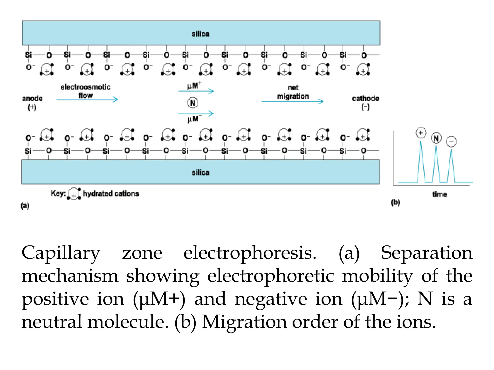
2. 2
Contents
Introduction
Electrophoretic Separation
Types of Electrophoresis
Capillary Electrophoresis
Paper Electrophoresis
2D Electrophoresis
Gel Electrophoresis
SDS-PAGE
3. 3
Introduction
Electrophoresis: It is defined as the
migration of particles under the
influence of electric field
It is a separation method based on the
differential rate of migration of
charged species in a buffer solution
across which a DC current field has
been applied.
4. 4
Introduction Contd…
Separations are based on differences in
charge-to-size ratio for various analytes
in the sample.
Larger this ratio, faster the ion migrates
in the electric field
5. 5
Introduction Contd…
This separation technique was first
developed by the Swedish chemist
Arne Tiselius in 1930s for the study of
serum protein
Awarded Noble Prize in 1948
6. 6
Electrophoretic Separation
It is performed by injecting a small
quantity of sample into an aqueous
buffer solution that is contained in a
narrow tube or on a gel.
A high DC potential is applied across
the length of the buffer by means of pair
of electrodes located at either end of
buffer.
7. 7
Electrophoretic Separation Contd…
The potential causes ions of the sample
to migrate towards one or the other
electrode.
Rate of migration depends on its
charge & also on its size
Separations are based on charge-to-size
ratio.
8. 8
Types of Electrophoresis
Slab Electrophoresis
Capillary Electrophoresis
Paper Electrophoresis
Gel Electrophoresis
2D Electrophoresis
Alternating-field Electrophoresis
10. 10
Capillary Electrophoresis (CE)
It is a high speed, high resolution
separations on exceptionally small
sample volumes (0.1 to 10nL)
Potentials of 20,000 to 60,000 V are
commonly used in capillary
electrophoresis
11. 11
Instrumentation for Capillary
Electrophoresis
A buffer-filled fused-silica capillary (10
to 100µm in ID & 40 to 100 cm long),
extends between two buffer reservoirs
that also holds platinum electrodes.
Sample introduction at one end and
detection at the other.
12.
13. 13
Sample Introduction
The most common sample introduction
methods are:
Electrokinetic Injection and
Pressure Injection
14. 14
Electrokinetic Injection
One end of capillary & its electrodes are
removed and placed in small cup
containing sample
potential is applied for measured time
15. 15
Pressure Injection
Pressure difference is used to drive the
sample and can be done by:
Applying vacuum at the detector end
By pressurizing the sample
By elevating the sample end
18. 18
Advantages of CE
High separation efficiency
Small sample size required (0.1-10 nL)
Fast separation (1 to 45 min)
Easy and predictable selectivity
Automation
Low operating cost
Reproducibility
Couple to mass spectrophotometer
19. 19
Limitations of CE
Not well suited for determination of
low molecular weight, volatile
compounds
Not well suited for determination on
nonionic, high molecular weight
polymers
Not as sensitive as HPLC
20.
21. 21
Modes of CE
Capillary Zone Electrophoresis
Capillary Gel Electrophoresis
Capillary Isoelectric Focusing
Capillary Isotachophoresis
22. 22
Capillary Zone Electrophoresis
Buffer composition is constant
throughout the separation region.
Applied potential causes the different
ionic mixture to migrate according to its
mobility to separate into zones
23. Capillary zone electrophoresis. (a) Separation
mechanism showing electrophoretic mobility of the
positive ion (μM+) and negative ion (μM−); N is a
neutral molecule. (b) Migration order of the ions.
24. 24
General Uses of CE
Separation and identification of
Polar & non-polar
Some elements, including non-ionic & ionic
compounds,
Inorganic anions & cations,
Macromulecules (proteins & oligonucleotides)
Chiral Compounds
25. 25
General Uses of CE Contd..
Quantitative and qualitative determination
of compounds and some elements in
mixture
Determination of molecular weights of
large biomolecules and isoelectric points of
proteins
Used in biochemical, clinical,
environmental, forensic, food and
Pharmaceutical applications
27. 27
2D Electrophoresis (2DE)
A separation is carried out in one direction
for a suitable time and then for an additional
time at right angles to the first separation
Often isoelectric focusing is combined with
SDS-PAGE in two-dimensional gel
electrophoresis
Proteins with similar isoelectric points or with
similar molecular weights can be separated
by this method
28. After isoelectric focusing in pH gradient of the
electric field the electrophoretic separation is
used in SDS-PAGE Electrophoresis
29. 29
Gel Electrophoresis (GE)
Generally performed in a porous gel
polymer matrix
Pores contains a buffer mixture in
which the separation is carried out
Most common type of gel used in
electrophoresis is a polyacrylamide
polymer.
30.
31.
32. 32
SDS-PAGE
SDS- Sodium Dodecyl Sulfate
PAGE- Polyacrylamide Gel
Electrophoresis
One of the most important
electrophoresis technique used to
measure molecular weights of proteins
33. 33
SDS-PAGE Contd…
SDS is a detergent which denatures
proteins by binding to the hydrophobic
regions
It coats the linear protein sequence with
the set of SDS molecules.
The SDS is negatively charged and thus
becomes the dominant charge of the
complex
34. 34
SDS-PAGE Contd…
Number of SDS molecules that bind is
proportional to size of protein
Then run through inert polymer,
Polyacrylamide
37. 37
References
Principles of Instrumental Analysis by
Skoog, Holler, Nieman, 5th Edition, 2001,
Page no. 778-792
Handbook of Instrumental Techniques for
Analytical Chemistry by Frank Settle, 2004
Page No. 165-180
Paper Chromatography and Electrophoresis
(Vol. I) by Gunter Zweing & John R.
Whitaker, 1967, Page No. 1-46
www.google.com