
More Related Content
What's hot
What's hot (20)
Viewers also liked
Viewers also liked (10)
Similar to Overview of Electrophoresis
Similar to Overview of Electrophoresis (20)
More from Marudhar Kesari Jain College for Women Vaniyambadi - 635 751, Tamil Nadu, INDIA.
More from Marudhar Kesari Jain College for Women Vaniyambadi - 635 751, Tamil Nadu, INDIA. (20)
Recently uploaded
Recently uploaded (20)
Overview of Electrophoresis
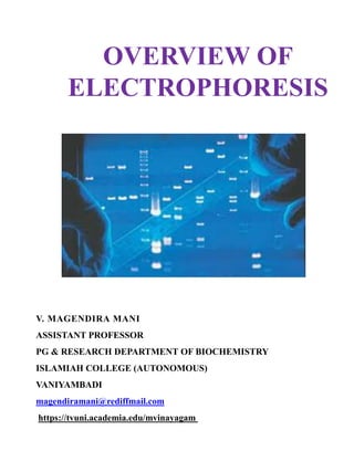
- 1. OVERVIEW OF ELECTROPHORESIS V. MAGENDIRA MANI ASSISTANT PROFESSOR PG & RESEARCH DEPARTMENT OF BIOCHEMISTRY ISLAMIAH COLLEGE (AUTONOMOUS) VANIYAMBADI magendiramani@rediffmail.com https://tvuni.academia.edu/mvinayagam
- 2. Electrophoresis Electrophoresis is defined as the migration of charged particles through a solution under influence of an electric field. Biological molecules such as amino acids, peptides, protein, nucleotides, and nucleic acids possess ionisable groups and are made to exist as electrically charged species either as cations or anions. Even carbohydrates can be given weak charges by derivatization such as borates or phosphates. In electrophoresis cation move towards cathode and anion move towards anode. The rate of migration is depends on The charge of the particle Applied electric field Temperature Nature of the suspended medium
- 4. SAMPLE Charge : Rate of migration increases with increase in net charge. It depends on pH. Size : Rate of migration decreases for larger molecules. It is due to increase frictional and electrostatics forces. Shape : Molecular have similar charge but differ in shape exhibit different migration rate.
- 5. ELECTRIC FIELD According to ohms law I=V/R Current = Voltage / Resistance Voltage : Increase in voltage leads to increase in rate of migration Current : Increase in current leads to Increase in voltage, so the migration also Increases Resistance: If resistance increase migration decreases.
- 6. BUFFER Buffer determines & stabilizes pH of the supporting medium Also affects the migration rate of compounds in a number of ways Composition of Buffer Acetate Barbiturate Citrate EDTA Formate Phosphate Pyridine buffers commonly used.
- 7. IONIC STRENGTH As ionic strength of buffer increases Proportion of current carried by buffer increases Proportion of current carried by the sample decreases and hence showing decrease in sample rate of migration. High ionic strength • Also increases overall current and hence heat is produced As ionic strength of buffer decreases Proportion of current carried by buffer decreases Proportion of current carried by the sample increases and hence showing increase in sample rate of migration. Low ionic strength • Also decrease in overall current and hence decrease in heat production
- 8. pH pH determines the ionization, if ionization of organic acid increases as pH increases, ionization of organic acid decreases as pH decrease. Therefore the degree of ionization is pH dependent. SUPPORTING MEDIUM Adsorption Adsorption is the retentio n of sample molecule by supporting medium. Adsorption causes tailing of sample so that it moves in the shape of a ‘comet’ rather than a distinct compact band Adsorption reduces both the rate of migration and resolution of separation of molecule.
- 9. Electro – endo-osmosis Electro – endo-osmosis due to the presence of charged groups on the surface of the supporting medium Eg. Paper - Carboxyl group (COO-) Agarose - Sulphate group (SO2-) Glass wall- Silanol (SiO-) .
- 10. Above the pH value of three these charged groups will have ionize and generates negatively charged sites. These ionized groups create an electrical double layer or region at supporting medium. When voltage is applied, cation in electrolyte near supporting medium migrate towards cathode pulling electrolyte solution with them. This creates a net Electro – endo-osmotic flow towards the cathode. The Electro – endo-osmosis will accelerate the movement of cations, but retard anion movements.
- 11. MOLECULAR SIEVING Gels have sieve like structure In agar, starch, and poly acryl amide gels the movement of large molecule is hindered by decreasing the pore size, since all the molecule has to transverse through pores. If sephadex gel is used, small molecules are tightly held by pores and large molecules are excluded by small pores causing movement outside the pores called molecular sieving.
- 13. Moving boundary electrophoresis Moving boundary electrophoresis technique was introduced by Swedish biochemist Arne Tiselius (1937) to separate proteins. The Tiselius apparatus is an U–tube in which the protein dissolved in buffer is taken in the lower part of the tube and plain buffer taken in the upper part of the tube. Then the limbs are connected to electrodes. Electrophoresis is carried out for 21/2 hours.at a reduced temperature (40 C). As current passed protein moved towards limbswhere only buffer is placed. Depending on the charge, the rate of movement differs and they will form boundaries. The movement of boundaries is observed in schieleran optical system. As protein moves, the system gives peaks. Each peak indicates one protein. Optical system works in the variation of refractive index of each boundary with the help of this diagram one can find the concentration & direction of various peaks.
- 14. Applications:- To separate proteins. To study protein–protein interaction. To measure homogenesity of protein. Homogenous protein give one peak. Disadvantages:- Slow technique. Requires complex optical system. Liable to disturbances by conventional and vibrational current. Cannot give complete separation of one protein.
- 15. SODIUMDODECYLSULPHATE POLY ACRYL AMIDE GEL ELECTROPHORESIS
- 16. Principle SDS PAGE is widely used method for separating protein mixture and for determining the molecular weights. SDS - is an anionic detergent Binds strongly to protein Causes their Denaturation Polymerization Cross linked poly acryl amide gel are formed by polymerization of acryl amide monomer and N, N’ Methylene Bisacryl amide in the presence of ammonium per sulphate and TEMED (Tetra Methylene Diamine). Ammonium per sulphate acts as free radical catalyst and TEMED acts as initiator.
- 17. Photopolymerization Photopolymerization is an alternative method that can be used to polymerise Acryl amide gels. Ammonium per sulphate, TEMED are replaced by Riboflavin. When the gel is poured it is placed in front of bright light for 2 – 3 hrs. Photopolymerization of riboflavin generates a free radical that initiates polymerization.
- 18. Pore size of the PAGE Pore size of the gel can be varied by changing the concentration of both Acryl amide and Methylene Bisacryl amide. PAGE can be made with content of 3 % to 30 %. Low percentage of gel 3 % have large pore size and used for separation of proteins and DNA. High percentage of gel 10 -20 % have smaller pore size and used for SDS PAGE.
- 19. Sample preparation The sample is boiled with the buffer containing β- mercaptoethanol and SDS for 5 min. β-mercaptoethanol reduces any disulfide bridges in protein that are holding tertiary structure of protein. SDS binds strongly to proteins and denature the protein On average one SDS molecule binds to every two amino acids. So the native charge of the molecule is completely swamped by SDS molecule (negative) Each protein molecule will be fully denatured, opens in to a rod shaped structure with a series of – ve charged SDS molecule along with polypeptide chain.
- 20. Gel preparation Gels used are vertical slabs, because it is more economical and more sample can be compared with each other when run under identical conditions (eg. 20 different samples) Gels are prepared in glass containers in which they are to be used. The two glass plates are held together but held apart from each other by plastic spacer, vertical slab gels are run along with the glass plate. Choice of percentage of gel to be used depends on the size of the protein sample. Separating gel used may vary from 10 % PAGE to 15 % PAGE. 15 % of gel used for separation of protein having molecular weight 10,000 to 1, 00,000 and 10 % of gel used for separation of protein having molecular weight 1, 50,000.
- 21. Sample application Dissolved samples can be applied using a micro syringe into wells of the gel. Sample buffer containing 10-15 % Sucrose or Glycerol, which increase the density of the buffer and ensures the sinking of the sample in to the wells. It also prevents the sample from mixing with buffer in the upper buffer reservoir. Sample buffer contain marker/tracker dye Bromophenol blue. It is a small molecule and it moves freely and indicates the electrophoretic migration. Urea, SDS, Disulfide reducing agent such as β-Mercaptoethanol are added to protein sample to facilitate their solubilisation. Protein sample can be loaded in the form of sharp band by using a staking gel over the separating gel. Only µ g of sample are used for analyzing.
- 24. Running the gel The gel slab sandwiched in between the glass plate is placed in the lower reservoir with the top of the gel in contact with the buffer in the upper reservoir. Thus the gel completes the electrical circuit between the lower and upper compartments. Although the buffer dissipates the heat generated, additional cooling may be needed. In sample small protein can more easily pass through the pores and larger proteins are successively retarded by frictional resistance due to sieving effect of the gel. Precise voltage and time required for the optimal separation Voltage: 30 mA; Time; 3 hrs
- 26. Detection When tracker dye reaches the bottom of the gel the current is turned off. Gel slabs are removed without any pressure, after removal gel is immersed in 7 % acetic acid to minimize diffusion of components. Then the gel is shaken well in an appropriate stain solution. Usually Commassive Brilliant Blue R250 is used and the gel is immersed for few hours. Then the gel is transferred in to a destain solution and kept for overnight. Destain solution removes unbound background dye from gel leaving stain protein visible as blue bands on a clear background.
- 28. Staining solution (100 ml) a. Coomassie Brilliant Blue R-250: 250 mg b. Methanol: 50 % c. Acetic acid: 10 % d. Distilled water: 40 % De-staining solution (100 ml) a. Methanol: 50 % b. Acetic acid: 10 % c. Distilled water: 40 % Time required for electrophoresis Gel preparation - 1– 1 ½ hrs Running the gel - 3 hrs Staining - 2– 3 hrs De-staining - overnight
- 29. Determination of Molecular weight (PROTEIN) by SDS PAGE. The Molecular weight can be determined comparing mobility of standard protein of known Molecular weight with of unknown Molecular weight that is run on the same gel. A calibration curve is constructed for standard protein of known Molecular weight by Distance migrated Vs Molecular weight x 104 The migration of unknown is measured are extrapolating this value in the calibration curve, the molecular weight of unknown can be determined.
- 32. Agarose gel electrophoresis is the easiest and most popular way of separating and analyzing DNA. Here DNA molecules are separated on the basis of charge by applying an electric field to the electrophoretic apparatus. Shorter molecules migrate more easily and move faster than longermolecules through the pores of the gel and this process is called sieving. The gel might be used to look at the DNA in order to quantify it or to isolate a particular band. The DNA can be visualized in the gel by the addition of ethidium bromide.
- 33. Agarose is a polysaccharide obtained from the red algae Porphyra umbilicalis. Its systematic name is (1 4)-3, 6- anhydro-a-L-galactopyranosyl-(1,3)-β-D-galactopyranan. Agarose makes an inert matrix. Most agarose gels are made between 0.7% and 2% of agarose. A 0.7% gel will show good separation for large DNA fragments (5-10kb) and a 2% gel will show good resolution for small fragments with size range of 0.2-1kb.
- 34. Materials Required: Gel casting trays, which are available in a variety of sizes and composed of Uv transparent plastic. Sample combs, around which molten agarose is poured to form sample wells in the gel. Electrophoresis buffer, usually Tris-acetate-EDTA (TAE) or Tris-borate-EDTA (TBE).
- 35. The migration rate of DNA fragments in both of these buffers is somewhat different due to the differences in ionic strength. These buffers provide the ions for supporting conductivity. Loading buffer, which contains something dense (e.g. glycerol) to allow the sample to "fall" into the sample wells, and one or two tracking dyes, which migrate in the gel and allow visual monitoring or how far the electrophoresis has proceeded.
- 36. Ethidium Bromide (EtBr) Ethidium bromide, a fluorescent dye used for staining nucleic acids. It is an intercalating agent which intercalates between nucleic acid bases and allows the convenient detection of DNA fragments in gel. When exposed to UV light, it will fluoresce with an orange colour. After the running of DNA through an EtBr-treated gel, any band containing more than ~20 ng DNA becomes distinctly visible under UV light. EtBr is a known "mutagen", however, safer alternatives are available. It can be incorporated with agarose gels or DNA samples before loading, for visualization of the fragments. Binding of Ethidium bromide to DNA alters its mass and rigidity, and thereby its mobility. Transilluminator (an ultraviolet light box), which is used to visualize ethidium bromide stained DNA in gels.
- 38. General procedure Casting of gel The gel is prepared by dissolving the agarose powder in an appropriate buffer, such as TAE or TBE, to be used in electrophoresis. The agarose is dispersed in the buffer before heating it to near-boiling point, but avoid boiling. The melted agarose is allowed to cool sufficiently before pouring the solution into a cast as the cast may warp or crack if the agarose solution is too hot. A comb is placed in the cast to create wells for loading sample, and the gel should be completely set before use. The concentration of gel affects the resolution of DNA separation. For a standard agarose gel electrophoresis, a 0.8% gives good separation or resolution of large 5-10kb DNA fragments, while 2% gel gives good resolution for small 0.2-1kb fragments. 1% gels are common for many applications
- 40. Loading of samples Once the gel has set, the comb is removed, leaving wells where DNA samples can be loaded. Loading buffer is mixed with the DNA sample before the mixture is loaded into the wells. The loading buffer contains a dense compound, which may be glycerol, sucrose, or Ficoll, that raises the density of the sample so that the DNA sample may sink to the bottom of the well. If the DNA sample contains residual ethanol after its preparation, it may float out of the well. The loading buffer also include colored dyes such as xylene cyanol and bromophenol blue used to monitor the progress of the electrophoresis. The DNA samples are loaded using a micropipette.
- 41. Electrophoresis Agarose gel electrophoresis is most commonly done horizontally in a submarine mode whereby the slab gel is completely submerged in buffer during electrophoresis. For optimal resolution of DNA greater than 2 kb in size in standard gel electrophoresis, 5 to 8 V/cm is recommended (the distance in cm refers to the distance between electrodes, therefore this recommended voltage would be 5 to 8 multiplied by the distance between the electrodes in cm). Voltage may also be limited by the fact that it heats the gel and may cause the gel to melt if it is run at high voltage for a prolonged period, especially if the gel used is LMP agarose gel.
- 42. Too high a voltage may also reduce resolution, as well as causing band streaking for large DNA molecules. Too low a voltage may lead to broadening of band for small DNA fragments due to dispersion and diffusion. A DNA marker is also run together for the estimation of the molecular weight of the DNA fragments.
- 43. Visualization / Staining DNA as well as RNA is normally visualized by staining with ethidium bromide, which intercalates into the major grooves of the DNA and fluoresces under UV light. The ethidium bromide may be added to the agarose solution before it gels, or the DNA gel may be stained later after electrophoresis. Destaining of the gel is not necessary but may produce better images.
- 44. Other methods of staining are available; examples are SYBR Green, GelRed, methylene blue, brilliant cresyl blue, Nile bluesulphate, and crystal violet. SYBR Green, GelRed and other similar commercial products are sold as safer alternatives to ethidium bromide as it has been shown to be mutagenic in Ames test, although the carcinogenicity of ethidium bromide has not actually been established. SYBR Green requires the use of a blue-light transilluminator. DNA stained with crystal violet can be viewed under natural light without the use of a UV transilluminator which is an advantage; however it may not produce a strong band.
- 45. When stained with ethidium bromide, the gel is viewed with an ultraviolet (UV) transilluminator. Standard transilluminators use wavelengths of 302/312-nm UV-B. The transilluminator apparatus may also contain image capture devices, such as a digital or Polaroid camera that allow an image of the gel to be taken or printed. Applications Estimation of the size of DNA molecules following restriction enzyme digestion, e.g. in restriction mapping of cloned DNA. Analysis of PCR products, e.g. in molecular genetic diagnosis or genetic fingerprinting Separation of restricted genomic DNA prior to Southern transfer or of RNA prior to Northern transfer.
- 46. Agarose gel electrophoresis is a method of gel electrophoresis used in biochemistry, molecular biology, and clinical chemistry to separate a mixed population of DNA or proteins in a matrix of agarose. The proteins may be separated by charge and/or size (IEF agarose, essentially size independent), and the DNA and RNA fragments by length. Biomolecules are separated by applying an electric field to move the charged molecules through an agarose matrix, and the biomolecules are separated by size in the agarose gel matrix.
- 47. V. MAGENDIRA MANI ASSISTANT PROFESSOR PG & RESEARCH DEPARTMENT OF BIOCHEMISTRY ISLAMIAH COLLEGE (AUTONOMOUS) VANIYAMBADI magendiramani@rediffmail.com https://tvuni.academia.edu/mvinayagam