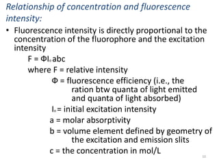
This document provides an overview of fluorometry, including basic concepts, instrumentation, and applications. It discusses how fluorescence occurs when a molecule absorbs light at one wavelength and reemits light at a longer wavelength. Factors that affect fluorescence such as temperature, pH, and dissolved oxygen are also covered. The relationship between fluorescence intensity and concentration is explained. Additionally, the document defines fluorescence polarization and describes various types of quenching including self-quenching, chemical quenching, and collisional quenching.
This presentation include the detailed explanation of various parts of a UV-Visible spectrophotometer and two types of UV-Visible spectrophotometers-Single beam and Doube beam. It also include the comparison between single beam and double beam spectrophotometers.
Ultraviolet-visible (UV-Vis) spectrophotometry is a technique used to measure light absorbance across the ultraviolet and visible ranges of the electromagnetic spectrum. When incident light strikes matter it can either be absorbed, reflected, or transmitted. The absorbance of radiation in the UV-Vis range causes atomic excitation, which refers to the transition of molecules from a low-energy ground state to an excited state.
Detectors are the brain of any chromatograhic system. It help us to record the chromatogram based on certain characteristics of the analyte and help us in identifying that compound both qualitatively and quantitatively.
Instrumentation of Thin Layer ChromatographyTanmoy Sarkar
Chromatography is a laboratory technique for the separation of a mixture. The mixture is dissolved in a fluid called the mobile phase, which carries it through a system on which a material called the stationary phase is fixed.
Thin-layer chromatography is a chromatography technique used to separate non-volatile mixtures. Thin-layer chromatography is performed on a sheet of an inert substrate such as glass, plastic, or aluminium foil, which is coated with a thin layer of adsorbent material, usually silica gel, aluminium oxide, or cellulose.
In this slide contains principle, instrumentation, methodology, and application of gel chromatography.
Presented by: SATHEES CHANDRA (Department of pharmaceutical analysis).
RIPER, anantapur
Spectrofluorimetry or fluorimetry (www.Redicals.com)Goa App
The term fluorescence comes from the mineral fluorspar (calcium fluoride) when Sir George G. Stokes observed in 1852 that fluorspar would give off visible light (fluoresce) when exposed to electromagnetic radiation in the ultraviolet wavelength.
spectrofluorometer is the instrument for recording fluorescence emission and absorption spectra When a beam of light is incident on certain substances they emit visible light or radiations. This is known as fluorescence. Fluorescence starts immediately after the absorption of light and stops as soon as the incident light is cut off. The substances showing this phenomenon are known as flourescent substances.
Fluorescence is the phenomenon whereby a molecule, after absorption radiation, emits radiation of a longer wavelength.
A compound absorbs radiation in the UV-rgion and emits visible light.
Absorption of uv/visible radiation causes transition of electrons from ground state (low energy) to excited state (high energy).
This increase in wavelength is known as the Stokes shift.
Tom Selleck Health: A Comprehensive Look at the Iconic Actor’s Wellness Journeygreendigital
Tom Selleck, an enduring figure in Hollywood. has captivated audiences for decades with his rugged charm, iconic moustache. and memorable roles in television and film. From his breakout role as Thomas Magnum in Magnum P.I. to his current portrayal of Frank Reagan in Blue Bloods. Selleck's career has spanned over 50 years. But beyond his professional achievements. fans have often been curious about Tom Selleck Health. especially as he has aged in the public eye.
Follow us on: Pinterest
Introduction
Many have been interested in Tom Selleck health. not only because of his enduring presence on screen but also because of the challenges. and lifestyle choices he has faced and made over the years. This article delves into the various aspects of Tom Selleck health. exploring his fitness regimen, diet, mental health. and the challenges he has encountered as he ages. We'll look at how he maintains his well-being. the health issues he has faced, and his approach to ageing .
Early Life and Career
Childhood and Athletic Beginnings
Tom Selleck was born on January 29, 1945, in Detroit, Michigan, and grew up in Sherman Oaks, California. From an early age, he was involved in sports, particularly basketball. which played a significant role in his physical development. His athletic pursuits continued into college. where he attended the University of Southern California (USC) on a basketball scholarship. This early involvement in sports laid a strong foundation for his physical health and disciplined lifestyle.
Transition to Acting
Selleck's transition from an athlete to an actor came with its physical demands. His first significant role in "Magnum P.I." required him to perform various stunts and maintain a fit appearance. This role, which he played from 1980 to 1988. necessitated a rigorous fitness routine to meet the show's demands. setting the stage for his long-term commitment to health and wellness.
Fitness Regimen
Workout Routine
Tom Selleck health and fitness regimen has evolved. adapting to his changing roles and age. During his "Magnum, P.I." days. Selleck's workouts were intense and focused on building and maintaining muscle mass. His routine included weightlifting, cardiovascular exercises. and specific training for the stunts he performed on the show.
Selleck adjusted his fitness routine as he aged to suit his body's needs. Today, his workouts focus on maintaining flexibility, strength, and cardiovascular health. He incorporates low-impact exercises such as swimming, walking, and light weightlifting. This balanced approach helps him stay fit without putting undue strain on his joints and muscles.
Importance of Flexibility and Mobility
In recent years, Selleck has emphasized the importance of flexibility and mobility in his fitness regimen. Understanding the natural decline in muscle mass and joint flexibility with age. he includes stretching and yoga in his routine. These practices help prevent injuries, improve posture, and maintain mobilit
HOT NEW PRODUCT! BIG SALES FAST SHIPPING NOW FROM CHINA!! EU KU DB BK substit...GL Anaacs
Contact us if you are interested:
Email / Skype : kefaya1771@gmail.com
Threema: PXHY5PDH
New BATCH Ku !!! MUCH IN DEMAND FAST SALE EVERY BATCH HAPPY GOOD EFFECT BIG BATCH !
Contact me on Threema or skype to start big business!!
Hot-sale products:
NEW HOT EUTYLONE WHITE CRYSTAL!!
5cl-adba precursor (semi finished )
5cl-adba raw materials
ADBB precursor (semi finished )
ADBB raw materials
APVP powder
5fadb/4f-adb
Jwh018 / Jwh210
Eutylone crystal
Protonitazene (hydrochloride) CAS: 119276-01-6
Flubrotizolam CAS: 57801-95-3
Metonitazene CAS: 14680-51-4
Payment terms: Western Union,MoneyGram,Bitcoin or USDT.
Deliver Time: Usually 7-15days
Shipping method: FedEx, TNT, DHL,UPS etc.Our deliveries are 100% safe, fast, reliable and discreet.
Samples will be sent for your evaluation!If you are interested in, please contact me, let's talk details.
We specializes in exporting high quality Research chemical, medical intermediate, Pharmaceutical chemicals and so on. Products are exported to USA, Canada, France, Korea, Japan,Russia, Southeast Asia and other countries.
New Drug Discovery and Development .....NEHA GUPTA
The "New Drug Discovery and Development" process involves the identification, design, testing, and manufacturing of novel pharmaceutical compounds with the aim of introducing new and improved treatments for various medical conditions. This comprehensive endeavor encompasses various stages, including target identification, preclinical studies, clinical trials, regulatory approval, and post-market surveillance. It involves multidisciplinary collaboration among scientists, researchers, clinicians, regulatory experts, and pharmaceutical companies to bring innovative therapies to market and address unmet medical needs.
Recomendações da OMS sobre cuidados maternos e neonatais para uma experiência pós-natal positiva.
Em consonância com os ODS – Objetivos do Desenvolvimento Sustentável e a Estratégia Global para a Saúde das Mulheres, Crianças e Adolescentes, e aplicando uma abordagem baseada nos direitos humanos, os esforços de cuidados pós-natais devem expandir-se para além da cobertura e da simples sobrevivência, de modo a incluir cuidados de qualidade.
Estas diretrizes visam melhorar a qualidade dos cuidados pós-natais essenciais e de rotina prestados às mulheres e aos recém-nascidos, com o objetivo final de melhorar a saúde e o bem-estar materno e neonatal.
Uma “experiência pós-natal positiva” é um resultado importante para todas as mulheres que dão à luz e para os seus recém-nascidos, estabelecendo as bases para a melhoria da saúde e do bem-estar a curto e longo prazo. Uma experiência pós-natal positiva é definida como aquela em que as mulheres, pessoas que gestam, os recém-nascidos, os casais, os pais, os cuidadores e as famílias recebem informação consistente, garantia e apoio de profissionais de saúde motivados; e onde um sistema de saúde flexível e com recursos reconheça as necessidades das mulheres e dos bebês e respeite o seu contexto cultural.
Estas diretrizes consolidadas apresentam algumas recomendações novas e já bem fundamentadas sobre cuidados pós-natais de rotina para mulheres e neonatos que recebem cuidados no pós-parto em unidades de saúde ou na comunidade, independentemente dos recursos disponíveis.
É fornecido um conjunto abrangente de recomendações para cuidados durante o período puerperal, com ênfase nos cuidados essenciais que todas as mulheres e recém-nascidos devem receber, e com a devida atenção à qualidade dos cuidados; isto é, a entrega e a experiência do cuidado recebido. Estas diretrizes atualizam e ampliam as recomendações da OMS de 2014 sobre cuidados pós-natais da mãe e do recém-nascido e complementam as atuais diretrizes da OMS sobre a gestão de complicações pós-natais.
O estabelecimento da amamentação e o manejo das principais intercorrências é contemplada.
Recomendamos muito.
Vamos discutir essas recomendações no nosso curso de pós-graduação em Aleitamento no Instituto Ciclos.
Esta publicação só está disponível em inglês até o momento.
Prof. Marcus Renato de Carvalho
www.agostodourado.com
Explore natural remedies for syphilis treatment in Singapore. Discover alternative therapies, herbal remedies, and lifestyle changes that may complement conventional treatments. Learn about holistic approaches to managing syphilis symptoms and supporting overall health.
Basavarajeeyam is an important text for ayurvedic physician belonging to andhra pradehs. It is a popular compendium in various parts of our country as well as in andhra pradesh. The content of the text was presented in sanskrit and telugu language (Bilingual). One of the most famous book in ayurvedic pharmaceutics and therapeutics. This book contains 25 chapters called as prakaranas. Many rasaoushadis were explained, pioneer of dhatu druti, nadi pareeksha, mutra pareeksha etc. Belongs to the period of 15-16 century. New diseases like upadamsha, phiranga rogas are explained.
Title: Sense of Taste
Presenter: Dr. Faiza, Assistant Professor of Physiology
Qualifications:
MBBS (Best Graduate, AIMC Lahore)
FCPS Physiology
ICMT, CHPE, DHPE (STMU)
MPH (GC University, Faisalabad)
MBA (Virtual University of Pakistan)
Learning Objectives:
Describe the structure and function of taste buds.
Describe the relationship between the taste threshold and taste index of common substances.
Explain the chemical basis and signal transduction of taste perception for each type of primary taste sensation.
Recognize different abnormalities of taste perception and their causes.
Key Topics:
Significance of Taste Sensation:
Differentiation between pleasant and harmful food
Influence on behavior
Selection of food based on metabolic needs
Receptors of Taste:
Taste buds on the tongue
Influence of sense of smell, texture of food, and pain stimulation (e.g., by pepper)
Primary and Secondary Taste Sensations:
Primary taste sensations: Sweet, Sour, Salty, Bitter, Umami
Chemical basis and signal transduction mechanisms for each taste
Taste Threshold and Index:
Taste threshold values for Sweet (sucrose), Salty (NaCl), Sour (HCl), and Bitter (Quinine)
Taste index relationship: Inversely proportional to taste threshold
Taste Blindness:
Inability to taste certain substances, particularly thiourea compounds
Example: Phenylthiocarbamide
Structure and Function of Taste Buds:
Composition: Epithelial cells, Sustentacular/Supporting cells, Taste cells, Basal cells
Features: Taste pores, Taste hairs/microvilli, and Taste nerve fibers
Location of Taste Buds:
Found in papillae of the tongue (Fungiform, Circumvallate, Foliate)
Also present on the palate, tonsillar pillars, epiglottis, and proximal esophagus
Mechanism of Taste Stimulation:
Interaction of taste substances with receptors on microvilli
Signal transduction pathways for Umami, Sweet, Bitter, Sour, and Salty tastes
Taste Sensitivity and Adaptation:
Decrease in sensitivity with age
Rapid adaptation of taste sensation
Role of Saliva in Taste:
Dissolution of tastants to reach receptors
Washing away the stimulus
Taste Preferences and Aversions:
Mechanisms behind taste preference and aversion
Influence of receptors and neural pathways
Impact of Sensory Nerve Damage:
Degeneration of taste buds if the sensory nerve fiber is cut
Abnormalities of Taste Detection:
Conditions: Ageusia, Hypogeusia, Dysgeusia (parageusia)
Causes: Nerve damage, neurological disorders, infections, poor oral hygiene, adverse drug effects, deficiencies, aging, tobacco use, altered neurotransmitter levels
Neurotransmitters and Taste Threshold:
Effects of serotonin (5-HT) and norepinephrine (NE) on taste sensitivity
Supertasters:
25% of the population with heightened sensitivity to taste, especially bitterness
Increased number of fungiform papillae
TEST BANK for Operations Management, 14th Edition by William J. Stevenson, Ve...kevinkariuki227
TEST BANK for Operations Management, 14th Edition by William J. Stevenson, Verified Chapters 1 - 19, Complete Newest Version.pdf
These simplified slides by Dr. Sidra Arshad present an overview of the non-respiratory functions of the respiratory tract.
Learning objectives:
1. Enlist the non-respiratory functions of the respiratory tract
2. Briefly explain how these functions are carried out
3. Discuss the significance of dead space
4. Differentiate between minute ventilation and alveolar ventilation
5. Describe the cough and sneeze reflexes
Study Resources:
1. Chapter 39, Guyton and Hall Textbook of Medical Physiology, 14th edition
2. Chapter 34, Ganong’s Review of Medical Physiology, 26th edition
3. Chapter 17, Human Physiology by Lauralee Sherwood, 9th edition
4. Non-respiratory functions of the lungs https://academic.oup.com/bjaed/article/13/3/98/278874
NVBDCP.pptx Nation vector borne disease control programSapna Thakur
NVBDCP was launched in 2003-2004 . Vector-Borne Disease: Disease that results from an infection transmitted to humans and other animals by blood-feeding arthropods, such as mosquitoes, ticks, and fleas. Examples of vector-borne diseases include Dengue fever, West Nile Virus, Lyme disease, and malaria.
2. INTRODUCTION:
• Luminescence is the emission of light by a substance. It
occurs when an electron returns to the electronic ground
state from an excited state and loses its excess energy as a
photon.
FLUORESCENCE: occurs when a molecule absorbs light at one
wavelength and reemits light at a longer wavelength
• An atom or molecule that fluoresces is termed a
Fluorophore
• Fluorometry is defined as the measurement of the emitted
fluorescence light
• Fluorometric analysis is a very sensitive and widely used
method of quantitative analysis in the chemical and
biological sciences
• Fluorescence starts immediately after the absorption of
light and stops as soon as the incident light is cut off 2
3. PHOSPHOFLUORESCENCE: This occurs when light
radiation is incident on certain substances and they emit
light continuously even after the incident light is cut off.
• Substances showing phosphorescence are
phosphorescent substances.
Fluorescence and Phosphoflourescence:
• A molecular electronic state in which all of the
electrons are paired are called singlet state.
• In a singlet state molecules are diamagnetic.
• Most of the molecules in their ground state are
paired.
• When such a molecule absorbs uv/visible radiation,
one or more of the paired electron raised to an
excited singlet state /excited triplet state. 3
4. • The electron spins in the excited state achieved by
absorption of radiation may either be parallel or
antiparallel. Accordingly, this may be a triplet
(parallel) or a singlet (antiparallel) state.
Singlet and Triplet States:
4
6. • Each molecule contains a series of closely spaced
energy levels
• Absorption of a quantum of light energy by a molecule
causes the transition of an electron from the singlet
ground state to one of the number of possible
vibrational levels of its first singlet state
• Once the molecule is in an excited state, it returns to
its original state in several ways. These are:
– Radiation-less vibrational equilibration
– The fluorescence process from the excited singlet state
– Quenching of the excited singlet state
– Radiation-less crossover to a triplet state
– Quenching of the first triplet state, and
– The phosphorescence process of light emission from the
triplet state. 6
7. • The vibrational equilibrium before fluorescence
results in some loss of the excitation energy,
• thus, the emitted fluorescence is of less energy
and longer wavelength than the excitation light
• The difference btw the max wavelength of the
excitation light and the max wavelength of the
emitted fluorescence lights is a constant – stokes
shift
• This constant is a measure of energy lost during
the lifetime of the excited state (RVD) before
returning to the ground singlet level (fluorescence
emission).
7
8. Time Relationship of Fluorescence Emission:
• There is a considerable time delay btw the (1)
absorption of light energy, (2) return to the lowest
excited state, (3) emission of fluorescence light.
8
9. • The time required for the emitted light to reach 1/e of its
initial intensity , where e is the Naperian base 2.718, is called
the average lifetime of the excited state of the molecule, or
the fluorescence decay time
• The advantage of time-resolved fluorometer (time delay btw
absorption of quanta of light and fluorescence) is the
elimination of background light scattering as a result of
Rayleigh and Raman signals and Short-lived fluorescence
background
• Time-resolved fluorometry depending on how the
fluorescence emission response is measured is categorized
as:
– Pulse flurometry: sample illuminated with as intense brief pulse
of light and the intensity of emission measured as a function of
time with a fast detector
– Phase fluorometry: continuous-wave laser illuminates the
sample, and the emission monitored for impulse and freq
response.
9
10. Relationship of concentration and fluorescence
intensity:
• Fluorescence intensity is directly proportional to the
concentration of the fluorophore and the excitation
intensity
F = ФIo abc
where F = relative intensity
Ф = fluorescence efficiency (i.e., the
ration btw quanta of light emitted
and quanta of light absorbed)
Io = initial excitation intensity
a = molar absorptivity
b = volume element defined by geometry of
the excitation and emission slits
c = the concentration in mol/L 10
11. • This relationship holds only for dilute solutions, where
absorbance is less than 2% of the exciting radiation.
Higher than 2%, the fluorescence intensity becomes
nonlinear – inner filter effect.
• The magnitude of fluorescence intensity of a
fluorophore is determined by:
– Its concentration
– The path length
– The intensity of the light source
• Fluorescence measurements are 100 – 1000 times
more sensitive than absorbance measurement due to:
– More intense light source
– Digital filtering techniques
– Sensitive emission photometers
11
12. • Fluorescence measurements are expressed in “relative”
intensity units because the intensity measured is not an
absolute quantity and its magnitude is defined by the
(instrument-related varriables):
– Instrument slit width
– Detector sensitivity
– Monochromator efficiency
– Excitation intensity
Fluorescence Polarization:
• This is defined by:
P = (Iv – Ih)/(Iv + Ih)
where Iv = intensity of emitted fluorescence light in vertical
plane
Ih = intensity of emitted fluorescence light in horizontal
plane
• Fluorescent polarization is measured by placing a
mechanically or electrically driven polarizer btw the sample
cuvet and detector
12
14. • In normal instrumentation mode, the sample is excited
with polarized light to obtain maximum sensitivity.
• The polarizer analyser is placed to measure the
intensity of the emitted fluorescence both in vertical
and horizontal planes before calculating for P.
• Fluorescence polarization is used to quantitate
analytes by use of the change in fluorescence
depolarization following immunological reactions
• This is done by adding a known quantity of
fluorescent-labelled analyte molecule to a reaction
solution containing an antibody specific to the analyte.
• The labeled analyte binds to the antibody resulting in a
change in its rotational relaxation time and
fluorescence polarization.
14
15. QUENCHING:
• Decrease in fluorescence intensity due to specific
effects of constituents of the solution e.g.,
concentration, ph, pressure of chemical
substances, temperature, viscosity, etc.
• Types of quenching include:
– Self quenching
– Chemical quenching
– Static quenching
– Collision quenching
Collisional Quenching:
• It reduces fluorescence by collision. where no. of
collisions increased hence quenching takes place.15
17. Chemical Quenching:
• Here decrease in fluorescence intensity due to the
factors like change in ph, presence of oxygen, halides
& heavy metals.
• pH- aniline at pH 5-13 gives fluorescence but at pH <5
&>13 it does not exhibit fluorescence.
• halides like chloride, bromide, iodide & electron
withdrawing groups like NO2, COOH etc. leads to
quenching.
• Heavy metals leads to quenching, because of
collisions of triplet ground state.
Static Quenching:
• This occurs due to complex formation e.g., caffeine
reduces the fluorescence of riboflavin by complex
formation.
17
18. PHOTODECOMPOSITION:
• Excitation of a weakly fluorescing or dilute solutions
with intense light sources will cause photochemical
decomposition of the analyte (Photobleaching). This
is minimized by:
– Use of the longest feasible wavelength for excitation that
does not introduce light-scattering effects.
– Measure the fluorescence immediately after excitation to
decrease the duration of excitation
– Protect unstable solutions from ambient light by storing
them in dark bottles
– Remove dissolved oxygen from the solution
• Decomposition introduces non-linear response curves
and loss of the majority of the sample fluorescence.18
19. Factors affecting Fluorescence and
Phosphorescence
Temperature/Viscosity:
• The change in temperature causes the viscosity of the
medium to change which in turn changes the number
of collisions of the molecules of the fluorophore with
solvent molecules.
• A rise in temperature is almost always accompanied by
a decrease in fluorescence.
• Increase in viscosity increases fluorescent intensity
• The increase in the number of collisions between
molecules in turn increases the probability for
deactivation by internal conversion and vibrational
relaxation (Collisional Quenching).
• Temperature of the reaction must be regulated to
within +/- 0.1’C 19
20. pH:
• Relatively small changes in pH can sometimes cause
substantial changes in the fluorescence intensity and spectral
characteristics of fluorescence.
– For example, serotonin shows a shift in fluorescence
emission maximum from 330 nm at neutral pH to 550 nm in
strong acid without any change in the absorption spectrum.
• In the molecules containing acidic or basic functional groups,
the changes in pH of the medium change the degree of
ionisation of the functional groups. This in turn may affect the
extent of conjugation or the aromaticity of the molecule which
affects its fluorescence.
– For example, aniline shows fluorescence while in acid
solution it does not show fluorescence due to the formation
of anilinium ion.
20
21. • Therefore, pH control is essential while working with
such molecules and suitable buffers should be
employed for the purpose.
Dissolved Oxygen:
• The paramagnetic substances like dissolved oxygen
and many transition metals with unpaired electrons
dramatically decrease fluorescence and cause
interference in fluorimetric determinations.
• The paramagnetic nature of molecular oxygen
promotes intersystem crossing from singlet to triplet
states in other molecules.
• The longer lifetimes of the triplet states increases the
opportunity for radiationless deactivation to occur.
• Presence of dissolved oxygen influences phospho-
rescence too and causes a large decrease in the
phosphorescence intensity. 21
22. • It is due to the fact that oxygen at the ground state
gets the energy from an electron in the triplet state
and gets excited.
• This is actually the oxygen emission and not the
phosphorescence. Therefore, it is advisable to make
phosphorescence measurement in the absence of
dissolved oxygen.
Solvent:
• The changes in the “polarity” or hydrogen bonding
ability of the solvent may also significantly affect the
fluorescent behaviour of the analyte.
• The difference in the effect of solvent on the
fluorescence is attributed to the difference in their
ability to stabilise the ground and excited states of the
fluorescent molecule. 22
23. • Besides solvent polarity, solvent viscosity and solvents with
heavy atoms also affect fluorescence and phosphorescence.
• Increased viscosity increases fluorescence as the
deactivation due to collisions is lowered.
• A higher fluorescence is observed when the solvents do not
contain heavy atoms while phosphorescence increases due
to the presence of heavy atoms in the solvent.
• Some solvents e.g Ethanol, also cause appreciable
fluorescence.
• Other sample matrix, e.g protiens and bilirubin are more
serious contributors to unwanted fluorescence
Adsorption:
• Extreme sensitiveness of the method requires very dilute
solution. Adsorption of the fluorescent substances on the
container wall create serious problems. Hence strong
solutions must be diluted.
• Certain quartz glass and Plastic materials that contain
ultraviolet adsorbers will fluoresce 23
24. Concentration Effects:
Concentration is proportional to the emitted light energy
absorbed . At the maximum concentration, fluorescence
peaks and may decrease thereafter.
• Concentration quenching
• Inner Filter Effect: refers to the deviation from a linear
relationship btw concentration and fluorescence as the
absorbance of the solution increases above 2% of the exciting
light
– Thus, as the concentration increases, the absorbance of the
excitation intensity increases, and the loss of the light as it travels
through the cuvet increases.
Light effects:
• Type of light: Monochromatic light is essential for the excitation
of fluorescence because the intensity will vary with wavelength.24
25. • Length of time of exposure: The number of molecules
excited increase with the time of exposure hence
decrease in fluorescence due to Photodecomposition
• Intensity of Incident light: Increase in intensity of light
incident on sample increases fluorescence intensity.
The intensity of light depends upon:
– Light emitted from the lamp.
– Excitation monochromaters.
– Excitation slit width
• Light scattering:
– Rayleigh scattering: Diffusion of radiation in the course
of its passage through a medium containing particles
the size of which is small compared with the
wavelength of the radiation
• occurs with no change of wavelength
– Raman scattering occurs with lengthening of
wavelength.
• It is a property of the solvent and difficult to eliminate at low
fluorophore concentration 25
26. Nature of Substituent:
• Electron donating group enhances fluorescence –
.g.NH2,OH etc.
• Electron withdrawing groups decrease or destroy
fluorescence. e.g.COOH,NO2, N=N etc.
• High atomic no: atom introduced into the electron
system decreases fluorescence.
Path length:
• The effective path length depends on both the
excitation and emission slit width.
• Use of microcuvette does not reduce the
fluorescence.
• Use of microcell may reduce interferences and
increases the measured fluorescence
26
28. Basic Components:
• Basic components of fluorometers and Spectrofluorometers
include:
– an excitation source, an excitation monochromator, a cuvet, an
emission monochromator, a detector.
• The optical geometry for fluorescence measurement is very
important:
– most commercial fluorometers use the right angle detector
approach because it minimizes the background signal that limits
analytical detection.
– The end-on approach allows the adaptation of a fluorescence
detector to existing 180 degrees absorption instrument.
– The front surface approach has a comparable limit of detection
to right angle detectors and it minimizes the inner filter effect,
but is more susceptible to background light scatter.
• To accommodate these different geometries, the sample
cell is oriented at different angles in relation to the
excitation source and the detector.
28
31. • If the emission slit is located near the front edge of the
sample cell, the inner filter effect is minimized.
• If the emission slit width is increased, the detector will
be more sensitive, but specificity may decrease.
31
32. Light Source:
• Mecury Arc Lamp:
– Produce intense line spectrum above 350nm.
– High pressure lamps give lines at 366,405, 436,
546,577,691,734nm.
– Low pressure lamps give additional radiation at 254nm.
• Xenon Arc Lamp:
– Intense radiation by passage of current through an
atmosphere of xenon.
– Spectrum is continuous over the range between over 250-
600nm,peak intensity about 470nm
• Tungsten Lamp:
– Intensity of the lamp is low.
– If excitation is done in the visible region this lamp is used.
– It does not offer UV radiation
• Turnable Dye Lasers:
– Pulsed nitrogen laser as the primary source
– Radiation in the range between 360 and 650 nm is
produced. 32
33. • Excitation monochromators:- isolates only the
radiation which is absorbed by the molecule.
• Emission monochromaters:- isolates only the radiation
emitted by the molecule.
• Primary filter:- absorbs visible light & transmits UV
light.
• Secondary filter:- absorbs UV radiations & transmits
visible light.
Cuvets:
• Cylindrical or rectangular cells fabricated of silica or
glass used.
• Path length is usually 10mm or 1cm.
• All the surfaces of the sample holder are polished in
fluorimetry.
Monochromators and Filters:
33
34. Detectors:
• These include:
– Photovoltaic Cell
– Phototube
– Photomultiplier tube: - best and accurate
• Photomultiplier Tube:
– Multiplication of photo electrons by secondary
emission of radiation.
– A photo cathode and series of dynodes are used.
– Each cathode is maintained at 75-100v higher than the
preceding one.
– Over all amplification of 106 is obtained.
34
35. Single-Beam Fluorometers:
• Tungsten lamp as source of light.
• The primary filter absorbs visible radiation and transmits
uv radiation.
• Emitted radiation measured at 90o by secondary filter.
• Secondary filter absorbs uv radiation and transmits visible
radiation.
Advantages:
• Simple in construction, easy to use and economical
Disadvantages:
• It is not possible to use reference solution & sample
solution at a time.
• Rapid scanning to obtain Exitation & emission spectrum of
the compound is not possible.
35
36. Double-Beam Fluorometer:
• Two incident beams of light source pass through
primary filters separately and fall on either sample
or reference solution
• The emitted radiation from sample or reference
pass separately through secondary filter.
Advantages:
• Sample and Reference solution can be analysed
simultaneously
Disadvantage:
• Rapid scanning is not possible due to use of filters.
36
37. APPLICATIONS:
• Determination of Vitamin B1 and B2
• Liquid Chromatography:
– It is an important method of determining compounds as
they appear at the end of chromatogram or capillary
electrophoresis column
• Organic Analysis:
– Qualitative and quantitative analysis of organic aromatic
compounds present in cigarette smoke, air pollutants,
automobile exhausts etc.
• Fluorescent indicators:
– Mainly used in Acid-Base titration e.g:
• Eosin: colourless – green
• Fluorescein: colourless – green
• Quinine sulphate: blue – violet
• Acridine: green – violet. 37
38. • Fluorometric reagent:
– Aromatic structure with two or more donor functional
groups
• Pharmaceutical Analysis:
38
39. • Other Applications include:
– Determination of inorganic substances: like Aluminum,
boron, cadmuim with 2-(2OH-phenyl)benzoxazole in
the presence of tartarate, ruthenuim
– Determination of Uranuim salts in Nuclear research
39
40. REFERENCES:
• Teitz Textbook of Clinical Chemistry and Molecular
diagnosis (5th Edition)
• Dr.B.K.Sharma, Instrumental methods of chemical
analysis.
• Gurdeep R Chatwal, Instrumental methods of
chemical analysis
• http://en.wikipedia.org/wiki/Fluorescence
• http://images.google.co.in/imghp?oe=UTF-
8&hl=en&tab=wi&q=fluorescence
• http://www.bertholdtech.com/ww/en
pub/bioanalytik/biomethods/fluor.cfm 40