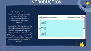
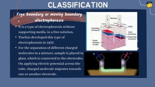
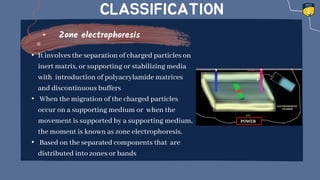
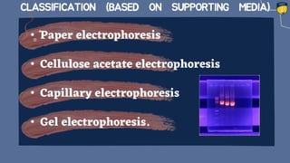
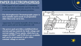
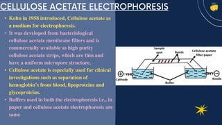
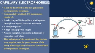
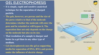
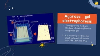
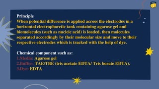
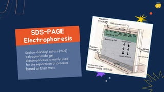
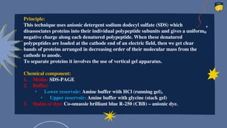
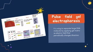
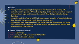
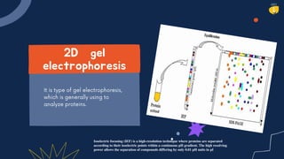
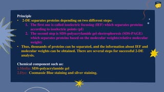
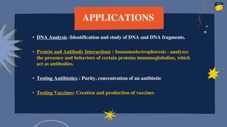
ELECTROPHORESIS
• INTRODUCTION
• FACTORS AFFECTING
ELECTROPHORETIC
MOBILITY
• TYPES
• APPLICATIONS
CONTENTS
INTRODUCTION
•
•
•
•
•
•
RATE OF
MIGRATION OF
CHARGED
PARTICLES
DEPENDS UPON
FOLLOWING
FACTORS
CLASSIFICATION
Free boundary or moving boundary
electrophoresis
• It is a type of electrophoresis without
supporting media, in a free solution.
• Tiselius developed this type of
electrophoresis in 1937.
• For the separation of different charged
molecules in a mixture, sample is placed in
glass, which is connected to the electrodes.
On applying electric potential across the
tube, charged molecule migrates towards
one or another electrode.
CLASSIFICATION
Zone electrophoresis
• It involves the separation of charged particles on
inert matrix, or supporting or stabilizing media
with introduction of polyacrylamide matrices
and discontinuous buffers
• When the migration of the charged particles
occur on a supporting medium or when the
movement is supported by a supporting medium,
the moment is known as zone electrophoresis.
• Based on the separated components that are
distributed into zones or bands
• Paper electrophoresis
• Cellulose acetate electrophoresis
• Capillary electrophoresis
• Gel electrophoresis.
CLASSIFICATION (BASED ON SUPPORTING MEDIA)
• Filter paper is very popular as a stabilizing
media and most commonly used for the study
of normal and normal plasma proteins.
• For electrophoresis Chromatography paper is
most suitable and it needs no preparation
other than to be cut to size.
• Apparatus: It consists basically of two items, a
power pack and an electrophoretic cell.
• The power pack provides a stabilized direct
current and has controls for both voltage and
current output. Power packs, which have an
output of 0-500 V and 0-150 rnA are available
and can be programmed to give either
constant voltage or current.
• Kohn in 1958 introduced, Cellulose acetate as
a medium for electrophoresis.
• It was developed from bacteriological
cellulose acetate membrane filters and is
commercially available as high purity
cellulose acetate strips, which are thin and
have a uniform micropore structure.
• Cellulose acetate is especially used for clinical
investigations such as separation of
hemoglobin’s from blood, lipoproteins and
glycoproteins.
• Buffers used in both the electrophoresis i.e., in
paper and cellulose acetate electrophoresis are
same
• It can be described as the new generation
electrophoretic technique.
• Commercially available CE instruments
consist of :
• An electrolyte-filled capillary, which passes
through the optical center of a detector
• A sample injector
• A high voltage power supply
• An auto-sampler. The entire instrument is
computer controlled.
• This technique of electrophoresis has become
very popular over the years because of the
many advantages that it has over conventional
electrophoresis techniques.
• Very high-level automation is possible.
• Fast analysis times.
• Detection of separated peaks is done online; thus, detection is a process that
goes hand in hand with separation and not post-separation as is the case with
conventional techniques.
• Heat generated inside the capillary is effectively dissipated through the walls
of the capillary; therefore, high voltages can be applied.
• High voltages mean a rapid separation.
• This faster separation along with online detection makes this technique
considerably faster as compared to the conventional techniques.
ADVANTAGES
• It is simple, rapid and sensitive analytical
technique for the separation of charged
particle.
• The gels, however, are porous and the size of
the pores relative to that of the molecule
determines whether the molecule will enter the
pore and be retarded or will bypass it. The
separation thus not only depends on the charge
on the molecule but also on its size.
• That resolution of a sample is sharper and
better in a gel than in any other type of
medium.
• Gel electrophoresis uses the gel as supporting
media for separation of DNA, RNA and protein
under the influence of electric charge.
Basically, gel
electrophoresis
apparatus is of
two kinds
1.Vertical gel apparatus:
It is commonly used for
the separation of
proteins in SDS-PAGE.
2. Horizontal gel apparatus:
It is used for immune
electrophoresis, iso-electric
focusing and electrophoresis
of DNA and RNA in the
agarose gel.
Type of gel
electrophoresis
Agarose gel electrophoresis
 SDS-PAGE
Pulse field gel electrophoresis
(PFGE)
2D gel electrophoresis
Principle
When potential difference is applied across the electrodes in a
horizontal electrophoretic tank containing agarose gel and
biomolecules (such as nucleic acid) is loaded, then molecules
separated accordingly by their molecular size and move to their
respective electrodes which is tracked with the help of dye.
Chemical component such as:
1.Media: Agarose gel
2.Buffer: TAE/TBE (tris acetate EDTA/ Tris borate EDTA).
3.Dye: EDTA
Principle:
This technique uses anionic detergent sodium dodecyl sulfate (SDS) which
disassociates proteins into their individual polypeptide subunits and gives a uniform
negative charge along each denatured polypeptide. When these denatured
polypeptides are loaded at the cathode end of an electric field, then we get clear
bands of proteins arranged in decreasing order of their molecular mass from the
cathode to anode.
To separate proteins it involves the use of vertical gel apparatus.
Chemical component:
1. Media: SDS-PAGE
2. Buffer:
• Lower reservoir: Amine buffer with HCl (running gel),
• Upper reservoir: Amine buffer with glycine (stack gel)
3. Stains or dye: Co-omassie brilliant blue R-250 (CBB) – anionic dye.
Principle
• It is a powerful genotyping technique used for the separation of large DNA
molecules (entire genomic DNA) after digesting it with unique restriction enzymes
and applying to a gel matrix under the electric field that periodically changes
direction.
• It permits analysis of bacterial DNA fragments over an order of magnitude larger
than that with conventional restriction enzyme analysis.
• It provides a good representation of the entire bacterial chromosome in a single
gel with a highly reproducible restriction profile, providing clearly distinct and
well-resolved DNA fragments.
Chemical component such as:
1.Media: Agarose gel
2.Buffer: EC Lysis buffer, TE (Tris-EDTA)buffer
3.Dye: Ethidium bromide solution
Isoelectric focusing (IEF) is a high-resolution technique where proteins are separated
according to their isoelectric points within a continuous pH gradient. The high resolving
power allows the separation of compounds differing by only 0.01 pH units in pI
Principle
• 2-DE separates proteins depending on two different steps:
1. The first one is called isoelectric focusing (IEF) which separates proteins
according to isoelectric points (pI)
2. The second step is SDS-polyacrylamide gel electrophoresis (SDS-PAGE)
which separates proteins based on the molecular weights(relative molecular
weight)
• Thus, thousands of proteins can be separated, and the information about IEF and
molecular weights can be obtained. There are several steps for successful 2-DE
analysis.
Chemical component such as:
1.Media: SDS-polyacrylamide gel
2.Dye: Coomassie Blue staining and silver staining.
APPLICATIONS
• DNA Analysis :Identification and study of DNA and DNA fragments.
• Protein and Antibody Interactions : Immunoelectrophoresis - analyzes
the presence and behaviors of certain proteins immunoglobulins, which
act as antibodies.
• Testing Antibiotics : Purity, concentration of an antibiotic
• Testing Vaccines: Creation and production of vaccines
23
REFERENCES
• Voet and Voet (1990). Biochemistry. John Wiley & Sons.
• Jahn, G.C.; D.W. Hall; S.G. Zam (1986). "A comparison of the life cycles of two Amblyospora
(Microspora: Amblyosporidae) in the mosquitoes Culex salinarius and Culex tarsalis
Coquillett". J. Florida Anti-Mosquito Assoc. 57: 24–27.
• Khattak, M.N.; R.C. Matthews (1993). "Genetic relatedness of Bordetella species as
determined by macrorestriction digests resolved by pulsed-field gel electrophoresis". Int. J.
Syst. Bacteriol. 43 (4): 659–64. doi:10.1099/00207713-43-4-659. PMID 8240949.
• Barz, D.P.J.; P. Ehrhard (2005). "Model and verification of electrokinetic flow and transport
in a micro-electrophoresis device". Lab Chip. 5 (9): 949–958. doi:10.1039/b503696h. PMID
16100579.
• Shim, J.; P. Dutta; C.F. Ivory (2007). "Modeling and simulation of IEF in 2-D
microgeometries". Electrophoresis. 28 (4): 527–586. doi:10.1002/elps.200600402. PMID
17253629. S2CID 23274096.
24
Electrophoresis

Electrophoresis

  • 1.
  • 2.
    • INTRODUCTION • FACTORSAFFECTING ELECTROPHORETIC MOBILITY • TYPES • APPLICATIONS CONTENTS
  • 3.
  • 4.
  • 5.
    CLASSIFICATION Free boundary ormoving boundary electrophoresis • It is a type of electrophoresis without supporting media, in a free solution. • Tiselius developed this type of electrophoresis in 1937. • For the separation of different charged molecules in a mixture, sample is placed in glass, which is connected to the electrodes. On applying electric potential across the tube, charged molecule migrates towards one or another electrode.
  • 6.
    CLASSIFICATION Zone electrophoresis • Itinvolves the separation of charged particles on inert matrix, or supporting or stabilizing media with introduction of polyacrylamide matrices and discontinuous buffers • When the migration of the charged particles occur on a supporting medium or when the movement is supported by a supporting medium, the moment is known as zone electrophoresis. • Based on the separated components that are distributed into zones or bands
  • 7.
    • Paper electrophoresis •Cellulose acetate electrophoresis • Capillary electrophoresis • Gel electrophoresis. CLASSIFICATION (BASED ON SUPPORTING MEDIA)
  • 8.
    • Filter paperis very popular as a stabilizing media and most commonly used for the study of normal and normal plasma proteins. • For electrophoresis Chromatography paper is most suitable and it needs no preparation other than to be cut to size. • Apparatus: It consists basically of two items, a power pack and an electrophoretic cell. • The power pack provides a stabilized direct current and has controls for both voltage and current output. Power packs, which have an output of 0-500 V and 0-150 rnA are available and can be programmed to give either constant voltage or current.
  • 9.
    • Kohn in1958 introduced, Cellulose acetate as a medium for electrophoresis. • It was developed from bacteriological cellulose acetate membrane filters and is commercially available as high purity cellulose acetate strips, which are thin and have a uniform micropore structure. • Cellulose acetate is especially used for clinical investigations such as separation of hemoglobin’s from blood, lipoproteins and glycoproteins. • Buffers used in both the electrophoresis i.e., in paper and cellulose acetate electrophoresis are same
  • 10.
    • It canbe described as the new generation electrophoretic technique. • Commercially available CE instruments consist of : • An electrolyte-filled capillary, which passes through the optical center of a detector • A sample injector • A high voltage power supply • An auto-sampler. The entire instrument is computer controlled. • This technique of electrophoresis has become very popular over the years because of the many advantages that it has over conventional electrophoresis techniques.
  • 11.
    • Very high-levelautomation is possible. • Fast analysis times. • Detection of separated peaks is done online; thus, detection is a process that goes hand in hand with separation and not post-separation as is the case with conventional techniques. • Heat generated inside the capillary is effectively dissipated through the walls of the capillary; therefore, high voltages can be applied. • High voltages mean a rapid separation. • This faster separation along with online detection makes this technique considerably faster as compared to the conventional techniques. ADVANTAGES
  • 12.
    • It issimple, rapid and sensitive analytical technique for the separation of charged particle. • The gels, however, are porous and the size of the pores relative to that of the molecule determines whether the molecule will enter the pore and be retarded or will bypass it. The separation thus not only depends on the charge on the molecule but also on its size. • That resolution of a sample is sharper and better in a gel than in any other type of medium. • Gel electrophoresis uses the gel as supporting media for separation of DNA, RNA and protein under the influence of electric charge.
  • 13.
    Basically, gel electrophoresis apparatus isof two kinds 1.Vertical gel apparatus: It is commonly used for the separation of proteins in SDS-PAGE. 2. Horizontal gel apparatus: It is used for immune electrophoresis, iso-electric focusing and electrophoresis of DNA and RNA in the agarose gel.
  • 14.
    Type of gel electrophoresis Agarosegel electrophoresis  SDS-PAGE Pulse field gel electrophoresis (PFGE) 2D gel electrophoresis
  • 16.
    Principle When potential differenceis applied across the electrodes in a horizontal electrophoretic tank containing agarose gel and biomolecules (such as nucleic acid) is loaded, then molecules separated accordingly by their molecular size and move to their respective electrodes which is tracked with the help of dye. Chemical component such as: 1.Media: Agarose gel 2.Buffer: TAE/TBE (tris acetate EDTA/ Tris borate EDTA). 3.Dye: EDTA
  • 18.
    Principle: This technique usesanionic detergent sodium dodecyl sulfate (SDS) which disassociates proteins into their individual polypeptide subunits and gives a uniform negative charge along each denatured polypeptide. When these denatured polypeptides are loaded at the cathode end of an electric field, then we get clear bands of proteins arranged in decreasing order of their molecular mass from the cathode to anode. To separate proteins it involves the use of vertical gel apparatus. Chemical component: 1. Media: SDS-PAGE 2. Buffer: • Lower reservoir: Amine buffer with HCl (running gel), • Upper reservoir: Amine buffer with glycine (stack gel) 3. Stains or dye: Co-omassie brilliant blue R-250 (CBB) – anionic dye.
  • 20.
    Principle • It isa powerful genotyping technique used for the separation of large DNA molecules (entire genomic DNA) after digesting it with unique restriction enzymes and applying to a gel matrix under the electric field that periodically changes direction. • It permits analysis of bacterial DNA fragments over an order of magnitude larger than that with conventional restriction enzyme analysis. • It provides a good representation of the entire bacterial chromosome in a single gel with a highly reproducible restriction profile, providing clearly distinct and well-resolved DNA fragments. Chemical component such as: 1.Media: Agarose gel 2.Buffer: EC Lysis buffer, TE (Tris-EDTA)buffer 3.Dye: Ethidium bromide solution
  • 21.
    Isoelectric focusing (IEF)is a high-resolution technique where proteins are separated according to their isoelectric points within a continuous pH gradient. The high resolving power allows the separation of compounds differing by only 0.01 pH units in pI
  • 22.
    Principle • 2-DE separatesproteins depending on two different steps: 1. The first one is called isoelectric focusing (IEF) which separates proteins according to isoelectric points (pI) 2. The second step is SDS-polyacrylamide gel electrophoresis (SDS-PAGE) which separates proteins based on the molecular weights(relative molecular weight) • Thus, thousands of proteins can be separated, and the information about IEF and molecular weights can be obtained. There are several steps for successful 2-DE analysis. Chemical component such as: 1.Media: SDS-polyacrylamide gel 2.Dye: Coomassie Blue staining and silver staining.
  • 23.
    APPLICATIONS • DNA Analysis:Identification and study of DNA and DNA fragments. • Protein and Antibody Interactions : Immunoelectrophoresis - analyzes the presence and behaviors of certain proteins immunoglobulins, which act as antibodies. • Testing Antibiotics : Purity, concentration of an antibiotic • Testing Vaccines: Creation and production of vaccines 23
  • 24.
    REFERENCES • Voet andVoet (1990). Biochemistry. John Wiley & Sons. • Jahn, G.C.; D.W. Hall; S.G. Zam (1986). "A comparison of the life cycles of two Amblyospora (Microspora: Amblyosporidae) in the mosquitoes Culex salinarius and Culex tarsalis Coquillett". J. Florida Anti-Mosquito Assoc. 57: 24–27. • Khattak, M.N.; R.C. Matthews (1993). "Genetic relatedness of Bordetella species as determined by macrorestriction digests resolved by pulsed-field gel electrophoresis". Int. J. Syst. Bacteriol. 43 (4): 659–64. doi:10.1099/00207713-43-4-659. PMID 8240949. • Barz, D.P.J.; P. Ehrhard (2005). "Model and verification of electrokinetic flow and transport in a micro-electrophoresis device". Lab Chip. 5 (9): 949–958. doi:10.1039/b503696h. PMID 16100579. • Shim, J.; P. Dutta; C.F. Ivory (2007). "Modeling and simulation of IEF in 2-D microgeometries". Electrophoresis. 28 (4): 527–586. doi:10.1002/elps.200600402. PMID 17253629. S2CID 23274096. 24