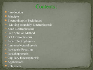
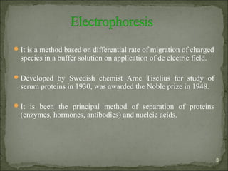
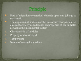
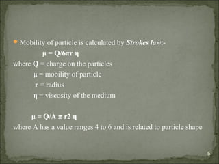
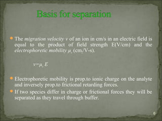
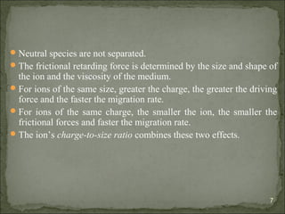
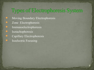
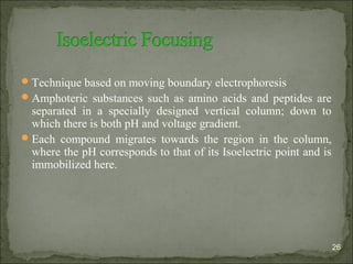
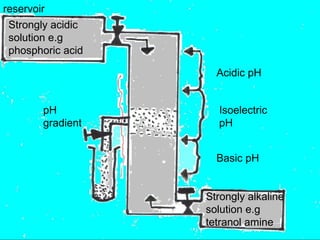
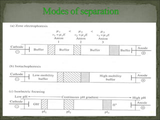
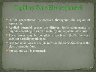
Definition, factors affecting electrophoresis, classification of electrophoresis in general, Iso-electric focusing in detail, IEF and its types (based on ampholytes), step wise procedure of IEF process, Problems involved and their remedies, Capillary iso electric focusing and its types, detection of analytes explained in animation (so watch it in slide show mode), advantages and applications of CIEF.
In this slide contains types, working principle, factors affecting, advantage and disadvantage of paper electrophoresis.
Presented by: G.Sai Swetha. (Department of pharmacology),
RIPER, anantapur.
Isoelectric focusing electrophoresis- Principle , procedure and applicationsJaskiranKaur72
IEF separates amphoteric compounds, such as proteins, with increased resolution in a medium possessing a stable pH gradient. The protein becomes “focused” at a point on the gel as it migrates to a zone where the pH of the gel matches the protein's pI. At this point, the charge of the protein becomes zero and its migration ceases.
Electrophoresis is an electrokinetic process which separates charged particles in a fluid using a field of electrical charge. It is most often used in life sciences to separate protein molecules or DNA and can be achieved through several different procedures depending on the type and size of the molecules. The procedures differ in some ways but all need a source for the electrical charge, a support medium and a buffer solution. Electrophoresis is used in laboratories for the separation of molecules based on size, density and purity.An electric field is applied to molecules and as they are electrically charged themselves it results in a force acting upon them. The greater the charge of the molecule the greater the force applied by the electrical field and therefore the further through the support medium the molecule will move relative to its mass.
Some example applications of electrophoresis include DNA and RNA analysis as well as protein electrophoresis which is a medical procedure used to analyse and separate the molecules found in a fluid sample (most commonly blood and urine samples).Different types of gels are usually used as the support medium for electrophoresis and this may be in slab or tube form depending on which is more beneficial. Gel slabs enable many samples to be run simultaneously and so are frequently used in laboratories. However, tube gels give a better resolution of the results so are often chosen for protein electrophoresis.
Agarose gel is commonly used for electrophoresis of DNA. It has a large pore structure allowing larger molecules to move easily but it is not suitable for sequencing smaller molecules.
Polyacrylamide gel electrophoresis (PAGE) has a clearer resolution than agarose gel making it more suitable for quantitative analysis. This makes it possible to identify how proteins bind to DNA. It can also be used to develop an understanding of how bacteria is becoming resistant to antibiotics through plasmid analysis.
This presentation contain the information about gel electrophoresis method , instruments & types.
Electrophoresis is a method through biological molecules are separated by applying an electric field.
Main purpose of this method is to determine the number , amount & mobility of biological component.
There are some internal & external factors that affects the process of electrophoresis.
The bio-molecules have charge on it & when we apply an electric field , the charge particles move to the opposite cathode. In this way, charge particles are separated
There are 3 types of gels that use in this process .
In this buffers are also used which provide ions that carry a current.
The technique of paper electrophoresis is simple and inexpensive and requires only micro quantities of plasma for separation.
The support medium is a filter paper
The electrophoresis apparatus in its simplest form consists of two troughs to contain buffer solution, through which electric current is passed.
Frequently used in isolating proteins, amino acids and oligopeptides.
INTRODUCTION, DEFINATION OF ELECTROPHORESIS, ELECTROPHORESIS PRINCIPLE, TYPES OF ELECTROPHORESIS, FREE ELECTROPHORESIS, ZONE ELECTROPHORESIS,PAPER ELECTROPHORESIS, WORKING OF PAPER ELECTROPHORESIS, PROCEDURE FOR PAPER ELECTROPHORESIS, VISUALISATION, FACTORS AFFECTING SEPARATION OF MOLECULES, APPLICATIONS, working of paper electrophoresis ,procedure for paper electrophoresis ,visualisation ,factors affecting separation of molecules ,applications ,forensics ,dna fingerprinting ,molecular biology ,microbiology information about the organisms ,biochemistry mapping of cellular components ,paper electrophoresis is also used in study of sic ,hemoglobin abnormalities ,separation of blood clotting factors ,serum plasma proteins from blood sample ,used in separation and identification of alkaloids ,used for testing water samples ,toxicity of water ,drug industry to determine presence of illelgal drUGS
It contains 2-3 acetyl groups per glucose unit and its adsorption capacity is less than that of paper.
It gives sharper bands.
Provides a good background for staining glycoproteins.
ADVANTAGE:
No tailing of proteins or hydrophilic materials.
Available in wide range of particle size and layer thickness.
Give sharp bands and offer good resolution.
High voltage can be applied which will enhance the resolution.
Multi-source connectivity as the driver of solar wind variability in the heli...Sérgio Sacani
The ambient solar wind that flls the heliosphere originates from multiple
sources in the solar corona and is highly structured. It is often described
as high-speed, relatively homogeneous, plasma streams from coronal
holes and slow-speed, highly variable, streams whose source regions are
under debate. A key goal of ESA/NASA’s Solar Orbiter mission is to identify
solar wind sources and understand what drives the complexity seen in the
heliosphere. By combining magnetic feld modelling and spectroscopic
techniques with high-resolution observations and measurements, we show
that the solar wind variability detected in situ by Solar Orbiter in March
2022 is driven by spatio-temporal changes in the magnetic connectivity to
multiple sources in the solar atmosphere. The magnetic feld footpoints
connected to the spacecraft moved from the boundaries of a coronal hole
to one active region (12961) and then across to another region (12957). This
is refected in the in situ measurements, which show the transition from fast
to highly Alfvénic then to slow solar wind that is disrupted by the arrival of
a coronal mass ejection. Our results describe solar wind variability at 0.5 au
but are applicable to near-Earth observatories.
Introduction:
RNA interference (RNAi) or Post-Transcriptional Gene Silencing (PTGS) is an important biological process for modulating eukaryotic gene expression.
It is highly conserved process of posttranscriptional gene silencing by which double stranded RNA (dsRNA) causes sequence-specific degradation of mRNA sequences.
dsRNA-induced gene silencing (RNAi) is reported in a wide range of eukaryotes ranging from worms, insects, mammals and plants.
This process mediates resistance to both endogenous parasitic and exogenous pathogenic nucleic acids, and regulates the expression of protein-coding genes.
What are small ncRNAs?
micro RNA (miRNA)
short interfering RNA (siRNA)
Properties of small non-coding RNA:
Involved in silencing mRNA transcripts.
Called “small” because they are usually only about 21-24 nucleotides long.
Synthesized by first cutting up longer precursor sequences (like the 61nt one that Lee discovered).
Silence an mRNA by base pairing with some sequence on the mRNA.
Discovery of siRNA?
The first small RNA:
In 1993 Rosalind Lee (Victor Ambros lab) was studying a non- coding gene in C. elegans, lin-4, that was involved in silencing of another gene, lin-14, at the appropriate time in the
development of the worm C. elegans.
Two small transcripts of lin-4 (22nt and 61nt) were found to be complementary to a sequence in the 3' UTR of lin-14.
Because lin-4 encoded no protein, she deduced that it must be these transcripts that are causing the silencing by RNA-RNA interactions.
Types of RNAi ( non coding RNA)
MiRNA
Length (23-25 nt)
Trans acting
Binds with target MRNA in mismatch
Translation inhibition
Si RNA
Length 21 nt.
Cis acting
Bind with target Mrna in perfect complementary sequence
Piwi-RNA
Length ; 25 to 36 nt.
Expressed in Germ Cells
Regulates trnasposomes activity
MECHANISM OF RNAI:
First the double-stranded RNA teams up with a protein complex named Dicer, which cuts the long RNA into short pieces.
Then another protein complex called RISC (RNA-induced silencing complex) discards one of the two RNA strands.
The RISC-docked, single-stranded RNA then pairs with the homologous mRNA and destroys it.
THE RISC COMPLEX:
RISC is large(>500kD) RNA multi- protein Binding complex which triggers MRNA degradation in response to MRNA
Unwinding of double stranded Si RNA by ATP independent Helicase
Active component of RISC is Ago proteins( ENDONUCLEASE) which cleave target MRNA.
DICER: endonuclease (RNase Family III)
Argonaute: Central Component of the RNA-Induced Silencing Complex (RISC)
One strand of the dsRNA produced by Dicer is retained in the RISC complex in association with Argonaute
ARGONAUTE PROTEIN :
1.PAZ(PIWI/Argonaute/ Zwille)- Recognition of target MRNA
2.PIWI (p-element induced wimpy Testis)- breaks Phosphodiester bond of mRNA.)RNAse H activity.
MiRNA:
The Double-stranded RNAs are naturally produced in eukaryotic cells during development, and they have a key role in regulating gene expression .
Richard's entangled aventures in wonderlandRichard Gill
Since the loophole-free Bell experiments of 2020 and the Nobel prizes in physics of 2022, critics of Bell's work have retreated to the fortress of super-determinism. Now, super-determinism is a derogatory word - it just means "determinism". Palmer, Hance and Hossenfelder argue that quantum mechanics and determinism are not incompatible, using a sophisticated mathematical construction based on a subtle thinning of allowed states and measurements in quantum mechanics, such that what is left appears to make Bell's argument fail, without altering the empirical predictions of quantum mechanics. I think however that it is a smoke screen, and the slogan "lost in math" comes to my mind. I will discuss some other recent disproofs of Bell's theorem using the language of causality based on causal graphs. Causal thinking is also central to law and justice. I will mention surprising connections to my work on serial killer nurse cases, in particular the Dutch case of Lucia de Berk and the current UK case of Lucy Letby.
The increased availability of biomedical data, particularly in the public domain, offers the opportunity to better understand human health and to develop effective therapeutics for a wide range of unmet medical needs. However, data scientists remain stymied by the fact that data remain hard to find and to productively reuse because data and their metadata i) are wholly inaccessible, ii) are in non-standard or incompatible representations, iii) do not conform to community standards, and iv) have unclear or highly restricted terms and conditions that preclude legitimate reuse. These limitations require a rethink on data can be made machine and AI-ready - the key motivation behind the FAIR Guiding Principles. Concurrently, while recent efforts have explored the use of deep learning to fuse disparate data into predictive models for a wide range of biomedical applications, these models often fail even when the correct answer is already known, and fail to explain individual predictions in terms that data scientists can appreciate. These limitations suggest that new methods to produce practical artificial intelligence are still needed.
In this talk, I will discuss our work in (1) building an integrative knowledge infrastructure to prepare FAIR and "AI-ready" data and services along with (2) neurosymbolic AI methods to improve the quality of predictions and to generate plausible explanations. Attention is given to standards, platforms, and methods to wrangle knowledge into simple, but effective semantic and latent representations, and to make these available into standards-compliant and discoverable interfaces that can be used in model building, validation, and explanation. Our work, and those of others in the field, creates a baseline for building trustworthy and easy to deploy AI models in biomedicine.
Bio
Dr. Michel Dumontier is the Distinguished Professor of Data Science at Maastricht University, founder and executive director of the Institute of Data Science, and co-founder of the FAIR (Findable, Accessible, Interoperable and Reusable) data principles. His research explores socio-technological approaches for responsible discovery science, which includes collaborative multi-modal knowledge graphs, privacy-preserving distributed data mining, and AI methods for drug discovery and personalized medicine. His work is supported through the Dutch National Research Agenda, the Netherlands Organisation for Scientific Research, Horizon Europe, the European Open Science Cloud, the US National Institutes of Health, and a Marie-Curie Innovative Training Network. He is the editor-in-chief for the journal Data Science and is internationally recognized for his contributions in bioinformatics, biomedical informatics, and semantic technologies including ontologies and linked data.
(May 29th, 2024) Advancements in Intravital Microscopy- Insights for Preclini...Scintica Instrumentation
Intravital microscopy (IVM) is a powerful tool utilized to study cellular behavior over time and space in vivo. Much of our understanding of cell biology has been accomplished using various in vitro and ex vivo methods; however, these studies do not necessarily reflect the natural dynamics of biological processes. Unlike traditional cell culture or fixed tissue imaging, IVM allows for the ultra-fast high-resolution imaging of cellular processes over time and space and were studied in its natural environment. Real-time visualization of biological processes in the context of an intact organism helps maintain physiological relevance and provide insights into the progression of disease, response to treatments or developmental processes.
In this webinar we give an overview of advanced applications of the IVM system in preclinical research. IVIM technology is a provider of all-in-one intravital microscopy systems and solutions optimized for in vivo imaging of live animal models at sub-micron resolution. The system’s unique features and user-friendly software enables researchers to probe fast dynamic biological processes such as immune cell tracking, cell-cell interaction as well as vascularization and tumor metastasis with exceptional detail. This webinar will also give an overview of IVM being utilized in drug development, offering a view into the intricate interaction between drugs/nanoparticles and tissues in vivo and allows for the evaluation of therapeutic intervention in a variety of tissues and organs. This interdisciplinary collaboration continues to drive the advancements of novel therapeutic strategies.
Slide 1: Title Slide
Extrachromosomal Inheritance
Slide 2: Introduction to Extrachromosomal Inheritance
Definition: Extrachromosomal inheritance refers to the transmission of genetic material that is not found within the nucleus.
Key Components: Involves genes located in mitochondria, chloroplasts, and plasmids.
Slide 3: Mitochondrial Inheritance
Mitochondria: Organelles responsible for energy production.
Mitochondrial DNA (mtDNA): Circular DNA molecule found in mitochondria.
Inheritance Pattern: Maternally inherited, meaning it is passed from mothers to all their offspring.
Diseases: Examples include Leber’s hereditary optic neuropathy (LHON) and mitochondrial myopathy.
Slide 4: Chloroplast Inheritance
Chloroplasts: Organelles responsible for photosynthesis in plants.
Chloroplast DNA (cpDNA): Circular DNA molecule found in chloroplasts.
Inheritance Pattern: Often maternally inherited in most plants, but can vary in some species.
Examples: Variegation in plants, where leaf color patterns are determined by chloroplast DNA.
Slide 5: Plasmid Inheritance
Plasmids: Small, circular DNA molecules found in bacteria and some eukaryotes.
Features: Can carry antibiotic resistance genes and can be transferred between cells through processes like conjugation.
Significance: Important in biotechnology for gene cloning and genetic engineering.
Slide 6: Mechanisms of Extrachromosomal Inheritance
Non-Mendelian Patterns: Do not follow Mendel’s laws of inheritance.
Cytoplasmic Segregation: During cell division, organelles like mitochondria and chloroplasts are randomly distributed to daughter cells.
Heteroplasmy: Presence of more than one type of organellar genome within a cell, leading to variation in expression.
Slide 7: Examples of Extrachromosomal Inheritance
Four O’clock Plant (Mirabilis jalapa): Shows variegated leaves due to different cpDNA in leaf cells.
Petite Mutants in Yeast: Result from mutations in mitochondrial DNA affecting respiration.
Slide 8: Importance of Extrachromosomal Inheritance
Evolution: Provides insight into the evolution of eukaryotic cells.
Medicine: Understanding mitochondrial inheritance helps in diagnosing and treating mitochondrial diseases.
Agriculture: Chloroplast inheritance can be used in plant breeding and genetic modification.
Slide 9: Recent Research and Advances
Gene Editing: Techniques like CRISPR-Cas9 are being used to edit mitochondrial and chloroplast DNA.
Therapies: Development of mitochondrial replacement therapy (MRT) for preventing mitochondrial diseases.
Slide 10: Conclusion
Summary: Extrachromosomal inheritance involves the transmission of genetic material outside the nucleus and plays a crucial role in genetics, medicine, and biotechnology.
Future Directions: Continued research and technological advancements hold promise for new treatments and applications.
Slide 11: Questions and Discussion
Invite Audience: Open the floor for any questions or further discussion on the topic.
3. It is a method based on differential rate of migration of charged
species in a buffer solution on application of dc electric field.
Developed by Swedish chemist Arne Tiselius for study of
serum proteins in 1930, was awarded the Noble prize in 1948.
It is been the principal method of separation of proteins
(enzymes, hormones, antibodies) and nucleic acids.
3
4. Rate of migration (separation) depends upon e/m (charge to
mass) ratio
The migration of particles or the rate of travel of particle, in
electrophoretic system depends on properties of the particles
as well as the instrumental system
1. Characteristic of particles
2. Property of electric field
3. Temperature
4. Nature of suspended medium
4
5. Mobility of particle is calculated by Strokes law:-
μ = Q/6πr η
where Q = charge on the particles
μ = mobility of particle
r = radius
η = viscosity of the medium
μ = Q/A π r2 η
where A has a value ranges 4 to 6 and is related to particle shape
5
6. The migration velocity v of an ion in cm/s in an electric field is
equal to the product of field strength E(V/cm) and the
electrophoretic mobility µe (cm2/V-s).
v=µe E
Electrophoretic mobility is prop.to ionic charge on the analyte
and inversely prop.to frictional retarding forces.
If two species differ in charge or frictional forces they will be
separated as they travel through buffer.
6
7. Neutral species are not separated.
The frictional retarding force is determined by the size and shape of
the ion and the viscosity of the medium.
For ions of the same size, greater the charge, the greater the driving
force and the faster the migration rate.
For ions of the same charge, the smaller the ion, the smaller the
frictional forces and faster the migration rate.
The ion’s charge-to-size ratio combines these two effects.
7
9. Principle: -
Method involves movement of charged particles in free moving
solution in absence of supporting medium.
9
10. Electro-Osmotic Flow : when a high potential is applied
across a capillary tube containing a buffer solution usually
results into migration of solvent toward the anode or cathode.
The cause of electro-osmotic flow is the electric double layer
that develops at the silica/solution interface.
In the presence of electro-osmosis, an ion’s velocity is the sum
of its migration velocity and the velocity of the electro-osmotic
flow.
10
11. Method:
The apparatus consists of a U shaped tube with provision for
introducing the cathode and anode electrodes into each of the
arms.
The sample solution is introduced and each arm is filled
carefully with a buffered solution.
If the sample consists of compounds with different mobilities,
their migration may be observed as several moving boundaries
11
13. Detection:-
Position of ions detected by measuring changes in refractive
index throughout the solution.
Advantages:-
Biologically active fractions can be recovered without using
denaturing agent.
As a reference method for measuring electro-mobility.
Gives information on isoelectric point and mobility of
compounds
13
14. Disadvantages:-
Complete separation is rarely achieved
Maintaining sharply defined boundaries (stabilization of
boundaries is needed)
Only fastest and slowest components can be separated in pure
form.
Not used for preparative and quantitative analysis.
Several problems are associated with the technique, including
stabilization of ion boundaries, boundaries anomalies, and the
need for specialized equipment
14
15. Zone electrophoresis makes use of a stabilizing medium to
minimize the problems associated with free-boundary
electrophoresis.
It involves migration of charged particles which are supported
on a relatively inert and homogenous solid or gel frame work.
Separated components are distributed into different zone in a
stabilizing media.
Make use of stabilizing media like paper, agar, cellulose,
starch, gels, polyacrylamide gels.
15
16. Types of supporting or stabilizing medium :
Free Solution Method
Gel Electrophoresis
Paper Electrophoresis
16
17. Rotating tube apparatus
Migration occurs in horizontal tube.
10 revolution / min
Micro syringe (small sample can be applied)
Profiles of zones are determined by scanning devices.
Advantages:-
Complete separation of electrophoretically different
components.
Small sample
17
19. Electrophoresis in compact gels, which depends at least in part
on size-exclusion effects to achieve separation, is used
frequently for the separations of proteins and nucleic acids
The overall migration in these gels is a combination of
movement under the influence of the electric field and size
separation by the pores of the gel
It is carried out by using
Agar
Starch
Polyacrylamide
19
20. The most frequently used techniques of polyacrylamide gel
electrophoresis (PAGE) Is the discontinuous buffer system
developed by Laemmli.
In this procedure sample is placed on a stacking gel with a low
level of cross linking and therefore a large pore size. During
movement through this gel, the sample is concentrated into a
narrow band and then deposited onto a separating gel that has a
higher cross linking and smaller pore size. The separation of
the solutes occurs in this phase.
20
21. In a special modification of this technique used for separation
of proteins, a detergent, such as sodium dodecylsulfate (SDS),
is introduced in the buffer.
This interacts with the proteins to produce particles of
consistent shape and uniform negative charge so that
separation occurs according to size alone.
This enables the simple determination of molecular weight
because the migration distance is proportional to the logarithm
of molecular weight, as in size exclusion chromatography.
21
22. One of the simplest procedures in electrophoresis involves
spotting a mixture of solutes in the middle of a paper strip,
moistening the paper with some electrolyte and placing it
between two sheets of glass. The ends of the paper strips are
immersed in beakers of electrolyte
Electrophoresis is allowed to continue for a period of several
hours.
22
24. Enzymatic and immunological methods also have been used
to detect proteins following electrophoresis in gels.
Immunochemical methods add an additional dimension to
protein identification. Following electrophoresis in an agar
gel backed with a microscope slide, an antibody is placed
into a trough cut parallel to the direction of electrophoresis.
24
25. The antibody and electrophoretically separated antigens
diffuse towards each other resulting in precipitin arcs where
antigen antibody complexes form. This technique has been
referred to as immunoelectrophoresis.
25
26. Technique based on moving boundary electrophoresis
Amphoteric substances such as amino acids and peptides are
separated in a specially designed vertical column; down to
which there is both pH and voltage gradient.
Each compound migrates towards the region in the column,
where the pH corresponds to that of its Isoelectric point and is
immobilized here.
26
28. Advantage:-
In separation and characterization of proteins in one step.
High resolution – (identifying iso enzymes)
Application:-
Useful for microanalysis of proteins
Identifying isoenzymes
28
29. Based on principle of moving boundary electrophoresis.
Separation is achieved either horizontally or vertically.
Solution in which the separation takes place is normally an
aqueous medium, which contains sucrose to provide a higher
density to the solution.
Where the separation by Isoelectric focusing depends on the
existence of a pH gradient in the system, the technique of
Isotachophoresis depends on the development of a potential
gradient.
A leading electrolyte (e.g. Chloride) with a higher mobility
than the analytes, and a trailing electrolyte (e.g. Glycinate)
with a lower mobility are used
29
30. Instrumental version in which electrophoresis is carried out in a
capillary and separated species is eluted out from one end of
capillary.
The long length and small cross-sectional area of the capillary
results into high resistance to solution. Because power dissipation
is inversely prop.to resistance, much higher potential (20,000-
60,000 V) can be applied.
This leads to corresponding improvements in speed and resolution.
Plate counts range from 1,00,000-2,00,000.
Yields high-speed and high resolution separations on small sample
volumes (0.1-10nL)
30
31. Order of elution in a typical capillary electrophoretic
separation is first the fastest cation followed by successive
slower cations, then all the neutrals in a single zone, and finally
the slowest anion followed by successively faster anions.
It is possible to revert the direction by adding cationic
surfactant to buffer.
The surfactant adsorbs on the capillary wall and makes the wall
positively charged, buffer anions congregate near the wall and
are swept toward the cathode or positive electrode.
31
32. A buffer filled fused silica capillary of 10-100µm in internal
diameter and 40-100cm long, extends between two buffer
reservoirs that also hold platinum electrodes.
Sample introduction is performed at one end and detection at
the other.
The polarity of the high-voltage power supply can be as
indicated or can be reversed for rapid separation of anions.
Sample introduction and detection is tedious because of small
volume of capillary µL.
32
33. Most commonly used methods are :
1. Electro-kinetic injection –
one end of capillary and electrode are removed from buffer
compartment and placed in a small cup containing sample.
A potential is then applied for a measured time causing
sample to enter the capillary by ionic migration and electro-
osmotic flow. It discriminates slower moving ions relative to
more mobile ions.
33
34. 2. Pressure injection :
• End of capillary is placed in the small cup containing
sample and pressure difference is then used to drive the
sample solution into the capillary. Pressure difference can be
achieved by applying vacuum at detector end or by
elevating to sample end. No discrimination due to mobility
of ions but cannot be used for gel filled capillaries.
• For both the volume injected is controlled by duration of
injection.
• Microinjection tips are also been constructed for volumes
such as pL (used in study of amino acids and
neurotransmitters)
34
35. In capillary electrophoresis each ion migrates at a rate
determined by its electrophoretic mobility. Thus analyte
band passes through the detector at different rates, which
result in peak areas that are dependent on retention times.
Absorbance methods
Electrochemical methods
Conductometry
Amperometry
Mass spectrometric methods
35
36. Absorbance methods : both fluorescence and absorbance
detectors are used but more common are absorbance detectors.
To keep the detection volume to nL or small it is carried on-
column done by taking a small section of capillary and
removing the polyimide coating from exterior by burning,
dissolution, or scraping.
This reduces the detection path lengths small with respect to
conc so small that it cannot be measured.
36
37. For increasing the sensitivity three main techniques are used
–
Bending the capillary to ‘Z’ shape
Bubble formation near the end of capillary.
Reflection of radiation (reflective coating of silver)
Indirect absorbance detection done by incorporation of ionic
chromophore in the buffer solution which makes the
detector receive constant signal and when analyte displaces
some of these the detector signal decreases.
37
38. Fluorescence detection –
Laser based instrumentation is used in order to focus the
excitation radiation on the small capillary and to achieve the
low detection limits available from intense sources.
Laser fluorescence detection have allowed detection up to
zeptomoles.
38
39. Mass Spectrometric Detection :
Small flow rates- 1 µL/min from electrophoretic capillaries
makes it feasible to couple the effluent to the ionization source
of a mass spectrometer.
Sample introduction/ionization interface currently used is
electrospray or fast atom bombardment is also used.
Widely used for detection of large molecules such as proteins,
DNA fragments and peptides.
Detection limits few tens of femtomoles for mol. Wt. 1,00,000
or more.
39
40. Capillary electrophoretic separations are performed in
several ways called modes.
These modes include-
Capillary zone electrophoresis (CZE)
Capillary gel electrophoresis (CGE)
Capillary isoelectric focusing (CIEF)
Capillary isotachophoresis (CITP)
40
42. Buffer concentration is constant throughout the region of
separation.
Applied potential causes the different ionic components to
migrate according to its own mobility and separate into zones.
These zones may be completely resolved (buffer between
each) or partially overlapped.
Best for small ions as analyte move in the same direction as the
electro-osmotic flow.
For cations wall is untreated.
42
43. For anions the electro-osmotic flow is usually reversed by
treating walls of capillary with an alkyl ammonium salt- cetyl
triethylammonium bromide.
+ly charged ammonium ions get attached to –ly charged silica
surface and create a –ly charged double layer of solution,
which is attracted towards anode thus reversing the electro-
osmotic flow.
43
44. Applications of CZE :
Separation of molecular species-
Synthetic herbicides
Pesticides-allizarin, methyl xanthine.
Pharmaceuticals such as anti-inflammatory drugs- naproxen,
ibuprofen, tolmetin.
Proteins, amino acids and carbohydrates.
Separation of smaller ions.
Advantages of CZE :
Lower equipment and maintenance cost.
Smaller sample size.
Greater speed and better resolution.
44
45. CGE performed in a porous gel matrix, the pores of which
contain a buffer mixture in which the separation is carried out.
This porous gel medium provides a molecular sieving action
that retards the migration of analyte species to various extents
depending upon the pore size of the analyte ions.
Most common type of gel used is a polyacrylamide (CH2=CH-
CO-NH2) polymer and cross-linking agent.
45
46. The pore size depends upon ratio of monomer to cross-linking
agent.
Increase in the amount of cross-linking agent results in smaller
pore size.
Other gels used are:
Agarose- polysaccharide extracted from marine algae.
Methyl cellulose
Polyethylene glycol
46
47. Applications
Separation of macromolecules such as proteins
differing in their sizes.
e.g. lactalbumin, carbonic anhydrase, ovalbumin,
BSA, phosphorylase B.
Separation of DNA fragments and oligonucleotides
that have substantially the same charge but differ in
their sizes.
47
48. Used to amphiprotic species.
An amphiprotic compound is a species capable of both
donating and accepting a proton.
When glycine is dissolved in water 3 equilibria operate-
The AA product bearing a +ve and a –ve charge is called a
zwitterion (no migration).
48
49. No net migration occurs in an electric field when the pH of the
solvent is such that the conc of anionic and cationic forms are
identical.
The pH at which no net migration occurs is called isoelectric
point (pI) and is an important physical constant for
characterizing amino acids.
The pI is related to the ionisation constant of the species. Thus
for glycine Ka and Kb are-
49
50. Electrophoresis is employed in biochemical and clinical field.
In the study of protein mixtures
Antigen antibody reactions
In fractioning protein.
In analysis of lipoprotein
Hemoglobin
In combination with autoradiography
Separation of organic acid, alkaloids, carbohydrates, amino
acids, alcohols, phenols, nucleic acids, insulin.
In food industry
50