Download to read offline

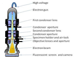
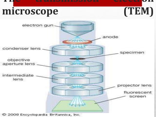
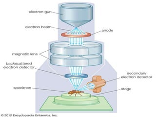
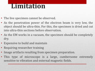
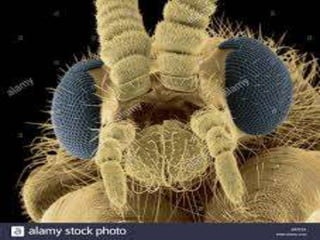
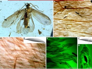
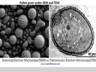
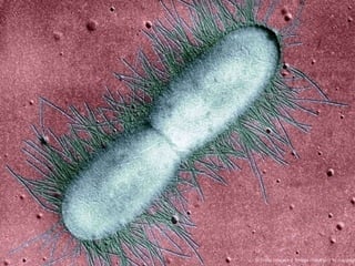

The document discusses electron microscopes. It defines electron microscopes as microscopes that use accelerated electrons rather than light to illuminate samples. Ernst Ruska built the first electron microscope in 1931. The document describes the working principles of transmission electron microscopes and scanning electron microscopes, including how each forms images. It also outlines the key components and parts of electron microscopes, their applications, advantages, and limitations.


![Polymer [ बहुलक ] Chemistry Notes PDF - Irfanullah Mehar - JJ Sir Chemistry.pdf](https://cdn.slidesharecdn.com/ss_thumbnails/polymerchemistrynotespdf-irfanullahmehar-jjsirchemistry-260210172118-3f9b37f7-thumbnail.jpg?width=640&height=640&fit=bounds)


