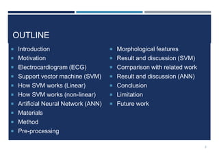
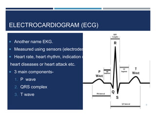
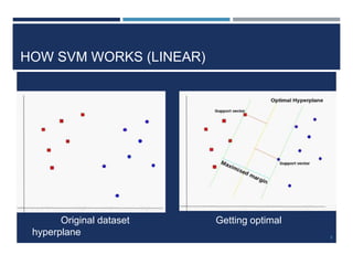
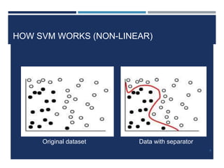
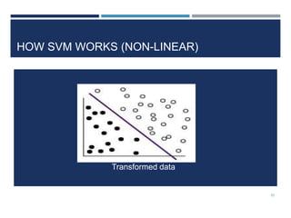
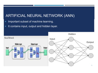
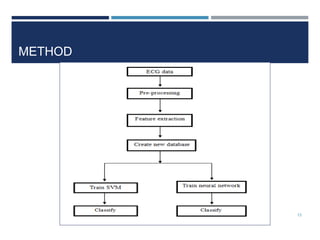
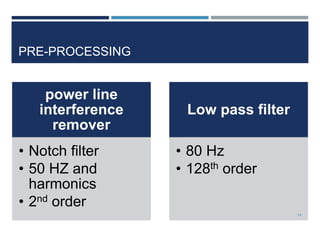
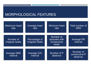
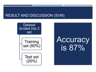
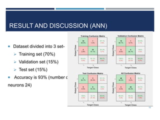
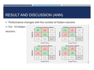
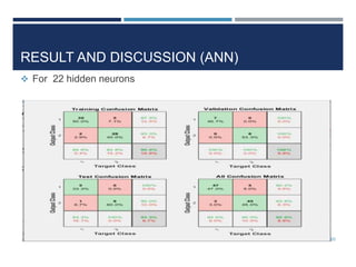
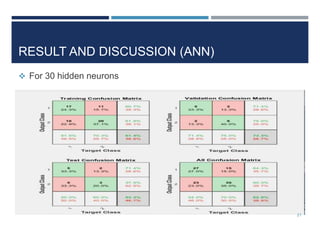
2. OUTLINE
Introduction
Motivation
Electrocardiogram (ECG)
Support vector machine (SVM)
How SVM works (Linear)
How SVM works (non-linear)
Artificial Neural Network (ANN)
Materials
Method
Pre-processing
Morphological features
Result and discussion (SVM)
Comparison with related work
Result and discussion (ANN)
Conclusion
Limitation
Future work
2
3. INTRODUCTION
ECG is a simple test
to visualize the
electrical activity of
the heart over a
period of time.
ECG classification is
done by machine
learning algorithm.
The machine
learning algorithm
are SVM and ANN
3
4. MOTIVATION
Number 1 cause
of death is
cardio vascular
diseases
Gain knowledge
and work with
machine
learning
Create effective
and easier
classifier
4
5. ELECTROCARDIOGRAM (ECG)
Another name EKG.
Measured using sensors (electrodes).
Heart rate, heart rhythm, indication of
heart diseases or heart attack etc.
3 main components-
1. P wave
2. QRS complex
3. T wave
5
7. SUPPORT VECTOR MACHINE (SVM)
Supervised machine learning algorithm.
Commonly use in classification and regression analysis.
Other applications -
Image classification,
Text classification,
Biological science etc.
7
8. HOW SVM WORKS (LINEAR)
Original dataset Getting optimal
hyperplane 8
9. HOW SVM WORKS (NON-LINEAR)
Original dataset Data with separator
9
15. MORPHOLOGICAL FEATURES
Maximum heart
rate
Average heart
rate
Minimum heart
rate
Total number of
QRS
Number of
irregular beats
Percentage of
irregular beats
Number of
episodes with
consecutive
beats
Average PR
interval
Average QRS
interval
Average QTc
interval
Number of P
absence
Number of
consecutive P
absence
15
16. RESULT AND DISCUSSION (SVM)
Dataset
divided into 2
set
Training
set (80%)
Test set
(20%)
Accuracy
is 87%
16
17. COMPARISON WITH RELATED WORK
Related Project Database Method Accuracy
ECG feature extraction
and classification using
wavelet transform and
support vector machines
MIT-BIH
arrhythmia
2 different feature extraction
methods-The wavelet
transform and
autoregressive modeling
(AR)
99.68%
Domain adaptation
methods for ECG
classification
MIT-BIH
arrhythmia
(divided in 2 set)
Two kinds of features:
1) ECG morphology
features and 2) ECG
wavelet features with
QRS width.
97% and
91%
ECG beat classification
method for ECG printout
with Principle components
analysis and support
vector machines
MIT-BIH
arrhythmia
Discrete wavelet transform
(DWT) and principle
components analysis (PCA)
99.6367%
with LIBSVM
This project
Fantasia, MIT-
BIH normal
sinus rhythm,
MIT-BIH
Morphology Feature
extraction method (12
features) 87%
17
18. RESULT AND DISCUSSION (ANN)
Dataset divided into 3 set-
Training set (70%)
Validation set (15%)
Test set (15%)
Accuracy is 93% (number of
neurons 24)
18
19. RESULT AND DISCUSSION (ANN)
Performance changes with the number of hidden neurons
For 10 hidden
neurons
19
23. LIMITATION
Can not work with raw data.
Choosing SVM parameter.
Problem with big data.
New area.
23
24. FUTURE WORK
Aim for more accuracy
More work with these features.
Finding most dominate features to work
with.
Work with advance neural network
(DNN and CNN)
24