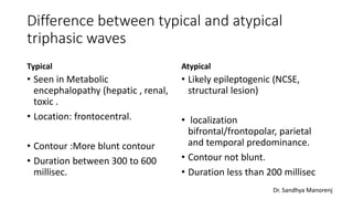
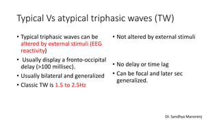
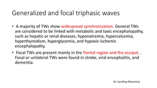
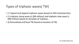
Triphasic waves in EEG
Dr Sandhya Manorenj
Associate Professor Neurology , Deccan College of Medical Sciences.
20/12/20
Introduction
• Triphasic waves (TW) are abnormal electroencephalogram (EEG)
waveforms seen in association with multiple clinical conditions,
including encephalopathy and structural brain lesions, among others.
• They were initially believed to be pathognomonic of hepatic
encephalopathy; however, they can be seen in any metabolic
encephalopathy (in fact, more common with renal than hepatic
conditions) and some other conditions. They may be considered a
subset of generalized periodic discharges (GPDs).
Dr. Sandhya Manorenj
Who described ?
• This EEG pattern was first described by Foley in 1950. It was an EEG
recording of a patient with hepatic encephalopathy.
• The term triphasic waves was first coined in 1955 by Bickford and
Butt, given the three-phase morphology.
Dr. Sandhya Manorenj
Pathophysiology
• Triphasic waves are not seen in a normal awake state and only seen in
patients with altered levels of consciousness.
• It is hypothesized that they occur due to structural or metabolic
abnormalities affecting the thalamocortical relays.
• The probable pathogenesis is dysfunction of the oscillatory system
between the cortex and thalamus for metabolic encephalopathy.
Extensive structural or metabolic injuries of the brain stem,
diencephalon, and subcortex may be caused by nonmetabolic factors.
Some studies have shown that TWs may be correlated with
intracranial hypertension.
Dr. Sandhya Manorenj
EEG characteristics of Triphasic waves
(typical)
• These waveforms have 3 phases with a total duration of 300 to 600
milliseconds.
• They are high amplitude sharply contoured waves, with the duration
of each phase longer than the next. The first phase is always negative;
the second wave is positive, displaying the highest amplitude (greater
than 70 microvolts), with a third slow negative deflection.
• The triphasic waves usually repeat periodically at a rate of 0.5 Hz to 2
Hz.
• They are seen diffusely with synchronous bifrontal and central
predominance and display a fronto-occipital time lag ( > 100 millisec).
Dr. Sandhya Manorenj
Images of Typical triphasic wave
Dr. Sandhya Manorenj
EEG characteristic of atypical Triphasic waves
• Atypical (likely epileptic) triphasic waves.
• Atypical features include localization : bifrontal/frontopolar, parietal
and temporal predominance in epileptiform triphasic waves
• Contour : Not a blunt contour
• Duration : less than 200 milliseconds with epileptiform or atypical
triphasic activity.
• Atypical triphasic waves not altered by external stimuli (EEG
reactivity) and
• Does not display a fronto-occipital delay
Dr. Sandhya Manorenj
Difference between typical and atypical
triphasic waves
Typical
• Seen in Metabolic
encephalopathy (hepatic , renal,
toxic .
• Location: frontocentral.
• Contour :More blunt contour
• Duration between 300 to 600
millisec.
Atypical
• Likely epileptogenic (NCSE,
structural lesion)
• localization
bifrontal/frontopolar, parietal
and temporal predominance.
• Contour not blunt.
• Duration less than 200 millisec
Dr. Sandhya Manorenj
Typical Vs atypical triphasic waves (TW)
• Typical triphasic waves can be
altered by external stimuli (EEG
reactivity)
• Usually display a fronto-occipital
delay (>100 millisec).
• Usually bilateral and generalized
• Classic TW is 1.5 to 2.5Hz
• Not altered by external stimuli
• No delay or time lag
• Can be focal and later sec
generalized.
Dr. Sandhya Manorenj
Generalized and focal triphasic waves
• A majority of TWs show widespread synchronization. General TWs
are considered to be linked with metabolic and toxic encephalopathy,
such as hepatic or renal diseases, hyponatremia, hypercalcemia,
hyperthyroidism, hyperglycemia, and hypoxic-ischemic
encephalopathy.
• Focal TWs are present mainly in the frontal region and the occiput. .
Focal or unilateral TWs were found in stroke, viral encephalitis, and
dementia.
Dr. Sandhya Manorenj
Types of triphasic waves( TW)
• 1 ) Typical and atypical triphasic waves based on EEG characteristics
• 2 ) triphasic sharp wave (≤ 200 millisec) and triphasic slow wave (>
200 millisec) based on duration of 3 phases .
• 3) Generalized and focal TW based on location of TW.
Dr. Sandhya Manorenj
Causes of triphasic waves
• Generalized TW usually in metabolic encephalopathy: associated with
slow background and causes are
• Hepatic encephalopathy, uremic encephalopathy, hyperthyroidism,
hypothyroidism,Hypernatremia,Hyponatremia,Hypercalcemia,Hypogl
ycemia,hyperosmolar state, azotemia.
• Toxic causes : Pregabalin toxicity, Naproxen, lithium toxicity,
Metrizamide, cefipime.
• Hypoxic ischemic insult
• Other conditions: Serotonin syndrome,Maple syrup urine disease.
• Ictal or post ictal state
Dr. Sandhya Manorenj
Triphasic waves due to structural lesion
• Usually focal and causes include
• Cerebral abscess
• Thalamic gliomas
• Craniopharyngioma
• Pontine stroke
• Cerebral carcinomatosis
• Multifocal cerebral lymphoma
• Creutzfeldt-Jakob disease (CJD)
• Alzheimer and other neurodegenerative diseases
• Head trauma
• Subdural hematoma
• Carcinomatous meningitis
Dr. Sandhya Manorenj
Triphasic waves ? Periodic discharges in CJD
• Typical and characteristic EEGs in Creutzfeldt–Jakob disease are
bisynchronous periodic waves.
• The EEG showed periodic biphasic and triphasic complex sharp waves
at 0.5 to 1.5 Hz predominantly in frontotemporal leads.
Dr. Sandhya Manorenj
Clinical significance of TW
• Triphasic waves can be used as a guide for treatment in addition to
laboratory workup.
• It is important to identify triphasic waves and differentiate them from
normal EEG variants and other abnormal EEG waveforms.
• The absence of asterixis in a patient with triphasic waveforms must
warrant neuroimaging to evaluate for structural abnormalities, which
can cause triphasic waves.
• These waveforms may also be seen in association with subclinical
status epilepticus.
Dr. Sandhya Manorenj
Clinical significance of TW
• When triphasic waves are present in an individual with acute
encephalopathy, it often indicates a more significant impairment and
prolonged hospital course.
• Response to stimulation methods routinely utilized in unresponsive
patients like nailbed pressure, etc., must be used when reviewing the EEG
tracings as it affects prognosis
• Non-reactive EEG patterns with triphasic activity in patients presenting
with acute encephalopathy are associated with increased odds of
mortality.
• When patients with severe encephalopathy have generalized periodic
discharges (GPDs), a longterm EEG monitoring should be considered at
times to exclude seizures.
Treatment
• Typical triphasic waves do not require treatment, but rather
treatment must be directed towards the etiology.
• With hepatic encephalopathy, treatment must be directed towards
identifying and treating precipitating factors that cause the hepatic
encephalopathy. In addition, medications like lactulose to lower
ammonia are important as it draws the ammonia into the colon and
promotes excretion.
• Metabolic correction : electrolyte imbalance, sugar control , azotemia
correction.
• If its epileptogenic triphasic / atypical TW, then AEDS indicated.
References
• https://www.ncbi.nlm.nih.gov/books/NBK557679/ . EEG Triphasic
waves.
• Liu, Chang ; Cheng, Shihuan ; Ma, Yue ; Liu, Caiyun ; Lv,
Yudan.Triphasic waves in electroencephalogram as a possible early
marker of carcinomatous meningitis: a case report.Medicine: August
14, 2020 - Volume 99 - Issue 33 - p e21735.
• Adriana Bermeo-Ovalle.Triphasic Waves: Swinging the Pendulum Back
in this
DiagnosticDilemma.https://www.ncbi.nlm.nih.gov/pmc/articles/PMC
5340556/pdf/i1535-7511-17-1-40.pdf

Triphasic waves in EEG

  • 1.
    Triphasic waves inEEG Dr Sandhya Manorenj Associate Professor Neurology , Deccan College of Medical Sciences. 20/12/20
  • 2.
    Introduction • Triphasic waves(TW) are abnormal electroencephalogram (EEG) waveforms seen in association with multiple clinical conditions, including encephalopathy and structural brain lesions, among others. • They were initially believed to be pathognomonic of hepatic encephalopathy; however, they can be seen in any metabolic encephalopathy (in fact, more common with renal than hepatic conditions) and some other conditions. They may be considered a subset of generalized periodic discharges (GPDs). Dr. Sandhya Manorenj
  • 3.
    Who described ? •This EEG pattern was first described by Foley in 1950. It was an EEG recording of a patient with hepatic encephalopathy. • The term triphasic waves was first coined in 1955 by Bickford and Butt, given the three-phase morphology. Dr. Sandhya Manorenj
  • 4.
    Pathophysiology • Triphasic wavesare not seen in a normal awake state and only seen in patients with altered levels of consciousness. • It is hypothesized that they occur due to structural or metabolic abnormalities affecting the thalamocortical relays. • The probable pathogenesis is dysfunction of the oscillatory system between the cortex and thalamus for metabolic encephalopathy. Extensive structural or metabolic injuries of the brain stem, diencephalon, and subcortex may be caused by nonmetabolic factors. Some studies have shown that TWs may be correlated with intracranial hypertension. Dr. Sandhya Manorenj
  • 5.
    EEG characteristics ofTriphasic waves (typical) • These waveforms have 3 phases with a total duration of 300 to 600 milliseconds. • They are high amplitude sharply contoured waves, with the duration of each phase longer than the next. The first phase is always negative; the second wave is positive, displaying the highest amplitude (greater than 70 microvolts), with a third slow negative deflection. • The triphasic waves usually repeat periodically at a rate of 0.5 Hz to 2 Hz. • They are seen diffusely with synchronous bifrontal and central predominance and display a fronto-occipital time lag ( > 100 millisec). Dr. Sandhya Manorenj
  • 6.
  • 7.
    Images of Typicaltriphasic wave Dr. Sandhya Manorenj
  • 8.
    EEG characteristic ofatypical Triphasic waves • Atypical (likely epileptic) triphasic waves. • Atypical features include localization : bifrontal/frontopolar, parietal and temporal predominance in epileptiform triphasic waves • Contour : Not a blunt contour • Duration : less than 200 milliseconds with epileptiform or atypical triphasic activity. • Atypical triphasic waves not altered by external stimuli (EEG reactivity) and • Does not display a fronto-occipital delay Dr. Sandhya Manorenj
  • 9.
    Difference between typicaland atypical triphasic waves Typical • Seen in Metabolic encephalopathy (hepatic , renal, toxic . • Location: frontocentral. • Contour :More blunt contour • Duration between 300 to 600 millisec. Atypical • Likely epileptogenic (NCSE, structural lesion) • localization bifrontal/frontopolar, parietal and temporal predominance. • Contour not blunt. • Duration less than 200 millisec Dr. Sandhya Manorenj
  • 10.
    Typical Vs atypicaltriphasic waves (TW) • Typical triphasic waves can be altered by external stimuli (EEG reactivity) • Usually display a fronto-occipital delay (>100 millisec). • Usually bilateral and generalized • Classic TW is 1.5 to 2.5Hz • Not altered by external stimuli • No delay or time lag • Can be focal and later sec generalized. Dr. Sandhya Manorenj
  • 11.
    Generalized and focaltriphasic waves • A majority of TWs show widespread synchronization. General TWs are considered to be linked with metabolic and toxic encephalopathy, such as hepatic or renal diseases, hyponatremia, hypercalcemia, hyperthyroidism, hyperglycemia, and hypoxic-ischemic encephalopathy. • Focal TWs are present mainly in the frontal region and the occiput. . Focal or unilateral TWs were found in stroke, viral encephalitis, and dementia. Dr. Sandhya Manorenj
  • 12.
    Types of triphasicwaves( TW) • 1 ) Typical and atypical triphasic waves based on EEG characteristics • 2 ) triphasic sharp wave (≤ 200 millisec) and triphasic slow wave (> 200 millisec) based on duration of 3 phases . • 3) Generalized and focal TW based on location of TW. Dr. Sandhya Manorenj
  • 13.
    Causes of triphasicwaves • Generalized TW usually in metabolic encephalopathy: associated with slow background and causes are • Hepatic encephalopathy, uremic encephalopathy, hyperthyroidism, hypothyroidism,Hypernatremia,Hyponatremia,Hypercalcemia,Hypogl ycemia,hyperosmolar state, azotemia. • Toxic causes : Pregabalin toxicity, Naproxen, lithium toxicity, Metrizamide, cefipime. • Hypoxic ischemic insult • Other conditions: Serotonin syndrome,Maple syrup urine disease. • Ictal or post ictal state Dr. Sandhya Manorenj
  • 14.
    Triphasic waves dueto structural lesion • Usually focal and causes include • Cerebral abscess • Thalamic gliomas • Craniopharyngioma • Pontine stroke • Cerebral carcinomatosis • Multifocal cerebral lymphoma • Creutzfeldt-Jakob disease (CJD) • Alzheimer and other neurodegenerative diseases • Head trauma • Subdural hematoma • Carcinomatous meningitis Dr. Sandhya Manorenj
  • 15.
    Triphasic waves ?Periodic discharges in CJD • Typical and characteristic EEGs in Creutzfeldt–Jakob disease are bisynchronous periodic waves. • The EEG showed periodic biphasic and triphasic complex sharp waves at 0.5 to 1.5 Hz predominantly in frontotemporal leads. Dr. Sandhya Manorenj
  • 16.
    Clinical significance ofTW • Triphasic waves can be used as a guide for treatment in addition to laboratory workup. • It is important to identify triphasic waves and differentiate them from normal EEG variants and other abnormal EEG waveforms. • The absence of asterixis in a patient with triphasic waveforms must warrant neuroimaging to evaluate for structural abnormalities, which can cause triphasic waves. • These waveforms may also be seen in association with subclinical status epilepticus. Dr. Sandhya Manorenj
  • 17.
    Clinical significance ofTW • When triphasic waves are present in an individual with acute encephalopathy, it often indicates a more significant impairment and prolonged hospital course. • Response to stimulation methods routinely utilized in unresponsive patients like nailbed pressure, etc., must be used when reviewing the EEG tracings as it affects prognosis • Non-reactive EEG patterns with triphasic activity in patients presenting with acute encephalopathy are associated with increased odds of mortality. • When patients with severe encephalopathy have generalized periodic discharges (GPDs), a longterm EEG monitoring should be considered at times to exclude seizures.
  • 18.
    Treatment • Typical triphasicwaves do not require treatment, but rather treatment must be directed towards the etiology. • With hepatic encephalopathy, treatment must be directed towards identifying and treating precipitating factors that cause the hepatic encephalopathy. In addition, medications like lactulose to lower ammonia are important as it draws the ammonia into the colon and promotes excretion. • Metabolic correction : electrolyte imbalance, sugar control , azotemia correction. • If its epileptogenic triphasic / atypical TW, then AEDS indicated.
  • 19.
    References • https://www.ncbi.nlm.nih.gov/books/NBK557679/ .EEG Triphasic waves. • Liu, Chang ; Cheng, Shihuan ; Ma, Yue ; Liu, Caiyun ; Lv, Yudan.Triphasic waves in electroencephalogram as a possible early marker of carcinomatous meningitis: a case report.Medicine: August 14, 2020 - Volume 99 - Issue 33 - p e21735. • Adriana Bermeo-Ovalle.Triphasic Waves: Swinging the Pendulum Back in this DiagnosticDilemma.https://www.ncbi.nlm.nih.gov/pmc/articles/PMC 5340556/pdf/i1535-7511-17-1-40.pdf