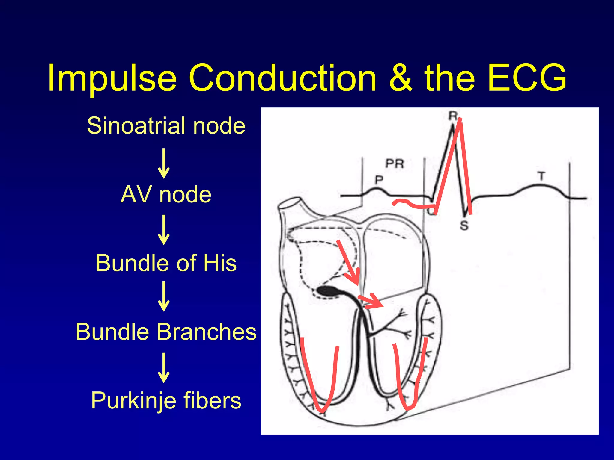
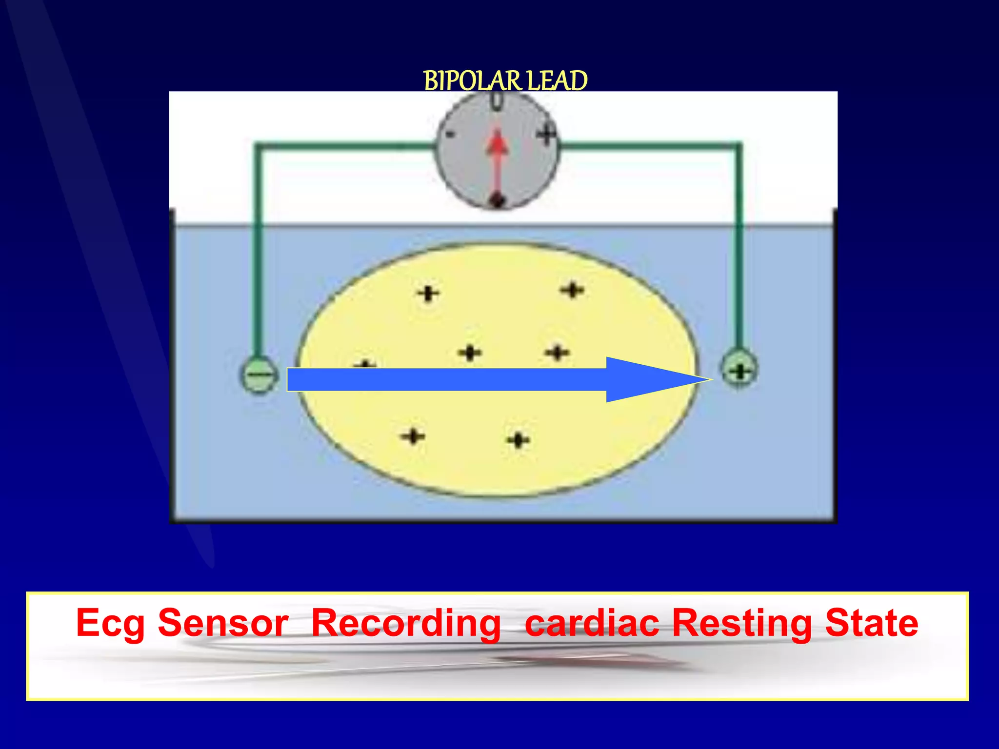
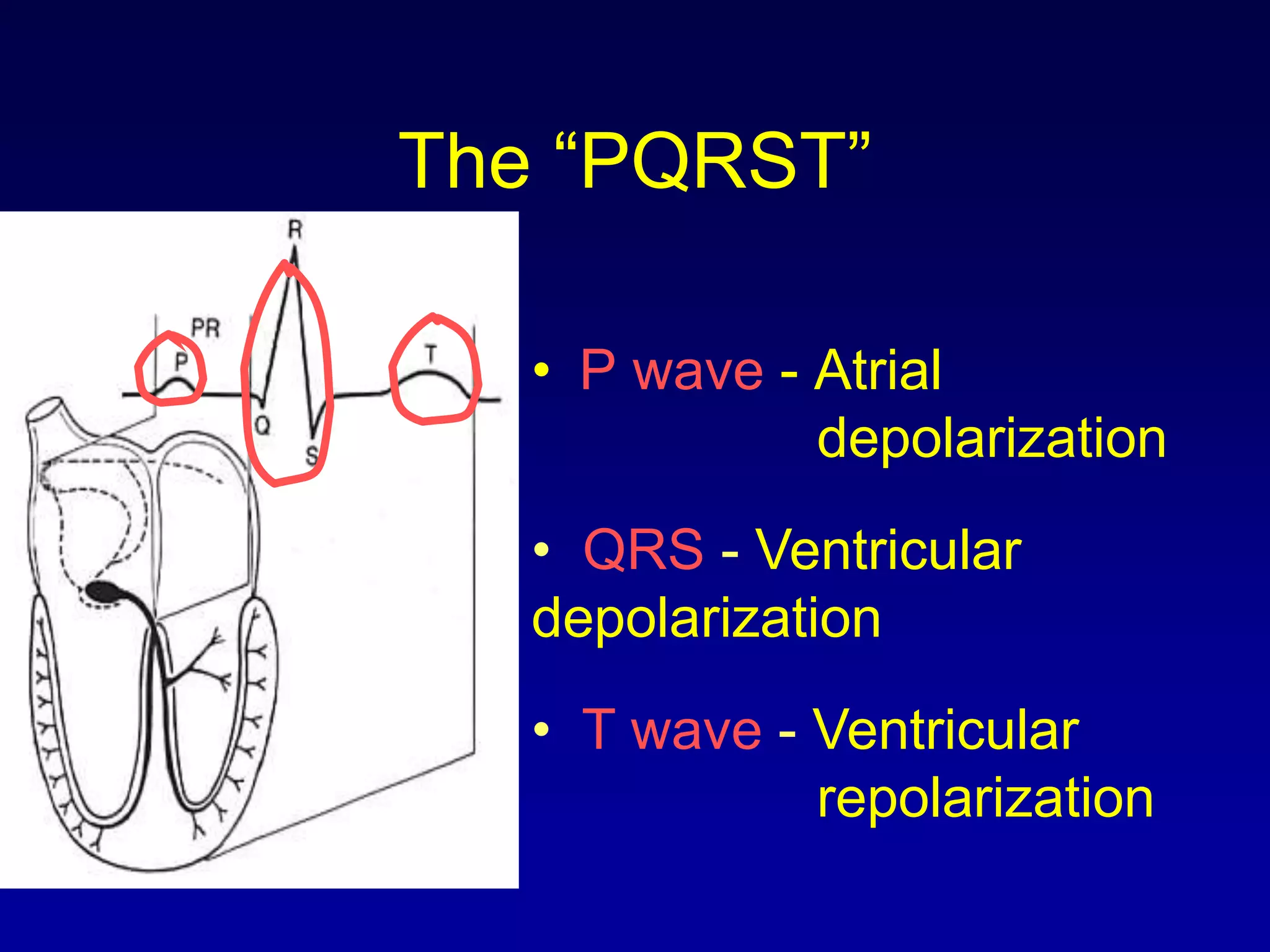
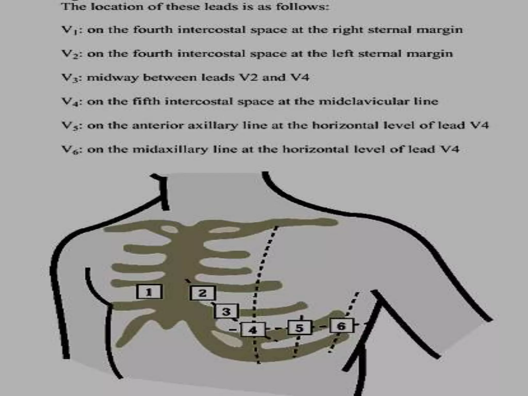
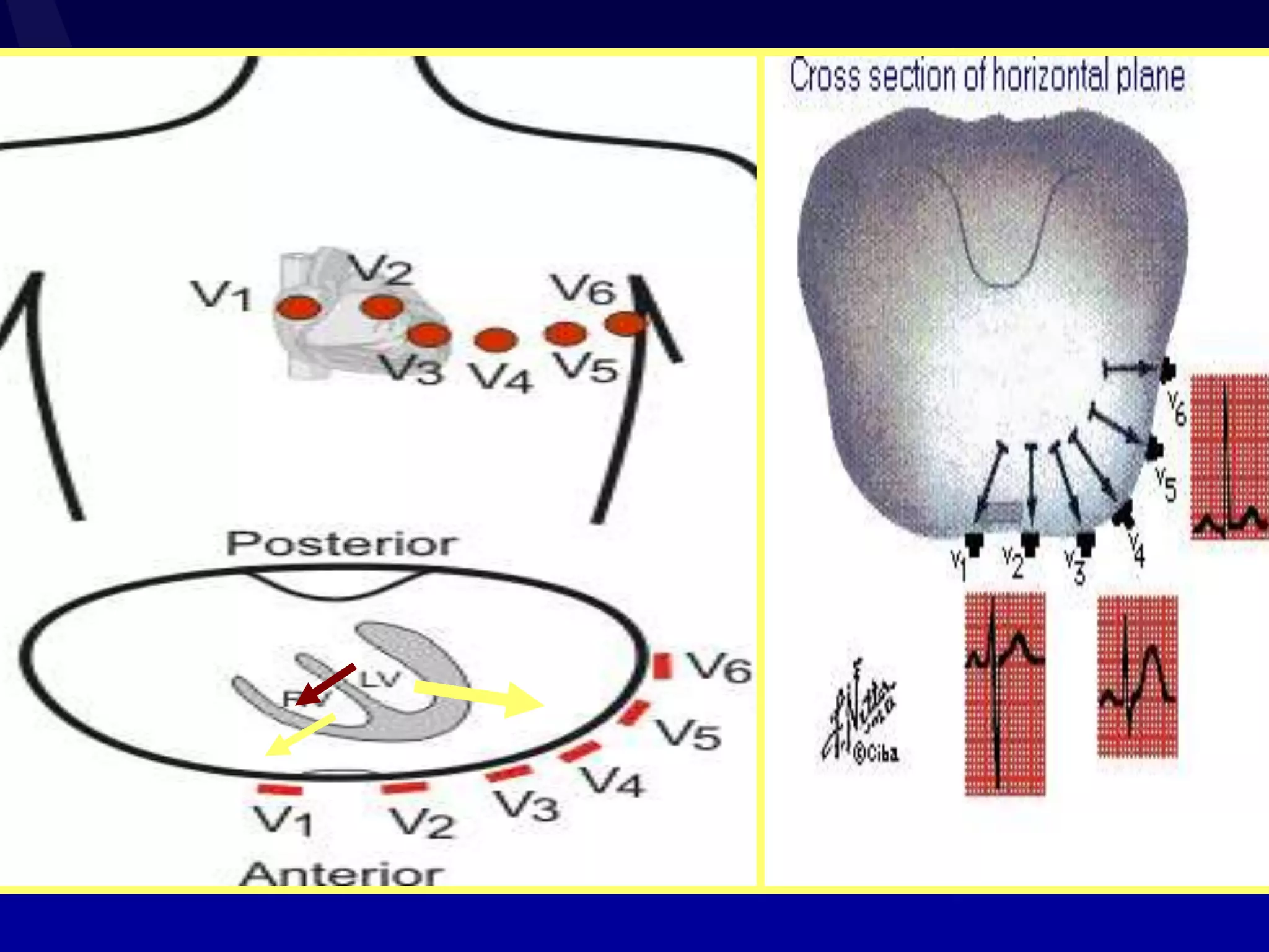
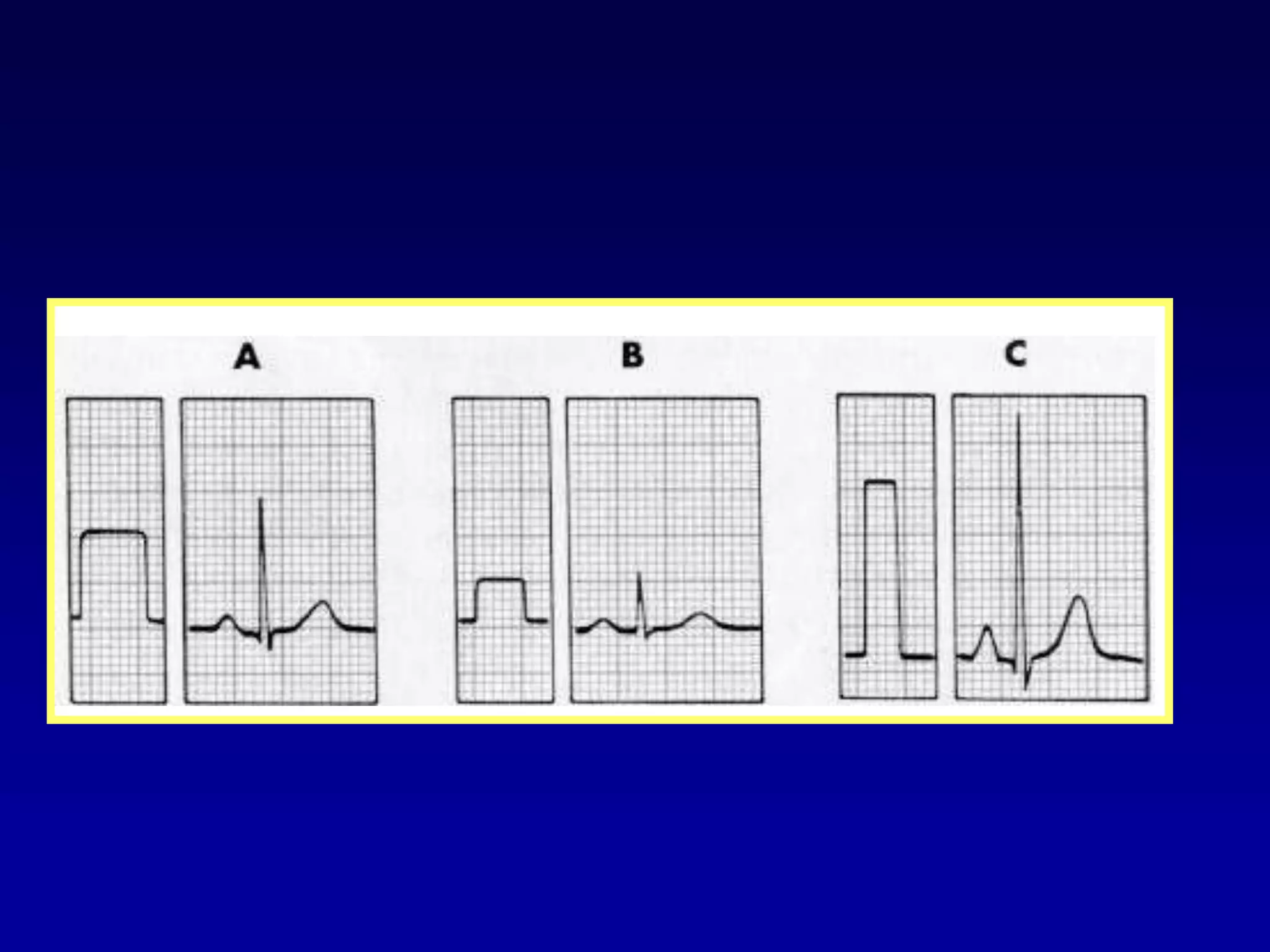
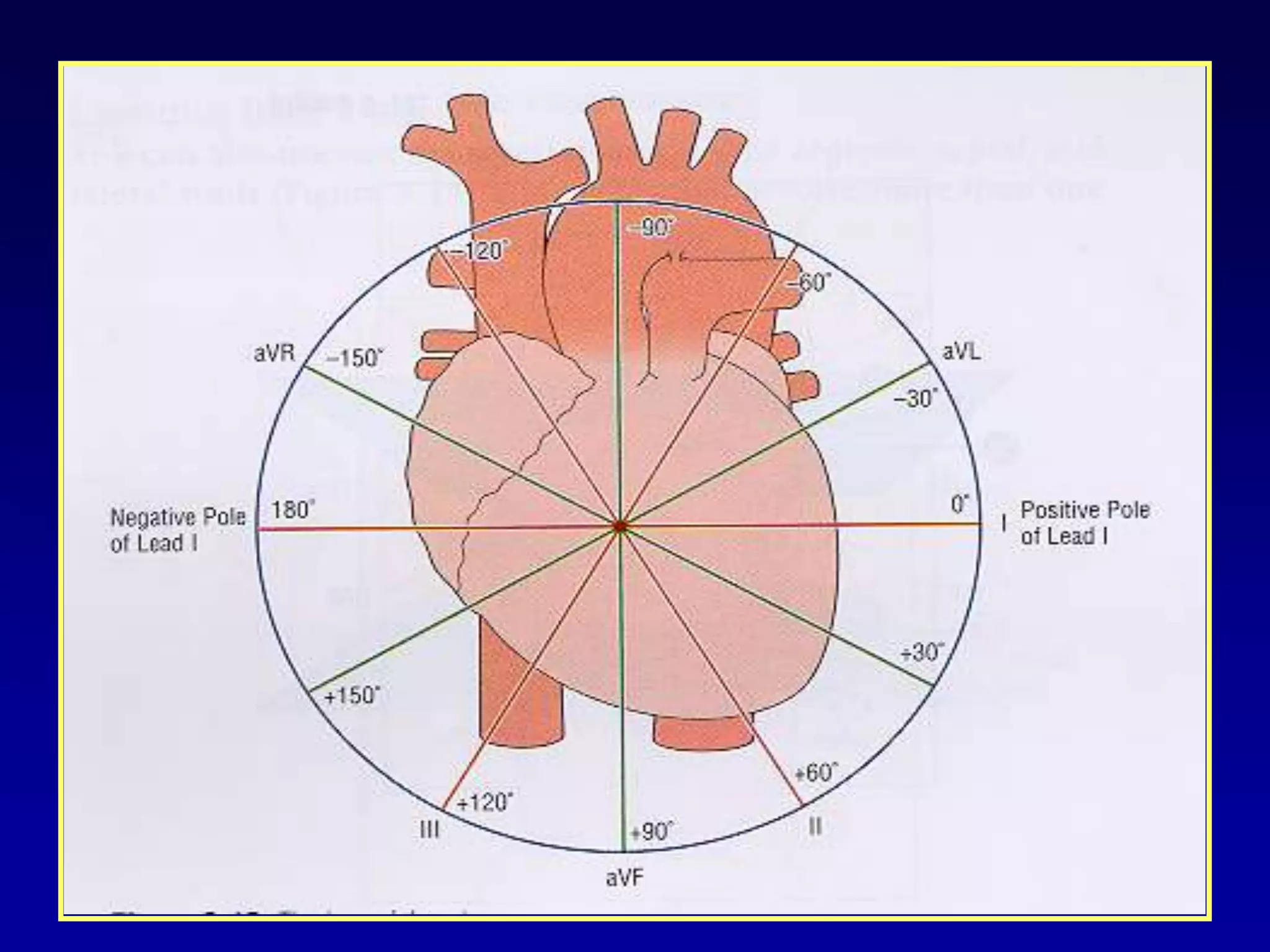
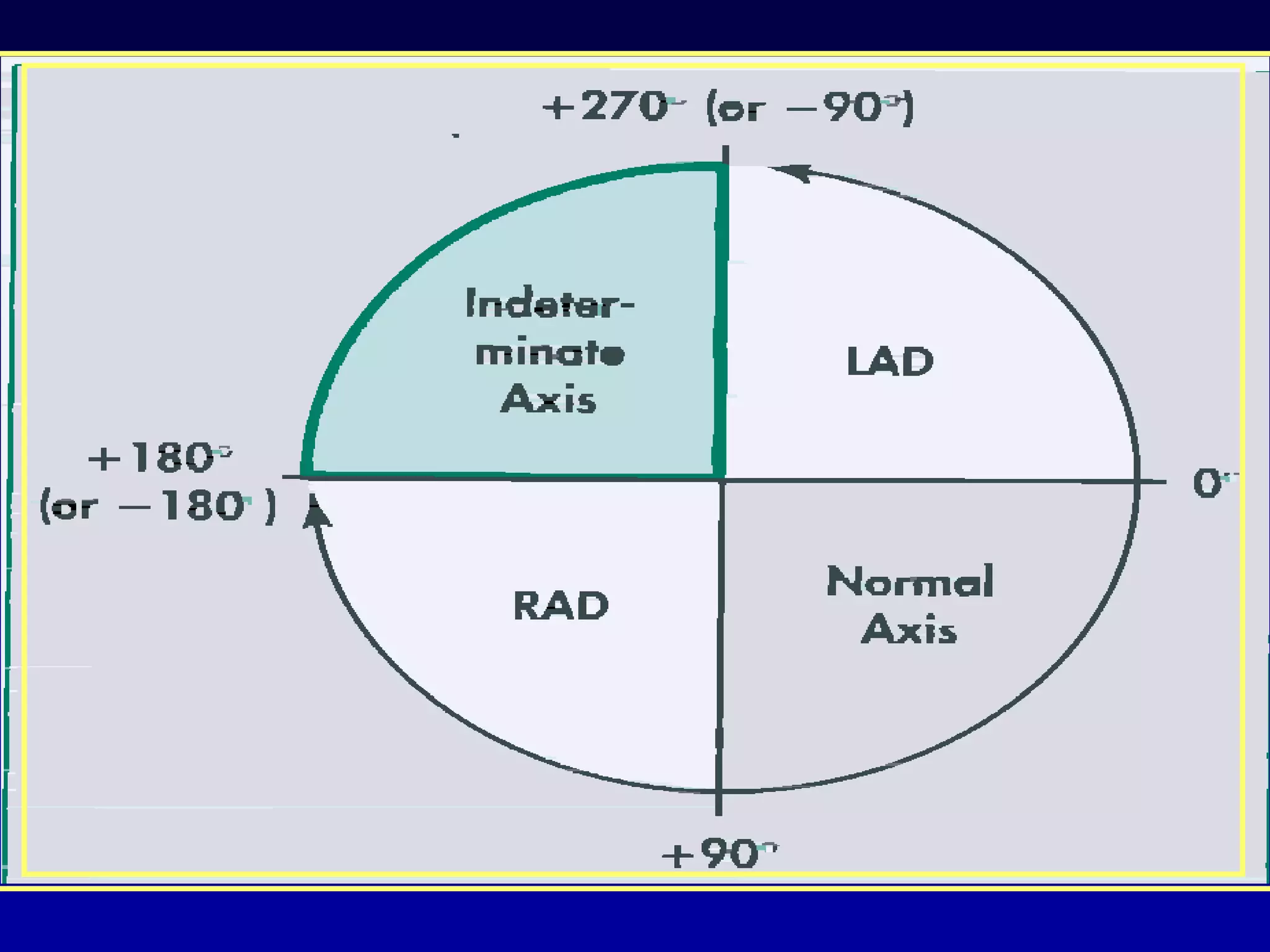
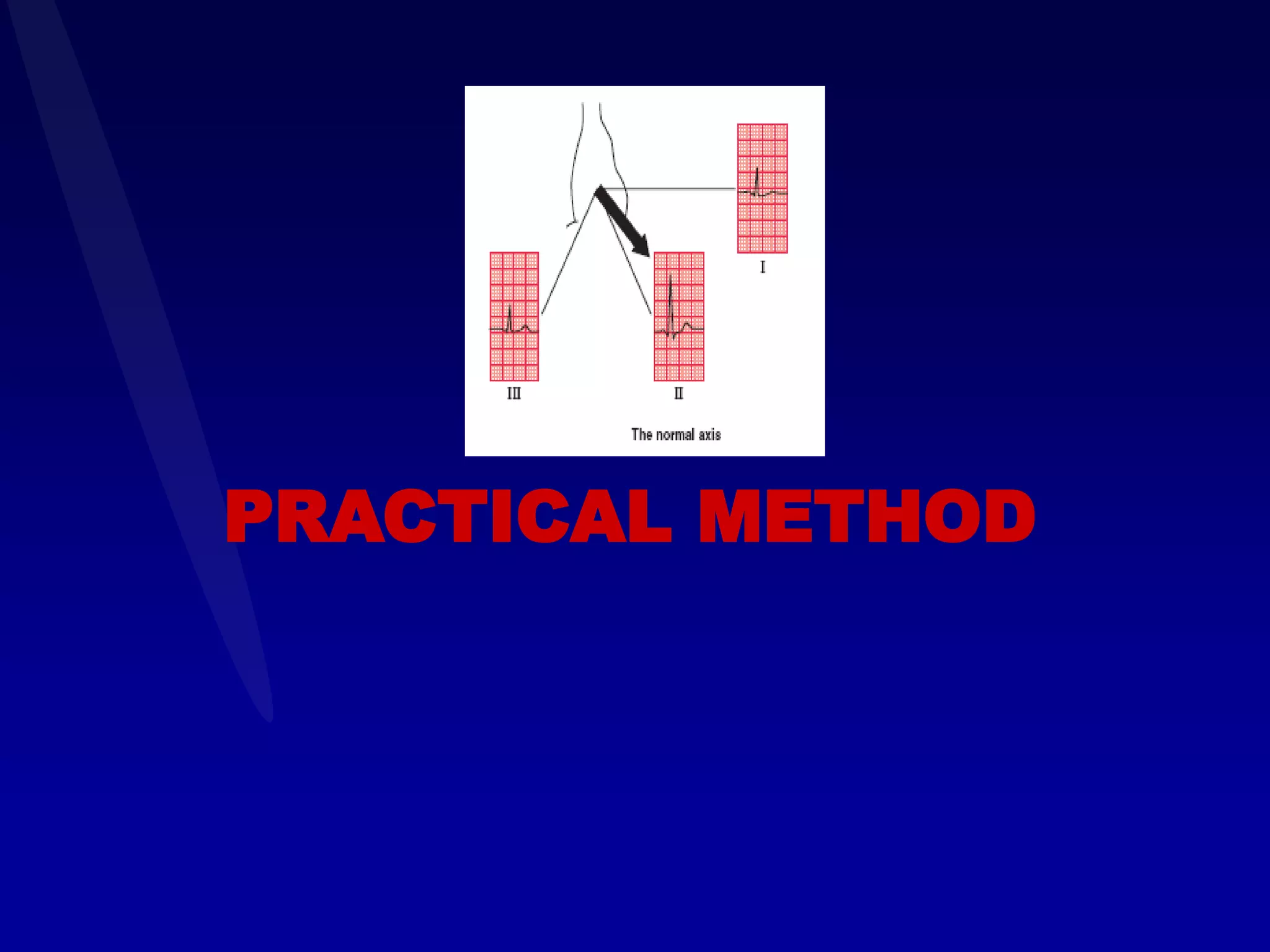
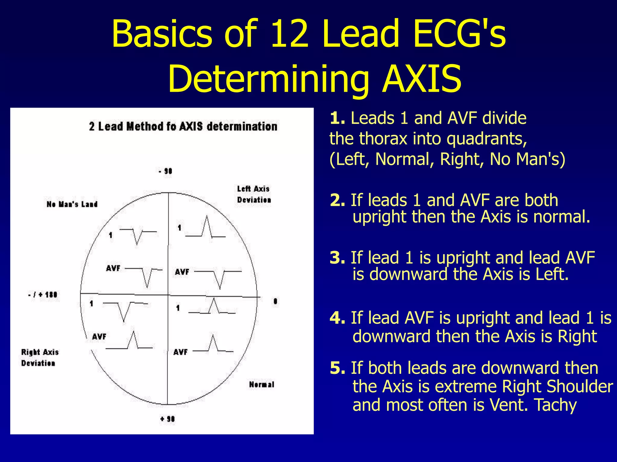
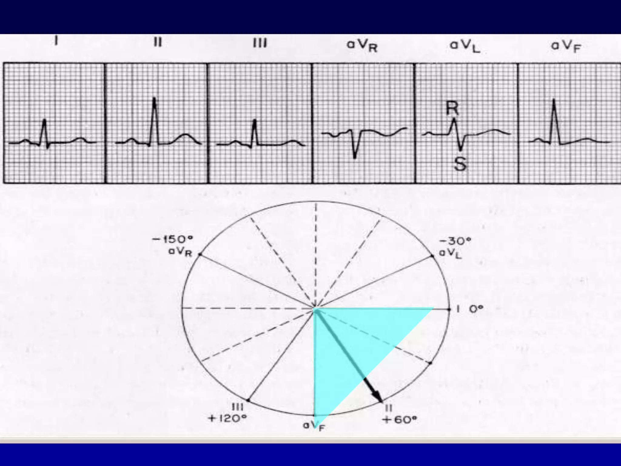
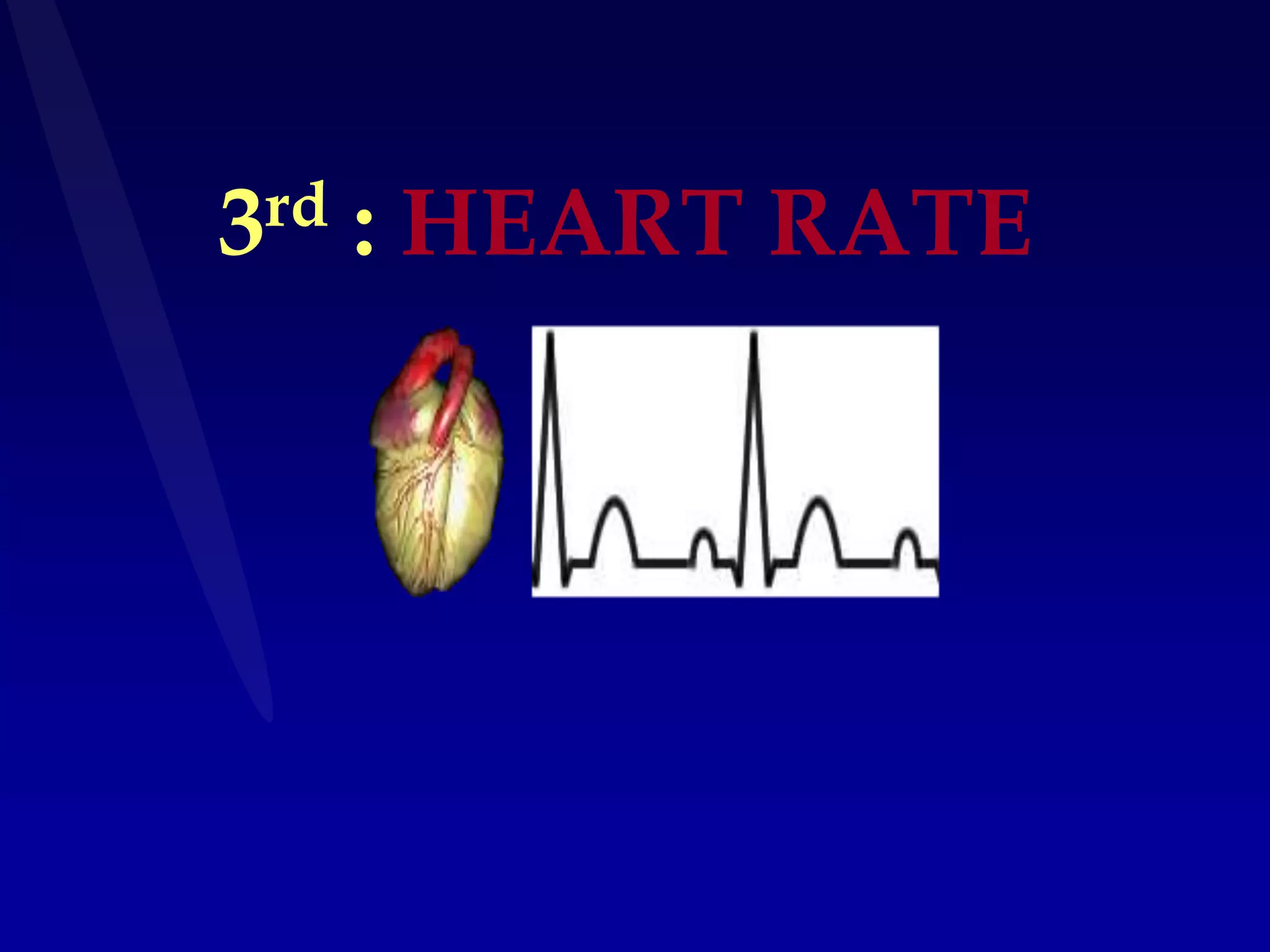
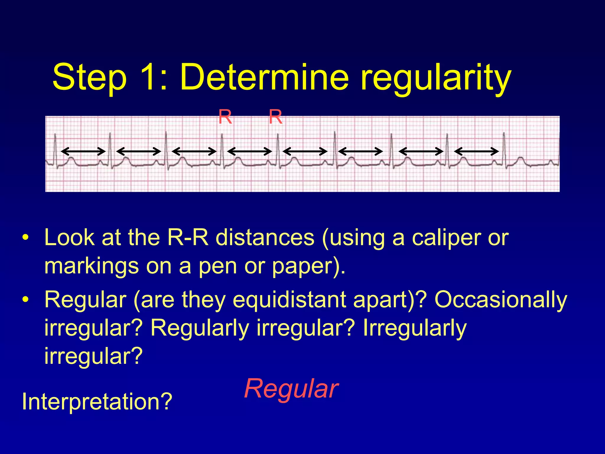
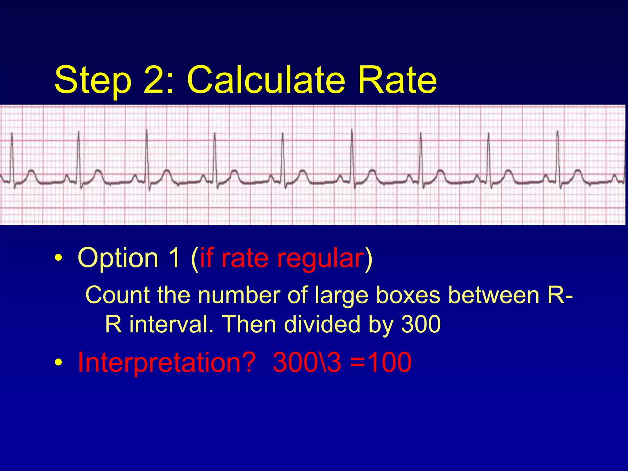
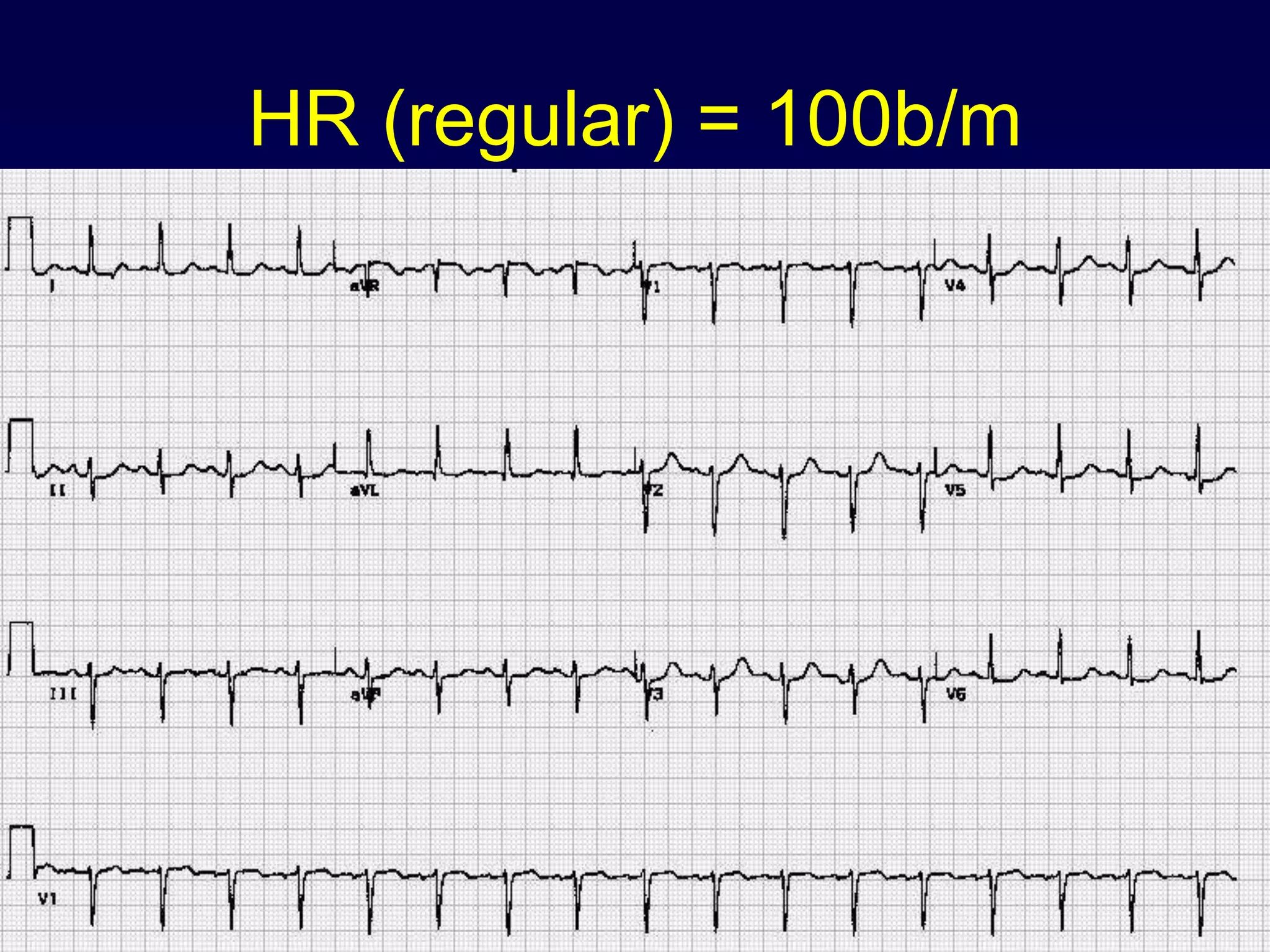
SlideShare a Scribd company logo
ECG
EARLY ECG
Conductive
System
Normal Impulse Conduction
Sinoatrial node
AV node
Bundle of His
Bundle Branches
Purkinje fibers
Impulse Conduction & the ECG
Sinoatrial node
AV node
Bundle of His
Bundle Branches
Purkinje fibers
Electrical
activity
Ecg Sensor Recording cardiac Resting State
BIPOLARLEAD
Spread of
depolarization
Repolarization
LEADII
+60
-120
Leads
and waves
The “PQRST”
• P wave - Atrial
depolarization
• T wave - Ventricular
repolarization
• QRS - Ventricular
depolarization
The PR Interval
Atrial depolarization
+
delay in AV junction
(AV node/Bundle of His)
(delay allows time for
the atria to contract
before the ventricles
contract)
The ECG Paper
• Horizontally
– One small box - 0.04 s
– One large box - 0.20 s
• Vertically
– One large box - 0.5 mV
The ECG Paper (cont)
• Every 3 seconds (15 large boxes) is
marked by a vertical line.
• This helps when calculating the heart
rate.
3 sec 3 sec
The 12-Lead ECG
• The 12-Lead ECG sees the heart
from 12 different views.
• Therefore, the 12-Lead ECG helps
you see what is happening in
different portions of the heart.
The 12-Leads
The 12-leads include:
–3 Limb leads
(I, II, III)
–3 Augmented leads
(aVR, aVL, aVF)
–6 Precordial leads
(V1- V6)
+ +
+
ECG LEADS
leads representing regions
ECG Interpretation
1st: ASSESS
CONNECTION & STANDERDIZATION
(voltage & timing)
2nd : CARDIAC AXIS
PRACTICAL METHOD
Basics of 12 Lead ECG's
Determining AXIS
1. Leads 1 and AVF divide
the thorax into quadrants,
(Left, Normal, Right, No Man's)
2. If leads 1 and AVF are both
upright then the Axis is normal.
3. If lead 1 is upright and lead AVF
is downward the Axis is Left.
4. If lead AVF is upright and lead 1 is
downward then the Axis is Right
5. If both leads are downward then
the Axis is extreme Right Shoulder
and most often is Vent. Tachy
• Lead I = left thumb and aVF = right thumb
– If both I & aVF are up = Normal Axis
– If I is up but aVF is down = LAD
– If I is down but aVF is up = RAD
– If both I & aVF are down = Extreme RAD
– Clue: If aVR is positive = extreme
RAD
EKG Axis for Dummies!
3rd : HEART RATE
Step 1: Determine regularity
• Look at the R-R distances (using a caliper or
markings on a pen or paper).
• Regular (are they equidistant apart)? Occasionally
irregular? Regularly irregular? Irregularly
irregular?
Interpretation?
Regular
R R
Step 2: Calculate Rate
• Option 1 (if rate regular)
Count the number of large boxes between R-
R interval. Then divided by 300
• Interpretation? 3003 =100
Step 2: Calculate Rate
• Option 2 (if rate irregular)
– Count the number of cardiac cycle(R –R
interval) in a 6 second rhythm strip, then
multiply by 10.
Interpretation? 9 x 10 = 90 bm
3 sec 3 sec
HR (regular) = 100b/m
HR(Irregular) = 70b/m
4th: ANALYSIS OF
waves, segments& intervals
12 LEAD ECG
The standard 12 Lead ECG
6 Limb Leads 6 Chest Leads (Precordial leads)
I, II, III, avR, avL, avF, V1, V2, V3, V4, V5 and V6
Rhythm Strip
Normal
ECG
PR interval0.12s-
0.2s(not>1 large sq)
QRS duration0.12s
(not>3 small squares)
PQRST
ST segmentisoelectric
T upright
EKG Basics: P Wave
• Normal
– Width < .1 secs
– Height .5 to 2.5 mm
– Morphology
• Flat
• Biphasic
EKG Basics: P Wave
• Abnormal
– Inversions
– Amplitude
• P-Pulmonale > 2.5 mm
– Duration
• P-Mitrale > .1 sec (or 2 ½ boxes)
– Absence
PR interval
• Normal: 0.12 - 0.20 seconds.
(3 - 5 small boxes)
Interpretation? 0.12 seconds
QRS
• Duration: .04 - .12 secs
• Amplitude: > 5mm;
< 20 mm in limb, < 25 in anterior leads
• Presence of Q waves < 0.04 msec and
< 2 mm
V1
V2
V3
V4
V5
V6
The R wave in the precordial leads must grow from V1 to at least V4
P
Q
T
S
All waves are negative in lead aVR
QRS Complex
May be too broad ( more than 0.12
seconds)
• A delay in the depolarisation of the
ventricles because the conduction
pathway is abnormal
Q Waves
Non Pathological Q waves
Q waves of less than 2mm are normal
Pathological Q waves
Q waves of more than 2mm
indicate full thickness myocardial
damage from an infarct
Late sign of MI (evolved)
ST Segment Depression
Can be characterised as:-
• Downsloping
• Upsloping
• Horizontal
Horizontal ST depression
ST Segment Depression
Downsloping ST segment depression:-
• Can be caused by digoxin.
Upward sloping ST segment depression:-
• Normal during exercise.
EKG Basics: T waves and
U waves
• T waves occur in
– Same direction as QRS
– Height: < 5 mm in limb leads, <10 mm in anterior
leads
– 0.18 – 0.22
• U waves
– After T wave
– Best seen in lead III
– Hypothermia/hypokalemia
QRS and T waves tend to have the same general direction in
the limb leads
The T wave must be upright in I, II, V2 to V6
I II III aVR aVL aVF V1 V2 V3 V4 V5 V6
The T wave must be upright in I, II, V2 to V6
T waves
Hyperacute T waves
CARDIAC CHAMBRE
ENLARGMENT
AA
RAA
LAA
Left Atrial Enlargement: Criteria
• P wave
• Notch in P wave
– Any lead
– Peaks > 0.04 secs
• V1
– Terminal portion of P wave > 1mm deep
and > 0.04 sec wide
P Wave: Left Atrial Enlargement
LVH & RVH
RBBB & LBBB
Right Bundle Branch Blocks
What QRS morphology is characteristic?
V1
For RBBB the wide QRS complex assumes a
unique, virtually diagnostic shape in those
leads overlying the right ventricle (V1 and V2).
“Rabbit Ears”
V1 V6
Myocardial infarction
Myocardial layers
Epicardial, Middle, Subendocardial and
Endocardial
 Ischemia is a relative condition that depends on
the balance among:
1. Coronary blood supply
2. Oxygenation of the blood
3. Myocardial workload
 The myocardial area affected by ischemia
depends on:
1. Proximity to intracavitary blood supply
2. Distance from the major coronary arteries
3. Workload
ECG Changes & the Evolving MI
There are two
distinct patterns
of ECG change
depending if the
infarction is:
–ST Elevation (Transmural or Q-wave), or
–Non-ST Elevation (Subendocardial or non-Q-wave)
Non-ST Elevation
ST Elevation
INDEX
sub
endocardial
vessels
sub epicardial
vessels
left
anterior
oblique
cross
section
of LV
Transmural
MI
SubendocardialMI
blockage
Origin of “pathological Q wave”
Stages of Ischemia & Infarction
• Ischemia during exercise: “ST-segment depression”
• usually indicative of subendocardial ischemia
J-point .08 seconds
Quantity or depth
of ST-segment
depression
Baseline
Reciprocal Changes
• Changes occurring on the opposite side
of the myocardium that is infarcting
leads representing regions
acute-inferior-MI
Inferior_MI
Acute antero-lateral MI
Acute Pericarditis
TESTS
Normal ECG
Quick Quiz
Mr Jones is diagnosed as having had an
anterior MI. On which leads would you
expect to see the main changes?
(a) II, III and avL.
(b) I and avL.
(c) V2 - V4.
Quick Quiz
Mr Jackson has ECG changes suggestive
of an MI on leads II, III and avF. Which
surface of his heart is affected?
(a) The anterior surface.
(b) The lateral surface.
(c) The inferior surface.
Quick Quiz
Mrs Brown requires PTCA to her Circumflex
artery after complaining of unstable angina
symptoms. Her 12 lead ECG shows ST
depression and T wave inversion in what
leads?
(a) I, avL, V5 and V6
(b) II, III and avL
(c) V3 and V4
A 55 year old man with 4 hours of “crushing” chest
pain.
Acute inferior myocardial infarction (with reciprocal changes)
 ST elevation in the inferior leads II, III and aVF
 reciprocal ST depression in the anterior leads
A 63 Year Old woman with 10 hours of chest pain and
sweating
Can you guess her diagnosis?
Acute anterior-lateral myocardial infarction
ST elevation in the anterior leads V1 - 6, I and aVL
reciprocal ST depression in the inferior leads
Normal ECG
LBBB + acute inferior MI
LVH + P mitrale
P pulmonale
RBBB + acute inferior MI + sinus
bradycardia (HR = 43 b/m)
Dysrhythmia
 Consider the following:
1- Heart rate (tachy or brady).
2- Rhythm (regular or irregular < regular – irregular>).
3- P wave (Presence - Shape).
4- QRS (Normal or abnormal shape and size).
5- A-V conduction.
(Is there P wave for each QRS?)
(PR interval)
Arrhythmia Formation
Arrhythmias can arise from problems in
the:
• Sinus node
• Atrial cells
• AV junction
• Ventricular cells
SA Node Problems
The SA Node can:
• fire too slow
• fire too fast
Sinus Bradycardia
Sinus Tachycardia
Atrial Cell Problems
Atrial cells can:
• fire occasionally
from a focus
• fire continuously
Atrial premature beat
P .S .V .T
Atrial Flutter
Atrial Cell Problems
Atrial cells can also:
• fire continuously
from multiple foci
Atrial Fibrillation
AV Junctional Problems
The AV junction can:
Fire (acting as
pacemaker)
• fire continuously
• block impulses
coming from the
SA Node
Premature nodal beat
Paroxysmal SVT
AV Junctional Blocks
Ventricular Cell Problems
Fire occasionally
from a focus
Fire contanously
from a focus
Fire continuously
from multiple foci
Premature Ventricular
Contractions (PVCs)
Ventricular Tachycardia
Ventricular fibrillation
Sinus Bradycardia
30 bpm• Rate?
• Regularity? regular
normal
0.10 s
• P waves?
• PR interval? 0.12 s
• QRS duration?
Interpretation? Sinus Bradycardia
Sinus Bradycardia
• Deviation from NSR
- Rate < 60 bpm
Sinus Tachycardia
130 bpm• Rate?
• Regularity? regular
normal
0.08 s
• P waves?
• PR interval? 0.16 s
• QRS duration?
Interpretation? Sinus Tachycardia
Sinus Tachycardia
• Deviation from NSR
- Rate > 100 bpm
Atrial premature beat
70 bpm• Rate?
• Regularity? occasionally irreg.
different contour
0.08
• P waves?
• PR interval? 0.14 s
• QRS duration?
Interpretation? NSR with Premature Atrial
Contractions
Atrial premature beat
• Deviation from NSR
–These ectopic beats come early than
the normal. QRS complex is normal
(narrow)= (0.04 - 0.12 s).
Atrial bigeminy
Sinus Rhythm with 1 PVC
60 bpm• Rate?
• Regularity? occasionally irreg.
none for 7th QRS
(wide)
• P waves?
• PR interval? 0.14 s
• QRS duration?
Interpretation? Sinus Rhythm with 1 PVC
PVCs
• Deviation from NSR
– Ectopic beats originate in the ventricles
resulting in wide and bizarre QRS
complexes.
– When there are more than 1 premature
beats and look alike, they are called
“uniform”. When they look different, they are
called “multiform”.
Junctional premature beats
Supraventricular Tachycardia
(PSVT)
74 150 bpm• Rate?
• Regularity? Regular
none
0.08 s
• P waves?
• PR interval? none
• QRS duration?
Interpretation? Paroxysmal Supraventricular
Tachycardia (PSVT)
70 bpm• Rate?
• Regularity? regular
flutter waves (Saw teeth)
0.06 s
• P waves?
• PR interval? none
• QRS duration?
Interpretation? Atrial Flutter
Atrial Flutter
Atrial Flutter
• Deviation from NSR
–No P waves. Instead flutter waves (note
“sawtooth” pattern) are formed at a rate
of 250 - 350 bpm.
–Only some impulses conduct through
the AV node (usually every other
impulse).
Atrial Fibrillation
100 bpm• Rate?
• Regularity? irregularly irregular
none
0.06 s
• P waves?
• PR interval? none
• QRS duration?
Interpretation? Atrial Fibrillation
Atrial Fibrillation
Deviation from NSR
–No organized atrial depolarization, so
no normal P waves (impulses are not
originating from the sinus node). R-R
interval is irregular bec. AV node
allows some of the impulses to pass
through at variable intervals (so rhythm
is irregularly irregular).QRS is normal
Ventricular Tachycardia
160 bpm• Rate?
• Regularity? regular
none
wide (> 0.12 sec)
• P waves?
• PR interval? none
• QRS duration?
Interpretation? Ventricular Tachycardia
Ventricular Tachycardia
• Deviation from NSR
–Impulse is originating in the ventricles
(no P waves, wide QRS).
Ventricular Fibrillation
none• Rate?
• Regularity? irregularly irreg.
none
wide, if recognizable
• P waves?
• PR interval? none
• QRS duration?
Interpretation? Ventricular Fibrillation
Ventricular Fibrillation
• Deviation from NSR
–Completely abnormal.
AV Nodal Blocks
• 1st Degree AV Block
• 2nd Degree AV Block, Type I
• 2nd Degree AV Block, Type II
• 3rd Degree AV Block
60 bpm• Rate?
• Regularity? regular
normal
0.08 s
• P waves?
• PR interval? 0.36 s
• QRS duration?
Interpretation? 1st Degree AV Block
1st Degree AV Block
1st Degree AV Block
• Deviation from NSR
–PR Interval > 0.20 s
(Prolonged conduction delay in the AV node).
2nd Degree AV Block, Type I
50 bpm• Rate?
• Regularity? regularly irregular
normal, but 4th no QRS
0.08 s
• P waves?
• PR interval? lengthens
• QRS duration?
Interpretation? 2nd Degree AV Block, Type I
2nd Degree AV Block, Type I
• Deviation from NSR
–PR interval progressively lengthens,
then the impulse is completely blocked
(P wave not followed by QRS).
2nd Degree AV Block, Type II
40 bpm• Rate?
• Regularity? regular
nl, 2 of 3 no QRS
0.08 s
• P waves?
• PR interval? 0.14 s
• QRS duration?
Interpretation? 2nd Degree AV Block, Type II
2nd Degree AV Block, Type II
• Deviation from NSR
–Occasional P waves are completely
blocked (P wave not followed by QRS).
3rd Degree AV Block
40 bpm• Rate?
• Regularity? regular
no relation to QRS
wide (> 0.12 s)
• P waves?
• PR interval? none
• QRS duration?
Interpretation? 3rd Degree AV Block
3rd Degree AV Block
• Etiology: There is complete block of
conduction in the AV junction, so the
atria and ventricles form impulses
independently of each other. Without
impulses from the atria, the ventricles
own intrinsic pacemaker fires in at
around 30 - 45 beats/minute.
Ecg final the best
Ecg final the best
Ecg final the best

More Related Content

What's hot

Bundle branch blocks
Bundle branch blocksBundle branch blocks
Bundle branch blocks
Sonukurian
 
SINOATRIAL (SA) node
SINOATRIAL (SA) node SINOATRIAL (SA) node
SINOATRIAL (SA) node
Malleswara rao Dangeti
 
Situs inversus totalis
Situs inversus totalisSitus inversus totalis
Situs inversus totalis
Aziza Alamri - UOD
 
Narrow complex tachycardias
Narrow complex tachycardiasNarrow complex tachycardias
Narrow complex tachycardias
salaheldin abusin
 
Basic ecg
Basic ecgBasic ecg
Basic ecg
A.Salam Sharif
 
Electrocardiogram in myocardial infarction
Electrocardiogram in myocardial infarction Electrocardiogram in myocardial infarction
Electrocardiogram in myocardial infarction
Ramachandra Barik
 
Systematic ECG Interpretation
Systematic ECG InterpretationSystematic ECG Interpretation
Systematic ECG Interpretation
SCGH ED CME
 
Ecg findings in life threatening conditions
Ecg findings in life threatening conditionsEcg findings in life threatening conditions
Ecg findings in life threatening conditions
MEEQAT HOSPITAL
 
Acute coronary syndrome
Acute coronary syndrome Acute coronary syndrome
Acute coronary syndrome
Dee Evardone
 
Antiarrhythmic therapy for supraventricular arrhythmias
Antiarrhythmic therapy for supraventricular arrhythmiasAntiarrhythmic therapy for supraventricular arrhythmias
Antiarrhythmic therapy for supraventricular arrhythmias
Kyaw Win
 
Lbbb + sgarbossa
Lbbb + sgarbossaLbbb + sgarbossa
Lbbb + sgarbossachricres
 
The secret of ECG
The secret of ECG The secret of ECG
The secret of ECG
Imtinan Barnawi
 
Atrial fibrillation -latest
Atrial fibrillation -latestAtrial fibrillation -latest
Atrial fibrillation -latest
Praveen Nagula
 
16 arrhythmias2009
16 arrhythmias200916 arrhythmias2009
16 arrhythmias2009internalmed
 
CARDIOGENIC SHOCK
CARDIOGENIC SHOCKCARDIOGENIC SHOCK
CARDIOGENIC SHOCK
Mahi Yeruva
 
ECG in Acute Myocardial Infarction
ECG in Acute Myocardial InfarctionECG in Acute Myocardial Infarction
ECG in Acute Myocardial Infarction
Chetan Ganteppanavar
 
Approach to heart failure cases
Approach to heart failure casesApproach to heart failure cases
Approach to heart failure cases
hospital
 
ECG in Emergency Department - Advances in ACS ECG
ECG in Emergency Department - Advances in ACS ECGECG in Emergency Department - Advances in ACS ECG
ECG in Emergency Department - Advances in ACS ECG
Dr.Mahmoud Abbas
 

What's hot (20)

Bundle branch blocks
Bundle branch blocksBundle branch blocks
Bundle branch blocks
 
SINOATRIAL (SA) node
SINOATRIAL (SA) node SINOATRIAL (SA) node
SINOATRIAL (SA) node
 
Situs inversus totalis
Situs inversus totalisSitus inversus totalis
Situs inversus totalis
 
Narrow complex tachycardias
Narrow complex tachycardiasNarrow complex tachycardias
Narrow complex tachycardias
 
Basic ecg
Basic ecgBasic ecg
Basic ecg
 
Electrocardiogram in myocardial infarction
Electrocardiogram in myocardial infarction Electrocardiogram in myocardial infarction
Electrocardiogram in myocardial infarction
 
Systematic ECG Interpretation
Systematic ECG InterpretationSystematic ECG Interpretation
Systematic ECG Interpretation
 
Ecg findings in life threatening conditions
Ecg findings in life threatening conditionsEcg findings in life threatening conditions
Ecg findings in life threatening conditions
 
Acute coronary syndrome
Acute coronary syndrome Acute coronary syndrome
Acute coronary syndrome
 
Antiarrhythmic therapy for supraventricular arrhythmias
Antiarrhythmic therapy for supraventricular arrhythmiasAntiarrhythmic therapy for supraventricular arrhythmias
Antiarrhythmic therapy for supraventricular arrhythmias
 
Lbbb + sgarbossa
Lbbb + sgarbossaLbbb + sgarbossa
Lbbb + sgarbossa
 
The secret of ECG
The secret of ECG The secret of ECG
The secret of ECG
 
Atrial fibrillation -latest
Atrial fibrillation -latestAtrial fibrillation -latest
Atrial fibrillation -latest
 
16 arrhythmias2009
16 arrhythmias200916 arrhythmias2009
16 arrhythmias2009
 
CARDIOGENIC SHOCK
CARDIOGENIC SHOCKCARDIOGENIC SHOCK
CARDIOGENIC SHOCK
 
ECG in Acute Myocardial Infarction
ECG in Acute Myocardial InfarctionECG in Acute Myocardial Infarction
ECG in Acute Myocardial Infarction
 
EP diagnosis of WIDE COMPLEX TACHYCARDIA
EP diagnosis of WIDE COMPLEX TACHYCARDIAEP diagnosis of WIDE COMPLEX TACHYCARDIA
EP diagnosis of WIDE COMPLEX TACHYCARDIA
 
Approach to heart failure cases
Approach to heart failure casesApproach to heart failure cases
Approach to heart failure cases
 
ECG: Atrial Bigeminy
ECG: Atrial BigeminyECG: Atrial Bigeminy
ECG: Atrial Bigeminy
 
ECG in Emergency Department - Advances in ACS ECG
ECG in Emergency Department - Advances in ACS ECGECG in Emergency Department - Advances in ACS ECG
ECG in Emergency Department - Advances in ACS ECG
 

Similar to Ecg final the best

ECG-2 RAMA.pptx
ECG-2 RAMA.pptxECG-2 RAMA.pptx
ECG-2 RAMA.pptx
manishadya
 
Ecg interpretation
Ecg interpretationEcg interpretation
Ecg interpretationSudhir Dev
 
ECG
ECGECG
Basics of ecg
Basics of ecgBasics of ecg
Basics of ecg
Mohammad Rehan
 
ECG
ECGECG
ECG
aanmol
 
base-110816084037-phpapp02.pdf
base-110816084037-phpapp02.pdfbase-110816084037-phpapp02.pdf
base-110816084037-phpapp02.pdf
ssuser61d4e0
 
ECG lecture
ECG lectureECG lecture
ECG lecture
Alric Mondragon
 
Ecg
EcgEcg
EKG Dasar medis michael johandadasdas.ppt
EKG Dasar medis michael johandadasdas.pptEKG Dasar medis michael johandadasdas.ppt
EKG Dasar medis michael johandadasdas.ppt
michaeljohan1211
 
‏‏Shafei ecg نسخة
‏‏Shafei ecg   نسخة‏‏Shafei ecg   نسخة
‏‏Shafei ecg نسخة
shafei lashin
 
Tutorial in ecg
Tutorial in ecgTutorial in ecg
Tutorial in ecg
Syahmi Mizan
 
Electrocardiography
ElectrocardiographyElectrocardiography
Electrocardiography
IshaShripad
 
ECG interpretation: the basics
ECG interpretation: the basicsECG interpretation: the basics
ECG interpretation: the basics
Jamie Ranse
 
Electrocardiogram And It's Interpretation
Electrocardiogram And It's InterpretationElectrocardiogram And It's Interpretation
Electrocardiogram And It's Interpretation
NeamatullahAhmed2
 
1382531 634694433533008750
1382531 6346944335330087501382531 634694433533008750
1382531 634694433533008750Ruth Smith
 
Ecg 5th year 2016
Ecg 5th year 2016Ecg 5th year 2016
Ecg 5th year 2016
Yousef Biuk
 
Ecg interpretation , Upgraded
Ecg interpretation , UpgradedEcg interpretation , Upgraded
Ecg interpretation , Upgraded
Abdullah Almazyad
 

Similar to Ecg final the best (20)

ECG-2 RAMA.pptx
ECG-2 RAMA.pptxECG-2 RAMA.pptx
ECG-2 RAMA.pptx
 
Ecg interpretation
Ecg interpretationEcg interpretation
Ecg interpretation
 
ECG
ECGECG
ECG
 
Basics of ecg
Basics of ecgBasics of ecg
Basics of ecg
 
ECG
ECGECG
ECG
 
base-110816084037-phpapp02.pdf
base-110816084037-phpapp02.pdfbase-110816084037-phpapp02.pdf
base-110816084037-phpapp02.pdf
 
Ecg
EcgEcg
Ecg
 
ECG lecture
ECG lectureECG lecture
ECG lecture
 
Ecg
EcgEcg
Ecg
 
EKG Dasar medis michael johandadasdas.ppt
EKG Dasar medis michael johandadasdas.pptEKG Dasar medis michael johandadasdas.ppt
EKG Dasar medis michael johandadasdas.ppt
 
‏‏Shafei ecg نسخة
‏‏Shafei ecg   نسخة‏‏Shafei ecg   نسخة
‏‏Shafei ecg نسخة
 
Tutorial in ecg
Tutorial in ecgTutorial in ecg
Tutorial in ecg
 
Electrocardiography
ElectrocardiographyElectrocardiography
Electrocardiography
 
Lec 6 ECG.pptx
Lec 6 ECG.pptxLec 6 ECG.pptx
Lec 6 ECG.pptx
 
ECG interpretation: the basics
ECG interpretation: the basicsECG interpretation: the basics
ECG interpretation: the basics
 
Electrocardiogram And It's Interpretation
Electrocardiogram And It's InterpretationElectrocardiogram And It's Interpretation
Electrocardiogram And It's Interpretation
 
1382531 634694433533008750
1382531 6346944335330087501382531 634694433533008750
1382531 634694433533008750
 
Ecg 5th year 2016
Ecg 5th year 2016Ecg 5th year 2016
Ecg 5th year 2016
 
Ecg assessment of ihd
Ecg assessment of ihdEcg assessment of ihd
Ecg assessment of ihd
 
Ecg interpretation , Upgraded
Ecg interpretation , UpgradedEcg interpretation , Upgraded
Ecg interpretation , Upgraded
 

Recently uploaded

Unit 2- Research Aptitude (UGC NET Paper I).pdf
Unit 2- Research Aptitude (UGC NET Paper I).pdfUnit 2- Research Aptitude (UGC NET Paper I).pdf
Unit 2- Research Aptitude (UGC NET Paper I).pdf
Thiyagu K
 
The Accursed House by Émile Gaboriau.pptx
The Accursed House by Émile Gaboriau.pptxThe Accursed House by Émile Gaboriau.pptx
The Accursed House by Émile Gaboriau.pptx
DhatriParmar
 
Francesca Gottschalk - How can education support child empowerment.pptx
Francesca Gottschalk - How can education support child empowerment.pptxFrancesca Gottschalk - How can education support child empowerment.pptx
Francesca Gottschalk - How can education support child empowerment.pptx
EduSkills OECD
 
BÀI TẬP BỔ TRỢ TIẾNG ANH GLOBAL SUCCESS LỚP 3 - CẢ NĂM (CÓ FILE NGHE VÀ ĐÁP Á...
BÀI TẬP BỔ TRỢ TIẾNG ANH GLOBAL SUCCESS LỚP 3 - CẢ NĂM (CÓ FILE NGHE VÀ ĐÁP Á...BÀI TẬP BỔ TRỢ TIẾNG ANH GLOBAL SUCCESS LỚP 3 - CẢ NĂM (CÓ FILE NGHE VÀ ĐÁP Á...
BÀI TẬP BỔ TRỢ TIẾNG ANH GLOBAL SUCCESS LỚP 3 - CẢ NĂM (CÓ FILE NGHE VÀ ĐÁP Á...
Nguyen Thanh Tu Collection
 
Honest Reviews of Tim Han LMA Course Program.pptx
Honest Reviews of Tim Han LMA Course Program.pptxHonest Reviews of Tim Han LMA Course Program.pptx
Honest Reviews of Tim Han LMA Course Program.pptx
timhan337
 
Language Across the Curriculm LAC B.Ed.
Language Across the  Curriculm LAC B.Ed.Language Across the  Curriculm LAC B.Ed.
Language Across the Curriculm LAC B.Ed.
Atul Kumar Singh
 
Sha'Carri Richardson Presentation 202345
Sha'Carri Richardson Presentation 202345Sha'Carri Richardson Presentation 202345
Sha'Carri Richardson Presentation 202345
beazzy04
 
Phrasal Verbs.XXXXXXXXXXXXXXXXXXXXXXXXXX
Phrasal Verbs.XXXXXXXXXXXXXXXXXXXXXXXXXXPhrasal Verbs.XXXXXXXXXXXXXXXXXXXXXXXXXX
Phrasal Verbs.XXXXXXXXXXXXXXXXXXXXXXXXXX
MIRIAMSALINAS13
 
Digital Tools and AI for Teaching Learning and Research
Digital Tools and AI for Teaching Learning and ResearchDigital Tools and AI for Teaching Learning and Research
Digital Tools and AI for Teaching Learning and Research
Vikramjit Singh
 
Synthetic Fiber Construction in lab .pptx
Synthetic Fiber Construction in lab .pptxSynthetic Fiber Construction in lab .pptx
Synthetic Fiber Construction in lab .pptx
Pavel ( NSTU)
 
Supporting (UKRI) OA monographs at Salford.pptx
Supporting (UKRI) OA monographs at Salford.pptxSupporting (UKRI) OA monographs at Salford.pptx
Supporting (UKRI) OA monographs at Salford.pptx
Jisc
 
CLASS 11 CBSE B.St Project AIDS TO TRADE - INSURANCE
CLASS 11 CBSE B.St Project AIDS TO TRADE - INSURANCECLASS 11 CBSE B.St Project AIDS TO TRADE - INSURANCE
CLASS 11 CBSE B.St Project AIDS TO TRADE - INSURANCE
BhavyaRajput3
 
Instructions for Submissions thorugh G- Classroom.pptx
Instructions for Submissions thorugh G- Classroom.pptxInstructions for Submissions thorugh G- Classroom.pptx
Instructions for Submissions thorugh G- Classroom.pptx
Jheel Barad
 
Thesis Statement for students diagnonsed withADHD.ppt
Thesis Statement for students diagnonsed withADHD.pptThesis Statement for students diagnonsed withADHD.ppt
Thesis Statement for students diagnonsed withADHD.ppt
EverAndrsGuerraGuerr
 
CACJapan - GROUP Presentation 1- Wk 4.pdf
CACJapan - GROUP Presentation 1- Wk 4.pdfCACJapan - GROUP Presentation 1- Wk 4.pdf
CACJapan - GROUP Presentation 1- Wk 4.pdf
camakaiclarkmusic
 
"Protectable subject matters, Protection in biotechnology, Protection of othe...
"Protectable subject matters, Protection in biotechnology, Protection of othe..."Protectable subject matters, Protection in biotechnology, Protection of othe...
"Protectable subject matters, Protection in biotechnology, Protection of othe...
SACHIN R KONDAGURI
 
Guidance_and_Counselling.pdf B.Ed. 4th Semester
Guidance_and_Counselling.pdf B.Ed. 4th SemesterGuidance_and_Counselling.pdf B.Ed. 4th Semester
Guidance_and_Counselling.pdf B.Ed. 4th Semester
Atul Kumar Singh
 
The geography of Taylor Swift - some ideas
The geography of Taylor Swift - some ideasThe geography of Taylor Swift - some ideas
The geography of Taylor Swift - some ideas
GeoBlogs
 
2024.06.01 Introducing a competency framework for languag learning materials ...
2024.06.01 Introducing a competency framework for languag learning materials ...2024.06.01 Introducing a competency framework for languag learning materials ...
2024.06.01 Introducing a competency framework for languag learning materials ...
Sandy Millin
 
1.4 modern child centered education - mahatma gandhi-2.pptx
1.4 modern child centered education - mahatma gandhi-2.pptx1.4 modern child centered education - mahatma gandhi-2.pptx
1.4 modern child centered education - mahatma gandhi-2.pptx
JosvitaDsouza2
 

Recently uploaded (20)

Unit 2- Research Aptitude (UGC NET Paper I).pdf
Unit 2- Research Aptitude (UGC NET Paper I).pdfUnit 2- Research Aptitude (UGC NET Paper I).pdf
Unit 2- Research Aptitude (UGC NET Paper I).pdf
 
The Accursed House by Émile Gaboriau.pptx
The Accursed House by Émile Gaboriau.pptxThe Accursed House by Émile Gaboriau.pptx
The Accursed House by Émile Gaboriau.pptx
 
Francesca Gottschalk - How can education support child empowerment.pptx
Francesca Gottschalk - How can education support child empowerment.pptxFrancesca Gottschalk - How can education support child empowerment.pptx
Francesca Gottschalk - How can education support child empowerment.pptx
 
BÀI TẬP BỔ TRỢ TIẾNG ANH GLOBAL SUCCESS LỚP 3 - CẢ NĂM (CÓ FILE NGHE VÀ ĐÁP Á...
BÀI TẬP BỔ TRỢ TIẾNG ANH GLOBAL SUCCESS LỚP 3 - CẢ NĂM (CÓ FILE NGHE VÀ ĐÁP Á...BÀI TẬP BỔ TRỢ TIẾNG ANH GLOBAL SUCCESS LỚP 3 - CẢ NĂM (CÓ FILE NGHE VÀ ĐÁP Á...
BÀI TẬP BỔ TRỢ TIẾNG ANH GLOBAL SUCCESS LỚP 3 - CẢ NĂM (CÓ FILE NGHE VÀ ĐÁP Á...
 
Honest Reviews of Tim Han LMA Course Program.pptx
Honest Reviews of Tim Han LMA Course Program.pptxHonest Reviews of Tim Han LMA Course Program.pptx
Honest Reviews of Tim Han LMA Course Program.pptx
 
Language Across the Curriculm LAC B.Ed.
Language Across the  Curriculm LAC B.Ed.Language Across the  Curriculm LAC B.Ed.
Language Across the Curriculm LAC B.Ed.
 
Sha'Carri Richardson Presentation 202345
Sha'Carri Richardson Presentation 202345Sha'Carri Richardson Presentation 202345
Sha'Carri Richardson Presentation 202345
 
Phrasal Verbs.XXXXXXXXXXXXXXXXXXXXXXXXXX
Phrasal Verbs.XXXXXXXXXXXXXXXXXXXXXXXXXXPhrasal Verbs.XXXXXXXXXXXXXXXXXXXXXXXXXX
Phrasal Verbs.XXXXXXXXXXXXXXXXXXXXXXXXXX
 
Digital Tools and AI for Teaching Learning and Research
Digital Tools and AI for Teaching Learning and ResearchDigital Tools and AI for Teaching Learning and Research
Digital Tools and AI for Teaching Learning and Research
 
Synthetic Fiber Construction in lab .pptx
Synthetic Fiber Construction in lab .pptxSynthetic Fiber Construction in lab .pptx
Synthetic Fiber Construction in lab .pptx
 
Supporting (UKRI) OA monographs at Salford.pptx
Supporting (UKRI) OA monographs at Salford.pptxSupporting (UKRI) OA monographs at Salford.pptx
Supporting (UKRI) OA monographs at Salford.pptx
 
CLASS 11 CBSE B.St Project AIDS TO TRADE - INSURANCE
CLASS 11 CBSE B.St Project AIDS TO TRADE - INSURANCECLASS 11 CBSE B.St Project AIDS TO TRADE - INSURANCE
CLASS 11 CBSE B.St Project AIDS TO TRADE - INSURANCE
 
Instructions for Submissions thorugh G- Classroom.pptx
Instructions for Submissions thorugh G- Classroom.pptxInstructions for Submissions thorugh G- Classroom.pptx
Instructions for Submissions thorugh G- Classroom.pptx
 
Thesis Statement for students diagnonsed withADHD.ppt
Thesis Statement for students diagnonsed withADHD.pptThesis Statement for students diagnonsed withADHD.ppt
Thesis Statement for students diagnonsed withADHD.ppt
 
CACJapan - GROUP Presentation 1- Wk 4.pdf
CACJapan - GROUP Presentation 1- Wk 4.pdfCACJapan - GROUP Presentation 1- Wk 4.pdf
CACJapan - GROUP Presentation 1- Wk 4.pdf
 
"Protectable subject matters, Protection in biotechnology, Protection of othe...
"Protectable subject matters, Protection in biotechnology, Protection of othe..."Protectable subject matters, Protection in biotechnology, Protection of othe...
"Protectable subject matters, Protection in biotechnology, Protection of othe...
 
Guidance_and_Counselling.pdf B.Ed. 4th Semester
Guidance_and_Counselling.pdf B.Ed. 4th SemesterGuidance_and_Counselling.pdf B.Ed. 4th Semester
Guidance_and_Counselling.pdf B.Ed. 4th Semester
 
The geography of Taylor Swift - some ideas
The geography of Taylor Swift - some ideasThe geography of Taylor Swift - some ideas
The geography of Taylor Swift - some ideas
 
2024.06.01 Introducing a competency framework for languag learning materials ...
2024.06.01 Introducing a competency framework for languag learning materials ...2024.06.01 Introducing a competency framework for languag learning materials ...
2024.06.01 Introducing a competency framework for languag learning materials ...
 
1.4 modern child centered education - mahatma gandhi-2.pptx
1.4 modern child centered education - mahatma gandhi-2.pptx1.4 modern child centered education - mahatma gandhi-2.pptx
1.4 modern child centered education - mahatma gandhi-2.pptx
 

Ecg final the best

  • 1. ECG
  • 3.
  • 5.
  • 6. Normal Impulse Conduction Sinoatrial node AV node Bundle of His Bundle Branches Purkinje fibers
  • 7. Impulse Conduction & the ECG Sinoatrial node AV node Bundle of His Bundle Branches Purkinje fibers
  • 8.
  • 10. Ecg Sensor Recording cardiac Resting State BIPOLARLEAD
  • 13.
  • 14.
  • 17.
  • 18.
  • 19. The “PQRST” • P wave - Atrial depolarization • T wave - Ventricular repolarization • QRS - Ventricular depolarization
  • 20. The PR Interval Atrial depolarization + delay in AV junction (AV node/Bundle of His) (delay allows time for the atria to contract before the ventricles contract)
  • 21. The ECG Paper • Horizontally – One small box - 0.04 s – One large box - 0.20 s • Vertically – One large box - 0.5 mV
  • 22. The ECG Paper (cont) • Every 3 seconds (15 large boxes) is marked by a vertical line. • This helps when calculating the heart rate. 3 sec 3 sec
  • 23. The 12-Lead ECG • The 12-Lead ECG sees the heart from 12 different views. • Therefore, the 12-Lead ECG helps you see what is happening in different portions of the heart.
  • 24. The 12-Leads The 12-leads include: –3 Limb leads (I, II, III) –3 Augmented leads (aVR, aVL, aVF) –6 Precordial leads (V1- V6)
  • 25.
  • 26. + + +
  • 27.
  • 28.
  • 29.
  • 33. 1st: ASSESS CONNECTION & STANDERDIZATION (voltage & timing)
  • 34.
  • 36.
  • 37.
  • 39. Basics of 12 Lead ECG's Determining AXIS 1. Leads 1 and AVF divide the thorax into quadrants, (Left, Normal, Right, No Man's) 2. If leads 1 and AVF are both upright then the Axis is normal. 3. If lead 1 is upright and lead AVF is downward the Axis is Left. 4. If lead AVF is upright and lead 1 is downward then the Axis is Right 5. If both leads are downward then the Axis is extreme Right Shoulder and most often is Vent. Tachy
  • 40.
  • 41.
  • 42.
  • 43. • Lead I = left thumb and aVF = right thumb – If both I & aVF are up = Normal Axis – If I is up but aVF is down = LAD – If I is down but aVF is up = RAD – If both I & aVF are down = Extreme RAD – Clue: If aVR is positive = extreme RAD EKG Axis for Dummies!
  • 44. 3rd : HEART RATE
  • 45. Step 1: Determine regularity • Look at the R-R distances (using a caliper or markings on a pen or paper). • Regular (are they equidistant apart)? Occasionally irregular? Regularly irregular? Irregularly irregular? Interpretation? Regular R R
  • 46. Step 2: Calculate Rate • Option 1 (if rate regular) Count the number of large boxes between R- R interval. Then divided by 300 • Interpretation? 3003 =100
  • 47. Step 2: Calculate Rate • Option 2 (if rate irregular) – Count the number of cardiac cycle(R –R interval) in a 6 second rhythm strip, then multiply by 10. Interpretation? 9 x 10 = 90 bm 3 sec 3 sec
  • 48. HR (regular) = 100b/m
  • 50. 4th: ANALYSIS OF waves, segments& intervals
  • 52. The standard 12 Lead ECG 6 Limb Leads 6 Chest Leads (Precordial leads) I, II, III, avR, avL, avF, V1, V2, V3, V4, V5 and V6 Rhythm Strip
  • 53. Normal ECG PR interval0.12s- 0.2s(not>1 large sq) QRS duration0.12s (not>3 small squares) PQRST ST segmentisoelectric T upright
  • 54.
  • 55. EKG Basics: P Wave • Normal – Width < .1 secs – Height .5 to 2.5 mm – Morphology • Flat • Biphasic
  • 56. EKG Basics: P Wave • Abnormal – Inversions – Amplitude • P-Pulmonale > 2.5 mm – Duration • P-Mitrale > .1 sec (or 2 ½ boxes) – Absence
  • 57.
  • 58. PR interval • Normal: 0.12 - 0.20 seconds. (3 - 5 small boxes) Interpretation? 0.12 seconds
  • 59.
  • 60. QRS • Duration: .04 - .12 secs • Amplitude: > 5mm; < 20 mm in limb, < 25 in anterior leads • Presence of Q waves < 0.04 msec and < 2 mm
  • 61. V1 V2 V3 V4 V5 V6 The R wave in the precordial leads must grow from V1 to at least V4
  • 62. P Q T S All waves are negative in lead aVR
  • 63. QRS Complex May be too broad ( more than 0.12 seconds) • A delay in the depolarisation of the ventricles because the conduction pathway is abnormal
  • 64. Q Waves Non Pathological Q waves Q waves of less than 2mm are normal Pathological Q waves Q waves of more than 2mm indicate full thickness myocardial damage from an infarct Late sign of MI (evolved)
  • 65.
  • 66.
  • 67. ST Segment Depression Can be characterised as:- • Downsloping • Upsloping • Horizontal
  • 69. ST Segment Depression Downsloping ST segment depression:- • Can be caused by digoxin. Upward sloping ST segment depression:- • Normal during exercise.
  • 70.
  • 71. EKG Basics: T waves and U waves • T waves occur in – Same direction as QRS – Height: < 5 mm in limb leads, <10 mm in anterior leads – 0.18 – 0.22 • U waves – After T wave – Best seen in lead III – Hypothermia/hypokalemia QRS and T waves tend to have the same general direction in the limb leads The T wave must be upright in I, II, V2 to V6
  • 72. I II III aVR aVL aVF V1 V2 V3 V4 V5 V6 The T wave must be upright in I, II, V2 to V6
  • 75.
  • 77. AA
  • 78. RAA
  • 79.
  • 80.
  • 81. LAA
  • 82. Left Atrial Enlargement: Criteria • P wave • Notch in P wave – Any lead – Peaks > 0.04 secs • V1 – Terminal portion of P wave > 1mm deep and > 0.04 sec wide
  • 83. P Wave: Left Atrial Enlargement
  • 84.
  • 86.
  • 87.
  • 88.
  • 89.
  • 90.
  • 91.
  • 92.
  • 93.
  • 94.
  • 95.
  • 96.
  • 98.
  • 99. Right Bundle Branch Blocks What QRS morphology is characteristic? V1 For RBBB the wide QRS complex assumes a unique, virtually diagnostic shape in those leads overlying the right ventricle (V1 and V2). “Rabbit Ears”
  • 100.
  • 101.
  • 102.
  • 103.
  • 104.
  • 105.
  • 106.
  • 107.
  • 108.
  • 109.
  • 110. V1 V6
  • 112. Myocardial layers Epicardial, Middle, Subendocardial and Endocardial  Ischemia is a relative condition that depends on the balance among: 1. Coronary blood supply 2. Oxygenation of the blood 3. Myocardial workload  The myocardial area affected by ischemia depends on: 1. Proximity to intracavitary blood supply 2. Distance from the major coronary arteries 3. Workload
  • 113. ECG Changes & the Evolving MI There are two distinct patterns of ECG change depending if the infarction is: –ST Elevation (Transmural or Q-wave), or –Non-ST Elevation (Subendocardial or non-Q-wave) Non-ST Elevation ST Elevation
  • 114. INDEX
  • 117. Stages of Ischemia & Infarction • Ischemia during exercise: “ST-segment depression” • usually indicative of subendocardial ischemia J-point .08 seconds Quantity or depth of ST-segment depression Baseline
  • 118.
  • 119.
  • 120.
  • 121.
  • 122.
  • 123.
  • 124.
  • 125.
  • 126.
  • 127. Reciprocal Changes • Changes occurring on the opposite side of the myocardium that is infarcting
  • 128.
  • 129.
  • 130.
  • 131.
  • 132.
  • 134.
  • 137.
  • 139.
  • 140.
  • 142. TESTS
  • 144.
  • 145.
  • 146.
  • 147.
  • 148. Quick Quiz Mr Jones is diagnosed as having had an anterior MI. On which leads would you expect to see the main changes? (a) II, III and avL. (b) I and avL. (c) V2 - V4.
  • 149. Quick Quiz Mr Jackson has ECG changes suggestive of an MI on leads II, III and avF. Which surface of his heart is affected? (a) The anterior surface. (b) The lateral surface. (c) The inferior surface.
  • 150. Quick Quiz Mrs Brown requires PTCA to her Circumflex artery after complaining of unstable angina symptoms. Her 12 lead ECG shows ST depression and T wave inversion in what leads? (a) I, avL, V5 and V6 (b) II, III and avL (c) V3 and V4
  • 151. A 55 year old man with 4 hours of “crushing” chest pain. Acute inferior myocardial infarction (with reciprocal changes)  ST elevation in the inferior leads II, III and aVF  reciprocal ST depression in the anterior leads
  • 152. A 63 Year Old woman with 10 hours of chest pain and sweating Can you guess her diagnosis? Acute anterior-lateral myocardial infarction ST elevation in the anterior leads V1 - 6, I and aVL reciprocal ST depression in the inferior leads
  • 154. LBBB + acute inferior MI
  • 155. LVH + P mitrale
  • 157. RBBB + acute inferior MI + sinus bradycardia (HR = 43 b/m)
  • 158.
  • 160.  Consider the following: 1- Heart rate (tachy or brady). 2- Rhythm (regular or irregular < regular – irregular>). 3- P wave (Presence - Shape). 4- QRS (Normal or abnormal shape and size). 5- A-V conduction. (Is there P wave for each QRS?) (PR interval)
  • 161.
  • 162.
  • 163. Arrhythmia Formation Arrhythmias can arise from problems in the: • Sinus node • Atrial cells • AV junction • Ventricular cells
  • 164. SA Node Problems The SA Node can: • fire too slow • fire too fast Sinus Bradycardia Sinus Tachycardia
  • 165. Atrial Cell Problems Atrial cells can: • fire occasionally from a focus • fire continuously Atrial premature beat P .S .V .T Atrial Flutter
  • 166. Atrial Cell Problems Atrial cells can also: • fire continuously from multiple foci Atrial Fibrillation
  • 167. AV Junctional Problems The AV junction can: Fire (acting as pacemaker) • fire continuously • block impulses coming from the SA Node Premature nodal beat Paroxysmal SVT AV Junctional Blocks
  • 168. Ventricular Cell Problems Fire occasionally from a focus Fire contanously from a focus Fire continuously from multiple foci Premature Ventricular Contractions (PVCs) Ventricular Tachycardia Ventricular fibrillation
  • 169. Sinus Bradycardia 30 bpm• Rate? • Regularity? regular normal 0.10 s • P waves? • PR interval? 0.12 s • QRS duration? Interpretation? Sinus Bradycardia
  • 170. Sinus Bradycardia • Deviation from NSR - Rate < 60 bpm
  • 171.
  • 172. Sinus Tachycardia 130 bpm• Rate? • Regularity? regular normal 0.08 s • P waves? • PR interval? 0.16 s • QRS duration? Interpretation? Sinus Tachycardia
  • 173. Sinus Tachycardia • Deviation from NSR - Rate > 100 bpm
  • 174.
  • 175. Atrial premature beat 70 bpm• Rate? • Regularity? occasionally irreg. different contour 0.08 • P waves? • PR interval? 0.14 s • QRS duration? Interpretation? NSR with Premature Atrial Contractions
  • 176. Atrial premature beat • Deviation from NSR –These ectopic beats come early than the normal. QRS complex is normal (narrow)= (0.04 - 0.12 s).
  • 178. Sinus Rhythm with 1 PVC 60 bpm• Rate? • Regularity? occasionally irreg. none for 7th QRS (wide) • P waves? • PR interval? 0.14 s • QRS duration? Interpretation? Sinus Rhythm with 1 PVC
  • 179. PVCs • Deviation from NSR – Ectopic beats originate in the ventricles resulting in wide and bizarre QRS complexes. – When there are more than 1 premature beats and look alike, they are called “uniform”. When they look different, they are called “multiform”.
  • 180.
  • 181.
  • 183.
  • 184. Supraventricular Tachycardia (PSVT) 74 150 bpm• Rate? • Regularity? Regular none 0.08 s • P waves? • PR interval? none • QRS duration? Interpretation? Paroxysmal Supraventricular Tachycardia (PSVT)
  • 185.
  • 186. 70 bpm• Rate? • Regularity? regular flutter waves (Saw teeth) 0.06 s • P waves? • PR interval? none • QRS duration? Interpretation? Atrial Flutter Atrial Flutter
  • 187. Atrial Flutter • Deviation from NSR –No P waves. Instead flutter waves (note “sawtooth” pattern) are formed at a rate of 250 - 350 bpm. –Only some impulses conduct through the AV node (usually every other impulse).
  • 188.
  • 189. Atrial Fibrillation 100 bpm• Rate? • Regularity? irregularly irregular none 0.06 s • P waves? • PR interval? none • QRS duration? Interpretation? Atrial Fibrillation
  • 190. Atrial Fibrillation Deviation from NSR –No organized atrial depolarization, so no normal P waves (impulses are not originating from the sinus node). R-R interval is irregular bec. AV node allows some of the impulses to pass through at variable intervals (so rhythm is irregularly irregular).QRS is normal
  • 191.
  • 192.
  • 193.
  • 194. Ventricular Tachycardia 160 bpm• Rate? • Regularity? regular none wide (> 0.12 sec) • P waves? • PR interval? none • QRS duration? Interpretation? Ventricular Tachycardia
  • 195. Ventricular Tachycardia • Deviation from NSR –Impulse is originating in the ventricles (no P waves, wide QRS).
  • 196.
  • 197. Ventricular Fibrillation none• Rate? • Regularity? irregularly irreg. none wide, if recognizable • P waves? • PR interval? none • QRS duration? Interpretation? Ventricular Fibrillation
  • 198. Ventricular Fibrillation • Deviation from NSR –Completely abnormal.
  • 199.
  • 200. AV Nodal Blocks • 1st Degree AV Block • 2nd Degree AV Block, Type I • 2nd Degree AV Block, Type II • 3rd Degree AV Block
  • 201. 60 bpm• Rate? • Regularity? regular normal 0.08 s • P waves? • PR interval? 0.36 s • QRS duration? Interpretation? 1st Degree AV Block 1st Degree AV Block
  • 202. 1st Degree AV Block • Deviation from NSR –PR Interval > 0.20 s (Prolonged conduction delay in the AV node).
  • 203. 2nd Degree AV Block, Type I 50 bpm• Rate? • Regularity? regularly irregular normal, but 4th no QRS 0.08 s • P waves? • PR interval? lengthens • QRS duration? Interpretation? 2nd Degree AV Block, Type I
  • 204. 2nd Degree AV Block, Type I • Deviation from NSR –PR interval progressively lengthens, then the impulse is completely blocked (P wave not followed by QRS).
  • 205.
  • 206.
  • 207. 2nd Degree AV Block, Type II 40 bpm• Rate? • Regularity? regular nl, 2 of 3 no QRS 0.08 s • P waves? • PR interval? 0.14 s • QRS duration? Interpretation? 2nd Degree AV Block, Type II
  • 208. 2nd Degree AV Block, Type II • Deviation from NSR –Occasional P waves are completely blocked (P wave not followed by QRS).
  • 209.
  • 210. 3rd Degree AV Block 40 bpm• Rate? • Regularity? regular no relation to QRS wide (> 0.12 s) • P waves? • PR interval? none • QRS duration? Interpretation? 3rd Degree AV Block
  • 211. 3rd Degree AV Block • Etiology: There is complete block of conduction in the AV junction, so the atria and ventricles form impulses independently of each other. Without impulses from the atria, the ventricles own intrinsic pacemaker fires in at around 30 - 45 beats/minute.