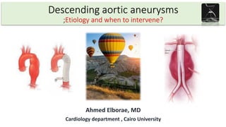
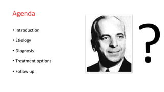
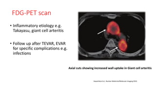
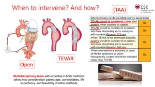
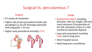
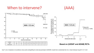
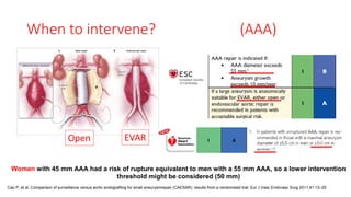
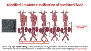
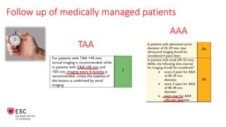
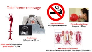
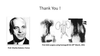
Descending aortic aneurysms
;Etiology and when to intervene?
Ahmed Elborae, MD
Cardiology department , Cairo University
Agenda
• Introduction
• Etiology
• Diagnosis
• Treatment options
• Follow up
Introduction
• Aorta carries about 200 million liters
of blood in average life time
• Also act as a second pump during
diastole
• Dealing with aortic tree as a whole
organ (25% of patients with AAA also
have TAA), 85% of femoral aneurysm
has AAA
• The 10-year risk of mortality from
other CVD (MI or stroke) is 15 times
the risk of aorta-related death
Introduction
• The descending aorta starts at
aortic isthmus distal Lt subclavian
• Normally with aging, aortic
expands about 0.9 mm in men and
0.7 mm in women each decade
• Aorta tapers in diameter distally
Definitions
• Increased diameter >50%, of
adjacent healthy segment
• Increased diameter ≥ 30 mm for
(Infra-aortic AAA)
• 80% of AAA are infra-renal
(Why???)
• No. of medial elastic lamellae
decreases as going distally
(From 55 layer in ascending
aorta to 26 at aortic bifurcation)
Etiology
Descending TAA:
• TAA encompasses a wide range of
etiologies, the most frequent
being degenerative (similar risk
factors as AAA)
• Hereditary: split into two
categories: syndromic e.g (Marfan
$ ,LD$,ED$) and non-syndromic,
e.g (TAAD)
• Also inflammatory causes e.g.
Takayasu and GCA were reported
Descending AAA:
• Age, male gender (x5), history of
atherosclerotic CVD, smoking
(X7) and hypertension
• A family history of AAA is a
powerful predictor (20%), ( up to
24 % in monozygotic twin)
Pathogenesis
• Signals from ↑hemodynamic
stress/genetic > chronic aortic wall
inflammation (↑expression of
proteinases) > degradation of matrix
proteins > mechanical failure of
medial elastin and adventitial
collagen > dilatation
• As infra-renal abdominal aorta lacks
vasa vasorum, so in the presence of
Intimal atherosclerosis> ↓ nutrients
diffusion> medial ischemia> SMC
apoptosis
Davis FM, et al. Mechanisms of aortic aneurysm formation: translating preclinical studies into clinical therapies. Heart. 2014;100:1498–1505.)
Clinical presentation
• Mostly silent, detected incidentally during abdominal imaging
for any indication
• Pressure symptoms: Cough, shortness of breath, hoarseness
of voice, difficult swallowing in TAA, or back pain in AAA
• Pulsatile mass below umbilicus
• Complications: Rupture (acute abdomen and shock),
thrombo-embolism
Imaging
• Ultrasonography is an excellent tool for
screening for AAA at low-cost
• Compared with CT, it underestimates AAA by
1–3 mm
Takagi H et al. A further meta-analysis of population-based screening for abdominal aortic aneurysm. J Vasc Surg 2010;52:1103–1108
AAA screening in men ≥ 65 years was associated with 45% decreased risk of AAA-related mortality at 10 years
Value of AAA screening
Imaging
Needed information:
• Aneurysm: Max. diameter,
extension (relationship to
the renal arteries)
• Landing zone: Max.
transverse diameter, length,
angulations
• Disadvantage: ionizing
radiation and contrast
• Inter- and intra-observer
variability of CT are < 5 mm
Thus, any change > 5 mm on
serial CT is considered
significant
CT aortography
Imaging
• MRI is an alternative option
especially for serial follow up
to avoid ionizing radiation
• Disadvantage: not sensitive
for calcifications, not ideal
for stainless steel stent graft
MRI
FDG-PET scan
• Inflammatory etiology e.g.
Takayasu, giant cell arteritis
• Follow up after TEVAR, EVAR
for specific complications e.g.
infections
Axial cuts showing increased wall uptake in Giant cell arteritis
Hayashida et al., Nuclear Medicine/Molecular Imaging 2010.
Natural history
Descending TAA:
• Descending TAAs grow faster (at 3
mm/year) in comparison to
ascending aorta (1 mm/year)
Descending AAA:
• Estimated rate of growth is 1–6
mm/year
• Accelerated by smoking (x2) , larger
dimeter
• Rupture is higher in women than in
men at similar diameters; women
present ruptured AAA on average
10 mm smaller than men
• Responsible for 150,000 to 200,000
deaths/ year worldwide
Basic physics
Prof. Laplace, France
Management
• Life style modification: Avoid competitive sports, counseling before
pregnancy
• Medical: Aiming to reduce shear stress (BP&HR) , control risk factors
• Intervention: Percutaneous (TEVAR/EVAR) Vs. surgical and when?
When to intervene? And how?
Multidisciplinary team with expertise in both methods,
taking into consideration patient age, comorbidities, life
expectancy, and feasibility of either methods
(TAA)
TEVAR
Open
Surgical Vs. percutaneous ?
Surgical
• Fit nearly all anatomies
• Higher rate of peri-procedural stroke and
paraplegia (↓ by CSF drainage especially
with long graft > 15 cm)
• Higher early procedural mortality 17 %
TEVAR
• Anatomical criteria: (Landing
diameter <40 mm, length >20 mm)
novel solutions (Transposition of
the left subclavian artery or a left
carotid-to-subclavian Bypass)
• Low early procedural mortality
5.6%, lost at long term
• Short hospital course
• Need long term surveillance
Cao P, et al. Comparison of surveillance versus aortic endografting for small aneurysmrepair (CAESAR): results from a randomised trial. Eur J Vasc Endovasc Surg 2011;41:13–25
When to intervene? (AAA)
Based on (UKSAT and ADAM) RCTs
AAA < 5.5 cm AAA > 5.5 cm
Cao P, et al. Comparison of surveillance versus aortic endografting for small aneurysmrepair (CAESAR): results from a randomised trial. Eur J Vasc Endovasc Surg 2011;41:13–25
Women with 45 mm AAA had a risk of rupture equivalent to men with a 55 mm AAA, so a lower intervention
threshold might be considered (50 mm)
When to intervene? (AAA)
EVAR
Open
Surgical Vs. percutaneous ?
Surgical
• Fit nearly all anatomies
• Long duration of AAA repair
procedures, need for aortic
clamping, blood loss and fluid
shifts are strong triggers ACS
• Procedural mortality about 4 %
EVAR
• Anatomical criteria: (Infra-renal,
landing diameter <30 mm,
length >15 mm, angle < 60, no
thrombus), novel solutions
• Low early procedural mortality
1.3%, lost at long term
• Short hospital course
• Need longterm surveillance
EVAR had significantly lower 30-day mortality Vs. open (1.8% vs. 4.3)
EVAR-1 trial
The mortality benefit was lost at 8 years FU due to higher re-intervention
Follow up after intervention
Nitinol stents
Endo-leak
25%
Modified Crawford classification of combined TAAA
Carries very high risk (mortality >10%) ,complex and usually performed via extensive thoraco-abdominal incision
and requires bypass to maintain perfusion of mesenteric vessels, and spinal fluid drainage to ↓ risk of paraplegia
“FEVAR”?
Follow up of medically managed patients
TAA
AAA
Take home message
Whole organ (Tandem lesions)
Whole patient (CVD risk)
Silent disease
U/S screening >65 years
Physics & epidemiology
Intervene > 55 mm
Control risk factors
Smoking x2 risk of rupture
MDT open Vs. percutaneous
Percutaneous better early outcome but need long surveillance
Thank You !
Prof. Charles Dubost, France
First AAA surgery using homograft On 29th March, 1951

Descending aortic aneurysm

  • 1.
    Descending aortic aneurysms ;Etiologyand when to intervene? Ahmed Elborae, MD Cardiology department , Cairo University
  • 2.
    Agenda • Introduction • Etiology •Diagnosis • Treatment options • Follow up
  • 3.
    Introduction • Aorta carriesabout 200 million liters of blood in average life time • Also act as a second pump during diastole • Dealing with aortic tree as a whole organ (25% of patients with AAA also have TAA), 85% of femoral aneurysm has AAA • The 10-year risk of mortality from other CVD (MI or stroke) is 15 times the risk of aorta-related death
  • 4.
    Introduction • The descendingaorta starts at aortic isthmus distal Lt subclavian • Normally with aging, aortic expands about 0.9 mm in men and 0.7 mm in women each decade • Aorta tapers in diameter distally
  • 5.
    Definitions • Increased diameter>50%, of adjacent healthy segment • Increased diameter ≥ 30 mm for (Infra-aortic AAA) • 80% of AAA are infra-renal (Why???) • No. of medial elastic lamellae decreases as going distally (From 55 layer in ascending aorta to 26 at aortic bifurcation)
  • 6.
    Etiology Descending TAA: • TAAencompasses a wide range of etiologies, the most frequent being degenerative (similar risk factors as AAA) • Hereditary: split into two categories: syndromic e.g (Marfan $ ,LD$,ED$) and non-syndromic, e.g (TAAD) • Also inflammatory causes e.g. Takayasu and GCA were reported Descending AAA: • Age, male gender (x5), history of atherosclerotic CVD, smoking (X7) and hypertension • A family history of AAA is a powerful predictor (20%), ( up to 24 % in monozygotic twin)
  • 7.
    Pathogenesis • Signals from↑hemodynamic stress/genetic > chronic aortic wall inflammation (↑expression of proteinases) > degradation of matrix proteins > mechanical failure of medial elastin and adventitial collagen > dilatation • As infra-renal abdominal aorta lacks vasa vasorum, so in the presence of Intimal atherosclerosis> ↓ nutrients diffusion> medial ischemia> SMC apoptosis Davis FM, et al. Mechanisms of aortic aneurysm formation: translating preclinical studies into clinical therapies. Heart. 2014;100:1498–1505.)
  • 8.
    Clinical presentation • Mostlysilent, detected incidentally during abdominal imaging for any indication • Pressure symptoms: Cough, shortness of breath, hoarseness of voice, difficult swallowing in TAA, or back pain in AAA • Pulsatile mass below umbilicus • Complications: Rupture (acute abdomen and shock), thrombo-embolism
  • 9.
    Imaging • Ultrasonography isan excellent tool for screening for AAA at low-cost • Compared with CT, it underestimates AAA by 1–3 mm
  • 10.
    Takagi H etal. A further meta-analysis of population-based screening for abdominal aortic aneurysm. J Vasc Surg 2010;52:1103–1108 AAA screening in men ≥ 65 years was associated with 45% decreased risk of AAA-related mortality at 10 years Value of AAA screening
  • 11.
    Imaging Needed information: • Aneurysm:Max. diameter, extension (relationship to the renal arteries) • Landing zone: Max. transverse diameter, length, angulations • Disadvantage: ionizing radiation and contrast • Inter- and intra-observer variability of CT are < 5 mm Thus, any change > 5 mm on serial CT is considered significant CT aortography
  • 12.
    Imaging • MRI isan alternative option especially for serial follow up to avoid ionizing radiation • Disadvantage: not sensitive for calcifications, not ideal for stainless steel stent graft MRI
  • 13.
    FDG-PET scan • Inflammatoryetiology e.g. Takayasu, giant cell arteritis • Follow up after TEVAR, EVAR for specific complications e.g. infections Axial cuts showing increased wall uptake in Giant cell arteritis Hayashida et al., Nuclear Medicine/Molecular Imaging 2010.
  • 14.
    Natural history Descending TAA: •Descending TAAs grow faster (at 3 mm/year) in comparison to ascending aorta (1 mm/year) Descending AAA: • Estimated rate of growth is 1–6 mm/year • Accelerated by smoking (x2) , larger dimeter • Rupture is higher in women than in men at similar diameters; women present ruptured AAA on average 10 mm smaller than men • Responsible for 150,000 to 200,000 deaths/ year worldwide
  • 15.
  • 16.
    Management • Life stylemodification: Avoid competitive sports, counseling before pregnancy • Medical: Aiming to reduce shear stress (BP&HR) , control risk factors • Intervention: Percutaneous (TEVAR/EVAR) Vs. surgical and when?
  • 17.
    When to intervene?And how? Multidisciplinary team with expertise in both methods, taking into consideration patient age, comorbidities, life expectancy, and feasibility of either methods (TAA) TEVAR Open
  • 18.
    Surgical Vs. percutaneous? Surgical • Fit nearly all anatomies • Higher rate of peri-procedural stroke and paraplegia (↓ by CSF drainage especially with long graft > 15 cm) • Higher early procedural mortality 17 % TEVAR • Anatomical criteria: (Landing diameter <40 mm, length >20 mm) novel solutions (Transposition of the left subclavian artery or a left carotid-to-subclavian Bypass) • Low early procedural mortality 5.6%, lost at long term • Short hospital course • Need long term surveillance
  • 19.
    Cao P, etal. Comparison of surveillance versus aortic endografting for small aneurysmrepair (CAESAR): results from a randomised trial. Eur J Vasc Endovasc Surg 2011;41:13–25 When to intervene? (AAA) Based on (UKSAT and ADAM) RCTs AAA < 5.5 cm AAA > 5.5 cm
  • 20.
    Cao P, etal. Comparison of surveillance versus aortic endografting for small aneurysmrepair (CAESAR): results from a randomised trial. Eur J Vasc Endovasc Surg 2011;41:13–25 Women with 45 mm AAA had a risk of rupture equivalent to men with a 55 mm AAA, so a lower intervention threshold might be considered (50 mm) When to intervene? (AAA) EVAR Open
  • 21.
    Surgical Vs. percutaneous? Surgical • Fit nearly all anatomies • Long duration of AAA repair procedures, need for aortic clamping, blood loss and fluid shifts are strong triggers ACS • Procedural mortality about 4 % EVAR • Anatomical criteria: (Infra-renal, landing diameter <30 mm, length >15 mm, angle < 60, no thrombus), novel solutions • Low early procedural mortality 1.3%, lost at long term • Short hospital course • Need longterm surveillance
  • 22.
    EVAR had significantlylower 30-day mortality Vs. open (1.8% vs. 4.3) EVAR-1 trial The mortality benefit was lost at 8 years FU due to higher re-intervention
  • 24.
    Follow up afterintervention Nitinol stents Endo-leak 25%
  • 25.
    Modified Crawford classificationof combined TAAA Carries very high risk (mortality >10%) ,complex and usually performed via extensive thoraco-abdominal incision and requires bypass to maintain perfusion of mesenteric vessels, and spinal fluid drainage to ↓ risk of paraplegia “FEVAR”?
  • 26.
    Follow up ofmedically managed patients TAA AAA
  • 27.
    Take home message Wholeorgan (Tandem lesions) Whole patient (CVD risk) Silent disease U/S screening >65 years Physics & epidemiology Intervene > 55 mm Control risk factors Smoking x2 risk of rupture MDT open Vs. percutaneous Percutaneous better early outcome but need long surveillance
  • 28.
    Thank You ! Prof.Charles Dubost, France First AAA surgery using homograft On 29th March, 1951