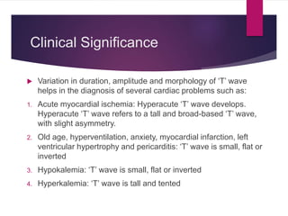
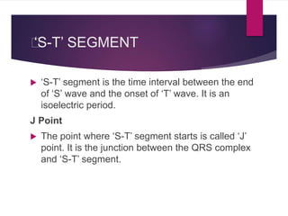
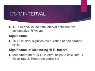
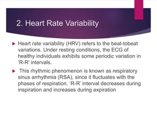
Electrocardiogram
(ECG)
MARIA IDREES; PT
Electrocardiography
 Electrocardiography is the technique by
which electrical activities of the heart are
studied.
Electrocardiograph
 Electrocardiograph is the instrument
(machine) by which electrical activities
of the heart are recorded.
Electrocardiogram
 Electrocardiogram (ECG or EKG from
electrokardiogram in Dutch) is the
record or graphical registration of
electrical activities of the heart, which
occur prior to the onset of mechanical
activities.
USES OF ECG
 Electrocardiogram is useful in determining and
diagnosing the following:
1. Heart rate
2. Heart rhythm
3. Abnormal electrical conduction
4. Poor blood flow to heart muscle (ischemia)
5. Heart attack
6. Coronary artery disease
7. Hypertrophy of heart chambers.
ELECTROCARDIOGRAPHIC
GRID
 The paper that is used for recording ECG is called
ECG paper.
 ECG machine amplifies the electrical signals
produced from the heart and records these signals on
a moving ECG paper.
 ECG paper has horizontal and vertical lines at regular
intervals of 1 mm.
 Every 5th line (5 mm) is thickened.
ECG LEADS
 ECG is recorded in 12 leads, which are
generally classified into two categories.
I. Bipolar leads
II. Unipolar leads.
BIPOLAR LIMB LEADS
UNIPOLAR LEADS
 Position of chest leads:
V1 : Over 4th intercostal space near right sternal margin
V2 : Over 4th intercostal space near left sternal margin
V3 : In between V2 and V4
V4 : Over left 5th intercostal space on the mid clavicular line
V5 : Over left 5th intercostal space on the anterior axillary line
V6 : Over left 5th intercostal space on the mid axillary line.
WAVES OF NORMAL ECG
 Normal ECG consists of waves, complexes,
intervals and segments.
 Waves of ECG recorded by limb lead II are
considered as the typical waves.
 Normal electrocardiogram has the following
waves, namely P, Q, R, S and T
Major Complexes in ECG
1. ‘P’ wave, the atrial complex
2. ‘QRS’ complex, the initial ventricular
complex
3. ‘T’ wave, the final ventricular complex
4. ‘QRST’, the ventricular complex.
„
‘P’ WAVE
 P’ wave is a positive wave and the first wave in ECG.
It is also called atrial complex.
 Cause ‘P’ wave is produced due to the depolarization
of atrial musculature.
 Depolarization spreads from SA node to all parts of
atrial musculature.
 Atrial repolarization is not recorded as a separate
wave in ECG because it merges with ventricular
repolarization (QRS complex).
Clinical Significance
 Variation in the duration, amplitude and morphology of
‘P’ wave helps in the diagnosis of several cardiac
problems such as:
1. Right atrial hypertrophy: ‘P’ wave is tall (more than 2.5
mm) in lead II. It is usually pointed
2. Left atrial dilatation or hypertrophy: It is tall and broad
based or M shaped
3. Atrial extrasystole: Small and shapeless ‘P’ wave,
followed by a small compensatory pause
4. Hyperkalemia: ‘P’ wave is absent or small
5. Atrial fibrillation: ‘P’ wave is absent
6. Middle AV nodal rhythm: ‘P’ wave is absent
7. Sinoatrial block: ‘P’ wave is inverted or absent
8. Atrial paroxysmal tachycardia: ‘P’ wave is inverted
9. Lower AV nodal rhythm: ‘P’ wave appears after QRS
complex.
‘QRS’ COMPLEX
 QRS’ complex is also called the initial
ventricular complex. ‘Q’ wave is a small
negative wave.
 It is continued as the tall ‘R’ wave, which is a
positive wave. ‘R’ wave is followed by a small
negative wave, the ‘S’ wave.
Cause
 ‘QRS’ complex is due to depolarization of
ventricular musculature. ‘Q’ wave is due to the
depolarization of basal portion of interventricular
septum.
 ‘R’ wave is due to the depolarization of apical
portion of interventricular septum and apical portion
of ventricular muscle.
 ‘S’ wave is due to the depolarization of basal portion
of ventricular muscle near the atrioventricular ring.
Clinical Significance
 Variation in the duration, amplitude and
morphology of ‘QRS’ complex helps in the
diagnosis of several cardiac problems such
as:
1. Bundle branch block: QRS is prolonged or
deformed
2. Hyperkalemia: QRS is prolonged.
„
‘T’ WAVE
 ‘T’ wave is the final ventricular complex and is a
positive wave.
Cause
 ‘T’ wave is due to the repolarization of ventricular
musculature.
Clinical Significance
 Variation in duration, amplitude and morphology of ‘T’ wave
helps in the diagnosis of several cardiac problems such as:
1. Acute myocardial ischemia: Hyperacute ‘T’ wave develops.
Hyperacute ‘T’ wave refers to a tall and broad-based ‘T’ wave,
with slight asymmetry.
2. Old age, hyperventilation, anxiety, myocardial infarction, left
ventricular hypertrophy and pericarditis: ‘T’ wave is small, flat or
inverted
3. Hypokalemia: ‘T’ wave is small, flat or inverted
4. Hyperkalemia: ‘T’ wave is tall and tented
‘U’ WAVE
 ‘U’ wave is not always seen. It is also an insignificant
wave in ECG. It is supposed to be due to
repolarization of papillary muscle.
Clinical Significance
 Appearance of ‘U’ wave in ECG indicates some
clinical conditions such as: 1. Hypercalcemia,
thyrotoxicosis and hypokalemia: ‘U’ wave appears. It
is very prominent in hypokalemia. 2. Myocardial
ischemia: Inverted ‘U’ wave appears.
INTERVALS AND SEGMENTS
OF ECG
„
‘P-R’ INTERVAL
 ‘P-R’ interval is the interval between the onset of ‘P’ wave and
onset of ‘Q’ wave. ‘P-R’ interval signifies the atrial depolarization
and conduction of impulses through AV node.
 It shows the duration of conduction of the impulses from the SA
node to ventricles through atrial muscle and AV node. ‘P’ wave
represents the atrial depolarization.
 Short isoelectric (zero voltage) period after the end of ‘P’ wave
represents the time taken for the passage of depolarization within
AV node.
Duration
 Normal duration of ‘P-R interval’ is 0.18 second
and varies between 0.12 and 0.2 second.
 If it is more than 0.2 second, it signifies the delay
in the conduction of impulse from SA node to the
ventricles.
 Usually, the delay occurs in the AV node. So it is
called the AV nodal delay
Clinical Significance
 Variation in the duration of ‘P-R’ intervals helps in the
diagnosis of several cardiac problems such as:
1. It is prolonged in bradycardia and first degree heart
block
2. It is shortened in tachycardia, Wolf-Parkinson White
syndrome, Lown-Ganong-Levine syndrome,
Duchenne muscular dystrophy and type II glycogen
storage disease.
„
‘Q-T’ INTERVAL
 ‘Q-T’ interval is the interval between the onset
of ‘Q’ wave and the end of ‘T’ wave.
 ‘Q-T’ interval indicates the ventricular
depolarization and ventricular repolarization,
i.e. it signifies the electrical activity in
ventricles.
Clinical Significance
1. ‘Q-T’ interval is prolonged in long ‘Q-T’
syndrome, myocardial infarction,
myocarditis, hypocalcemia and
hypothyroidism
2. ‘Q-T’ interval is shortened in short ‘Q-T’
syndrome and hypercalcemia.
„
‘S-T’ SEGMENT
 ‘S-T’ segment is the time interval between the end
of ‘S’ wave and the onset of ‘T’ wave. It is an
isoelectric period.
J Point
 The point where ‘S-T’ segment starts is called ‘J’
point. It is the junction between the QRS complex
and ‘S-T’ segment.
Clinical Significance
 Variation in the duration of ‘S-T’ segment and its
deviation from isoelectric base indicates the
pathological conditions such as:
1. Elevation of ‘S-T’ segment occurs in anterior or inferior
myocardial infarction, left bundle branch block and
acute pericarditis. In athletes, ‘S-T’ segment is usually
elevated
2. Depression of ‘S-T’ segment occurs in acute
myocardial ischemia, posterior myocardial infarction,
ventricular hypertrophy and hypokalemia
„
‘R-R’ INTERVAL
 ‘R-R’ interval is the time interval between two
consecutive ‘R’ waves.
Significance
 ‘R-R’ interval signifies the duration of one cardiac
cycle.
Significance of Measuring ‘R-R’ Interval
 Measurement of ‘R-R’ interval helps to calculate: 1.
Heart rate 2. Heart rate variability.
1. Heart Rate
Calculation of heart rate
 Time is plotted horizontally (X-axis). On X-axis,
interval between two thick lines is 0.2 sec (see above).
Time duration for 30 thick lines is 6 seconds.
 Number of ‘R’ waves (QRS complexes) in 6 seconds
(30 thick lines) is counted and multiplied by 10 to
obtain heart rate.
 For the sake of convenience, the ECG paper has
special time marking at every 3 seconds. So it is easy
to find the time duration of 6 seconds.
2. Heart Rate Variability
 Heart rate variability (HRV) refers to the beat-tobeat
variations. Under resting conditions, the ECG of
healthy individuals exhibits some periodic variation in
‘R-R’ intervals.
 This rhythmic phenomenon is known as respiratory
sinus arrhythmia (RSA), since it fluctuates with the
phases of respiration. ‘R-R’ interval decreases during
inspiration and increases during expiration
Significance of Heart Rate
Variability
 HRV decreases in many clinical conditions
like:
1. Cardiovascular dysfunctions such as
hypertension
2. Diabetes mellitus 3. Psychiatric problems
such as panic and anxiety.

Ecg

  • 1.
  • 2.
    Electrocardiography  Electrocardiography isthe technique by which electrical activities of the heart are studied.
  • 3.
    Electrocardiograph  Electrocardiograph isthe instrument (machine) by which electrical activities of the heart are recorded.
  • 4.
    Electrocardiogram  Electrocardiogram (ECGor EKG from electrokardiogram in Dutch) is the record or graphical registration of electrical activities of the heart, which occur prior to the onset of mechanical activities.
  • 5.
    USES OF ECG Electrocardiogram is useful in determining and diagnosing the following: 1. Heart rate 2. Heart rhythm 3. Abnormal electrical conduction 4. Poor blood flow to heart muscle (ischemia) 5. Heart attack 6. Coronary artery disease 7. Hypertrophy of heart chambers.
  • 6.
    ELECTROCARDIOGRAPHIC GRID  The paperthat is used for recording ECG is called ECG paper.  ECG machine amplifies the electrical signals produced from the heart and records these signals on a moving ECG paper.  ECG paper has horizontal and vertical lines at regular intervals of 1 mm.  Every 5th line (5 mm) is thickened.
  • 7.
    ECG LEADS  ECGis recorded in 12 leads, which are generally classified into two categories. I. Bipolar leads II. Unipolar leads.
  • 8.
  • 9.
  • 10.
     Position ofchest leads: V1 : Over 4th intercostal space near right sternal margin V2 : Over 4th intercostal space near left sternal margin V3 : In between V2 and V4 V4 : Over left 5th intercostal space on the mid clavicular line V5 : Over left 5th intercostal space on the anterior axillary line V6 : Over left 5th intercostal space on the mid axillary line.
  • 11.
    WAVES OF NORMALECG  Normal ECG consists of waves, complexes, intervals and segments.  Waves of ECG recorded by limb lead II are considered as the typical waves.  Normal electrocardiogram has the following waves, namely P, Q, R, S and T
  • 12.
    Major Complexes inECG 1. ‘P’ wave, the atrial complex 2. ‘QRS’ complex, the initial ventricular complex 3. ‘T’ wave, the final ventricular complex 4. ‘QRST’, the ventricular complex.
  • 13.
    „ ‘P’ WAVE  P’wave is a positive wave and the first wave in ECG. It is also called atrial complex.  Cause ‘P’ wave is produced due to the depolarization of atrial musculature.  Depolarization spreads from SA node to all parts of atrial musculature.  Atrial repolarization is not recorded as a separate wave in ECG because it merges with ventricular repolarization (QRS complex).
  • 14.
    Clinical Significance  Variationin the duration, amplitude and morphology of ‘P’ wave helps in the diagnosis of several cardiac problems such as: 1. Right atrial hypertrophy: ‘P’ wave is tall (more than 2.5 mm) in lead II. It is usually pointed 2. Left atrial dilatation or hypertrophy: It is tall and broad based or M shaped 3. Atrial extrasystole: Small and shapeless ‘P’ wave, followed by a small compensatory pause
  • 15.
    4. Hyperkalemia: ‘P’wave is absent or small 5. Atrial fibrillation: ‘P’ wave is absent 6. Middle AV nodal rhythm: ‘P’ wave is absent 7. Sinoatrial block: ‘P’ wave is inverted or absent 8. Atrial paroxysmal tachycardia: ‘P’ wave is inverted 9. Lower AV nodal rhythm: ‘P’ wave appears after QRS complex.
  • 16.
    ‘QRS’ COMPLEX  QRS’complex is also called the initial ventricular complex. ‘Q’ wave is a small negative wave.  It is continued as the tall ‘R’ wave, which is a positive wave. ‘R’ wave is followed by a small negative wave, the ‘S’ wave.
  • 17.
    Cause  ‘QRS’ complexis due to depolarization of ventricular musculature. ‘Q’ wave is due to the depolarization of basal portion of interventricular septum.  ‘R’ wave is due to the depolarization of apical portion of interventricular septum and apical portion of ventricular muscle.  ‘S’ wave is due to the depolarization of basal portion of ventricular muscle near the atrioventricular ring.
  • 20.
    Clinical Significance  Variationin the duration, amplitude and morphology of ‘QRS’ complex helps in the diagnosis of several cardiac problems such as: 1. Bundle branch block: QRS is prolonged or deformed 2. Hyperkalemia: QRS is prolonged.
  • 21.
    „ ‘T’ WAVE  ‘T’wave is the final ventricular complex and is a positive wave. Cause  ‘T’ wave is due to the repolarization of ventricular musculature.
  • 22.
    Clinical Significance  Variationin duration, amplitude and morphology of ‘T’ wave helps in the diagnosis of several cardiac problems such as: 1. Acute myocardial ischemia: Hyperacute ‘T’ wave develops. Hyperacute ‘T’ wave refers to a tall and broad-based ‘T’ wave, with slight asymmetry. 2. Old age, hyperventilation, anxiety, myocardial infarction, left ventricular hypertrophy and pericarditis: ‘T’ wave is small, flat or inverted 3. Hypokalemia: ‘T’ wave is small, flat or inverted 4. Hyperkalemia: ‘T’ wave is tall and tented
  • 23.
    ‘U’ WAVE  ‘U’wave is not always seen. It is also an insignificant wave in ECG. It is supposed to be due to repolarization of papillary muscle. Clinical Significance  Appearance of ‘U’ wave in ECG indicates some clinical conditions such as: 1. Hypercalcemia, thyrotoxicosis and hypokalemia: ‘U’ wave appears. It is very prominent in hypokalemia. 2. Myocardial ischemia: Inverted ‘U’ wave appears.
  • 24.
    INTERVALS AND SEGMENTS OFECG „ ‘P-R’ INTERVAL  ‘P-R’ interval is the interval between the onset of ‘P’ wave and onset of ‘Q’ wave. ‘P-R’ interval signifies the atrial depolarization and conduction of impulses through AV node.  It shows the duration of conduction of the impulses from the SA node to ventricles through atrial muscle and AV node. ‘P’ wave represents the atrial depolarization.  Short isoelectric (zero voltage) period after the end of ‘P’ wave represents the time taken for the passage of depolarization within AV node.
  • 25.
    Duration  Normal durationof ‘P-R interval’ is 0.18 second and varies between 0.12 and 0.2 second.  If it is more than 0.2 second, it signifies the delay in the conduction of impulse from SA node to the ventricles.  Usually, the delay occurs in the AV node. So it is called the AV nodal delay
  • 26.
    Clinical Significance  Variationin the duration of ‘P-R’ intervals helps in the diagnosis of several cardiac problems such as: 1. It is prolonged in bradycardia and first degree heart block 2. It is shortened in tachycardia, Wolf-Parkinson White syndrome, Lown-Ganong-Levine syndrome, Duchenne muscular dystrophy and type II glycogen storage disease.
  • 27.
    „ ‘Q-T’ INTERVAL  ‘Q-T’interval is the interval between the onset of ‘Q’ wave and the end of ‘T’ wave.  ‘Q-T’ interval indicates the ventricular depolarization and ventricular repolarization, i.e. it signifies the electrical activity in ventricles.
  • 28.
    Clinical Significance 1. ‘Q-T’interval is prolonged in long ‘Q-T’ syndrome, myocardial infarction, myocarditis, hypocalcemia and hypothyroidism 2. ‘Q-T’ interval is shortened in short ‘Q-T’ syndrome and hypercalcemia.
  • 29.
    „ ‘S-T’ SEGMENT  ‘S-T’segment is the time interval between the end of ‘S’ wave and the onset of ‘T’ wave. It is an isoelectric period. J Point  The point where ‘S-T’ segment starts is called ‘J’ point. It is the junction between the QRS complex and ‘S-T’ segment.
  • 30.
    Clinical Significance  Variationin the duration of ‘S-T’ segment and its deviation from isoelectric base indicates the pathological conditions such as: 1. Elevation of ‘S-T’ segment occurs in anterior or inferior myocardial infarction, left bundle branch block and acute pericarditis. In athletes, ‘S-T’ segment is usually elevated 2. Depression of ‘S-T’ segment occurs in acute myocardial ischemia, posterior myocardial infarction, ventricular hypertrophy and hypokalemia
  • 31.
    „ ‘R-R’ INTERVAL  ‘R-R’interval is the time interval between two consecutive ‘R’ waves. Significance  ‘R-R’ interval signifies the duration of one cardiac cycle. Significance of Measuring ‘R-R’ Interval  Measurement of ‘R-R’ interval helps to calculate: 1. Heart rate 2. Heart rate variability.
  • 32.
    1. Heart Rate Calculationof heart rate  Time is plotted horizontally (X-axis). On X-axis, interval between two thick lines is 0.2 sec (see above). Time duration for 30 thick lines is 6 seconds.  Number of ‘R’ waves (QRS complexes) in 6 seconds (30 thick lines) is counted and multiplied by 10 to obtain heart rate.  For the sake of convenience, the ECG paper has special time marking at every 3 seconds. So it is easy to find the time duration of 6 seconds.
  • 33.
    2. Heart RateVariability  Heart rate variability (HRV) refers to the beat-tobeat variations. Under resting conditions, the ECG of healthy individuals exhibits some periodic variation in ‘R-R’ intervals.  This rhythmic phenomenon is known as respiratory sinus arrhythmia (RSA), since it fluctuates with the phases of respiration. ‘R-R’ interval decreases during inspiration and increases during expiration
  • 34.
    Significance of HeartRate Variability  HRV decreases in many clinical conditions like: 1. Cardiovascular dysfunctions such as hypertension 2. Diabetes mellitus 3. Psychiatric problems such as panic and anxiety.