Downloaded 124 times

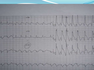
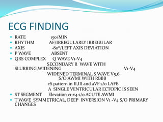
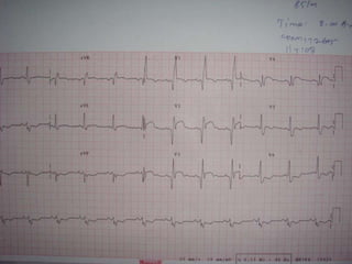
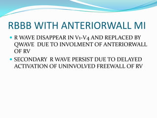
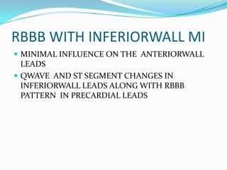
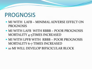


This ECG shows a patient with atrial fibrillation, left axis deviation, absent P waves, widened QRS complex with RBBB pattern, ST elevation in leads V1-V4 consistent with anterior wall myocardial infarction. There is also secondary R wave slurring and widening in V1-V4 and widened terminal S waves in V5-V6. The diagnosis is acute anterior wall myocardial infarction with bifascicular block (RBBB with left anterior fascicular block). The prognosis is poor, as myocardial infarction with bifascicular block can increase mortality by 4-5 times.