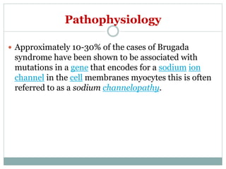
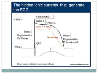
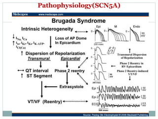
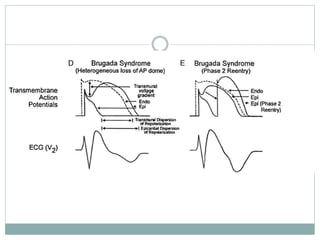
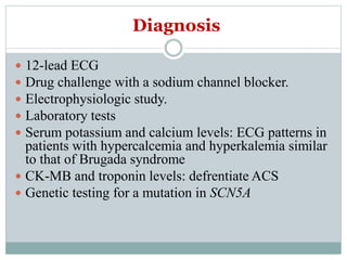
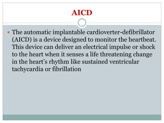
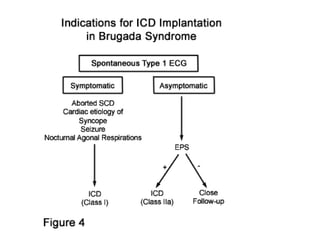
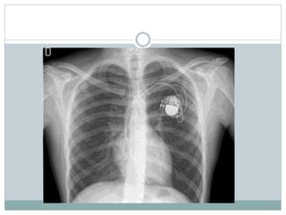
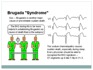
Brugada syndrome is a genetic heart condition characterized by abnormal ECG patterns and risk of sudden cardiac death. It is caused by mutations in genes encoding sodium channels. The condition is diagnosed through ECG showing ST segment elevation in leads V1-V3. Treatment involves implanting an ICD to detect and treat lethal arrhythmias with shocks. Prognosis depends on risk stratification and treatment.
The so Called Brugada Syndrome The True HistoryBortolo Martini
The syndrome of sudden Death, right bundle branch block and ST elevation was firstly described by A.Nava and B. Martini in 1988-1989, and only five years later by the Brugada Brothers. The ECG pattern is due to a conduction disturbance of the RVOT, caused by fibrofatty substitution of that structure.
Brugada Syndrome is a inherited sodium channel disorder leading to life threatening ventricular fibrillation in young population. diagnosis and ICD therapy could be life saving.
Brugada syndrome is the most common cause of sudden death in men < 40 years of age in south asia with no pre existing cardiac abnormalities, this presentation will help you understand brugada syndrome ...
#flozins
🫀DAPA 🆚placebo in HFpEF
Now we have a positive trial!
⬇️18% in CV☠️ death or
worsening HF among LVEF>40%
⬇️ 21%heart failure
💥Results same for LVEF> 60% 🆚LVEF<60%
Wolff–Parkinson–White syndrome (WPW) is one of several disorders of the conduction system of the heart that are commonly referred to as pre-excitation syndromes. WPW is caused by the presence of an abnormal accessory electrical conduction pathway between the atria and the ventricles. Electrical signals traveling down this abnormal pathway (known as the bundle of Kent) may stimulate the ventricles to contract prematurely, resulting in a unique type of supra-ventricular tachycardia referred to as an atrio-ventricular reciprocating tachycardia.
ST-segment Depression: All are Not Created Equal!asclepiuspdfs
ST depression on an electrocardiography can be from various causes including ischemia, acute coronary syndrome, electrolyte imbalance, posterior myocardial infarction, pulmonary embolism and others. Making the right diagnosis and therefore the right treatment is of paramount importance. This article goes into depth explaining why all ST-segment depressions are not created equal.
Title: Sense of Taste
Presenter: Dr. Faiza, Assistant Professor of Physiology
Qualifications:
MBBS (Best Graduate, AIMC Lahore)
FCPS Physiology
ICMT, CHPE, DHPE (STMU)
MPH (GC University, Faisalabad)
MBA (Virtual University of Pakistan)
Learning Objectives:
Describe the structure and function of taste buds.
Describe the relationship between the taste threshold and taste index of common substances.
Explain the chemical basis and signal transduction of taste perception for each type of primary taste sensation.
Recognize different abnormalities of taste perception and their causes.
Key Topics:
Significance of Taste Sensation:
Differentiation between pleasant and harmful food
Influence on behavior
Selection of food based on metabolic needs
Receptors of Taste:
Taste buds on the tongue
Influence of sense of smell, texture of food, and pain stimulation (e.g., by pepper)
Primary and Secondary Taste Sensations:
Primary taste sensations: Sweet, Sour, Salty, Bitter, Umami
Chemical basis and signal transduction mechanisms for each taste
Taste Threshold and Index:
Taste threshold values for Sweet (sucrose), Salty (NaCl), Sour (HCl), and Bitter (Quinine)
Taste index relationship: Inversely proportional to taste threshold
Taste Blindness:
Inability to taste certain substances, particularly thiourea compounds
Example: Phenylthiocarbamide
Structure and Function of Taste Buds:
Composition: Epithelial cells, Sustentacular/Supporting cells, Taste cells, Basal cells
Features: Taste pores, Taste hairs/microvilli, and Taste nerve fibers
Location of Taste Buds:
Found in papillae of the tongue (Fungiform, Circumvallate, Foliate)
Also present on the palate, tonsillar pillars, epiglottis, and proximal esophagus
Mechanism of Taste Stimulation:
Interaction of taste substances with receptors on microvilli
Signal transduction pathways for Umami, Sweet, Bitter, Sour, and Salty tastes
Taste Sensitivity and Adaptation:
Decrease in sensitivity with age
Rapid adaptation of taste sensation
Role of Saliva in Taste:
Dissolution of tastants to reach receptors
Washing away the stimulus
Taste Preferences and Aversions:
Mechanisms behind taste preference and aversion
Influence of receptors and neural pathways
Impact of Sensory Nerve Damage:
Degeneration of taste buds if the sensory nerve fiber is cut
Abnormalities of Taste Detection:
Conditions: Ageusia, Hypogeusia, Dysgeusia (parageusia)
Causes: Nerve damage, neurological disorders, infections, poor oral hygiene, adverse drug effects, deficiencies, aging, tobacco use, altered neurotransmitter levels
Neurotransmitters and Taste Threshold:
Effects of serotonin (5-HT) and norepinephrine (NE) on taste sensitivity
Supertasters:
25% of the population with heightened sensitivity to taste, especially bitterness
Increased number of fungiform papillae
The prostate is an exocrine gland of the male mammalian reproductive system
It is a walnut-sized gland that forms part of the male reproductive system and is located in front of the rectum and just below the urinary bladder
Function is to store and secrete a clear, slightly alkaline fluid that constitutes 10-30% of the volume of the seminal fluid that along with the spermatozoa, constitutes semen
A healthy human prostate measures (4cm-vertical, by 3cm-horizontal, 2cm ant-post ).
It surrounds the urethra just below the urinary bladder. It has anterior, median, posterior and two lateral lobes
It’s work is regulated by androgens which are responsible for male sex characteristics
Generalised disease of the prostate due to hormonal derangement which leads to non malignant enlargement of the gland (increase in the number of epithelial cells and stromal tissue)to cause compression of the urethra leading to symptoms (LUTS
These simplified slides by Dr. Sidra Arshad present an overview of the non-respiratory functions of the respiratory tract.
Learning objectives:
1. Enlist the non-respiratory functions of the respiratory tract
2. Briefly explain how these functions are carried out
3. Discuss the significance of dead space
4. Differentiate between minute ventilation and alveolar ventilation
5. Describe the cough and sneeze reflexes
Study Resources:
1. Chapter 39, Guyton and Hall Textbook of Medical Physiology, 14th edition
2. Chapter 34, Ganong’s Review of Medical Physiology, 26th edition
3. Chapter 17, Human Physiology by Lauralee Sherwood, 9th edition
4. Non-respiratory functions of the lungs https://academic.oup.com/bjaed/article/13/3/98/278874
ARTIFICIAL INTELLIGENCE IN HEALTHCARE.pdfAnujkumaranit
Artificial intelligence (AI) refers to the simulation of human intelligence processes by machines, especially computer systems. It encompasses tasks such as learning, reasoning, problem-solving, perception, and language understanding. AI technologies are revolutionizing various fields, from healthcare to finance, by enabling machines to perform tasks that typically require human intelligence.
Factory Supply Best Quality Pmk Oil CAS 28578–16–7 PMK Powder in Stockrebeccabio
Factory Supply Best Quality Pmk Oil CAS 28578–16–7 PMK Powder in Stock
Telegram: bmksupplier
signal: +85264872720
threema: TUD4A6YC
You can contact me on Telegram or Threema
Communicate promptly and reply
Free of customs clearance, Double Clearance 100% pass delivery to USA, Canada, Spain, Germany, Netherland, Poland, Italy, Sweden, UK, Czech Republic, Australia, Mexico, Russia, Ukraine, Kazakhstan.Door to door service
Hot Selling Organic intermediates
Ethanol (CH3CH2OH), or beverage alcohol, is a two-carbon alcohol
that is rapidly distributed in the body and brain. Ethanol alters many
neurochemical systems and has rewarding and addictive properties. It
is the oldest recreational drug and likely contributes to more morbidity,
mortality, and public health costs than all illicit drugs combined. The
5th edition of the Diagnostic and Statistical Manual of Mental Disorders
(DSM-5) integrates alcohol abuse and alcohol dependence into a single
disorder called alcohol use disorder (AUD), with mild, moderate,
and severe subclassifications (American Psychiatric Association, 2013).
In the DSM-5, all types of substance abuse and dependence have been
combined into a single substance use disorder (SUD) on a continuum
from mild to severe. A diagnosis of AUD requires that at least two of
the 11 DSM-5 behaviors be present within a 12-month period (mild
AUD: 2–3 criteria; moderate AUD: 4–5 criteria; severe AUD: 6–11 criteria).
The four main behavioral effects of AUD are impaired control over
drinking, negative social consequences, risky use, and altered physiological
effects (tolerance, withdrawal). This chapter presents an overview
of the prevalence and harmful consequences of AUD in the U.S.,
the systemic nature of the disease, neurocircuitry and stages of AUD,
comorbidities, fetal alcohol spectrum disorders, genetic risk factors, and
pharmacotherapies for AUD.
Lung Cancer: Artificial Intelligence, Synergetics, Complex System Analysis, S...Oleg Kshivets
RESULTS: Overall life span (LS) was 2252.1±1742.5 days and cumulative 5-year survival (5YS) reached 73.2%, 10 years – 64.8%, 20 years – 42.5%. 513 LCP lived more than 5 years (LS=3124.6±1525.6 days), 148 LCP – more than 10 years (LS=5054.4±1504.1 days).199 LCP died because of LC (LS=562.7±374.5 days). 5YS of LCP after bi/lobectomies was significantly superior in comparison with LCP after pneumonectomies (78.1% vs.63.7%, P=0.00001 by log-rank test). AT significantly improved 5YS (66.3% vs. 34.8%) (P=0.00000 by log-rank test) only for LCP with N1-2. Cox modeling displayed that 5YS of LCP significantly depended on: phase transition (PT) early-invasive LC in terms of synergetics, PT N0—N12, cell ratio factors (ratio between cancer cells- CC and blood cells subpopulations), G1-3, histology, glucose, AT, blood cell circuit, prothrombin index, heparin tolerance, recalcification time (P=0.000-0.038). Neural networks, genetic algorithm selection and bootstrap simulation revealed relationships between 5YS and PT early-invasive LC (rank=1), PT N0—N12 (rank=2), thrombocytes/CC (3), erythrocytes/CC (4), eosinophils/CC (5), healthy cells/CC (6), lymphocytes/CC (7), segmented neutrophils/CC (8), stick neutrophils/CC (9), monocytes/CC (10); leucocytes/CC (11). Correct prediction of 5YS was 100% by neural networks computing (area under ROC curve=1.0; error=0.0).
CONCLUSIONS: 5YS of LCP after radical procedures significantly depended on: 1) PT early-invasive cancer; 2) PT N0--N12; 3) cell ratio factors; 4) blood cell circuit; 5) biochemical factors; 6) hemostasis system; 7) AT; 8) LC characteristics; 9) LC cell dynamics; 10) surgery type: lobectomy/pneumonectomy; 11) anthropometric data. Optimal diagnosis and treatment strategies for LC are: 1) screening and early detection of LC; 2) availability of experienced thoracic surgeons because of complexity of radical procedures; 3) aggressive en block surgery and adequate lymph node dissection for completeness; 4) precise prediction; 5) adjuvant chemoimmunoradiotherapy for LCP with unfavorable prognosis.
These lecture slides, by Dr Sidra Arshad, offer a quick overview of physiological basis of a normal electrocardiogram.
Learning objectives:
1. Define an electrocardiogram (ECG) and electrocardiography
2. Describe how dipoles generated by the heart produce the waveforms of the ECG
3. Describe the components of a normal electrocardiogram of a typical bipolar leads (limb II)
4. Differentiate between intervals and segments
5. Enlist some common indications for obtaining an ECG
Study Resources:
1. Chapter 11, Guyton and Hall Textbook of Medical Physiology, 14th edition
2. Chapter 9, Human Physiology - From Cells to Systems, Lauralee Sherwood, 9th edition
3. Chapter 29, Ganong’s Review of Medical Physiology, 26th edition
4. Electrocardiogram, StatPearls - https://www.ncbi.nlm.nih.gov/books/NBK549803/
5. ECG in Medical Practice by ABM Abdullah, 4th edition
6. ECG Basics, http://www.nataliescasebook.com/tag/e-c-g-basics
Title: Sense of Smell
Presenter: Dr. Faiza, Assistant Professor of Physiology
Qualifications:
MBBS (Best Graduate, AIMC Lahore)
FCPS Physiology
ICMT, CHPE, DHPE (STMU)
MPH (GC University, Faisalabad)
MBA (Virtual University of Pakistan)
Learning Objectives:
Describe the primary categories of smells and the concept of odor blindness.
Explain the structure and location of the olfactory membrane and mucosa, including the types and roles of cells involved in olfaction.
Describe the pathway and mechanisms of olfactory signal transmission from the olfactory receptors to the brain.
Illustrate the biochemical cascade triggered by odorant binding to olfactory receptors, including the role of G-proteins and second messengers in generating an action potential.
Identify different types of olfactory disorders such as anosmia, hyposmia, hyperosmia, and dysosmia, including their potential causes.
Key Topics:
Olfactory Genes:
3% of the human genome accounts for olfactory genes.
400 genes for odorant receptors.
Olfactory Membrane:
Located in the superior part of the nasal cavity.
Medially: Folds downward along the superior septum.
Laterally: Folds over the superior turbinate and upper surface of the middle turbinate.
Total surface area: 5-10 square centimeters.
Olfactory Mucosa:
Olfactory Cells: Bipolar nerve cells derived from the CNS (100 million), with 4-25 olfactory cilia per cell.
Sustentacular Cells: Produce mucus and maintain ionic and molecular environment.
Basal Cells: Replace worn-out olfactory cells with an average lifespan of 1-2 months.
Bowman’s Gland: Secretes mucus.
Stimulation of Olfactory Cells:
Odorant dissolves in mucus and attaches to receptors on olfactory cilia.
Involves a cascade effect through G-proteins and second messengers, leading to depolarization and action potential generation in the olfactory nerve.
Quality of a Good Odorant:
Small (3-20 Carbon atoms), volatile, water-soluble, and lipid-soluble.
Facilitated by odorant-binding proteins in mucus.
Membrane Potential and Action Potential:
Resting membrane potential: -55mV.
Action potential frequency in the olfactory nerve increases with odorant strength.
Adaptation Towards the Sense of Smell:
Rapid adaptation within the first second, with further slow adaptation.
Psychological adaptation greater than receptor adaptation, involving feedback inhibition from the central nervous system.
Primary Sensations of Smell:
Camphoraceous, Musky, Floral, Pepperminty, Ethereal, Pungent, Putrid.
Odor Detection Threshold:
Examples: Hydrogen sulfide (0.0005 ppm), Methyl-mercaptan (0.002 ppm).
Some toxic substances are odorless at lethal concentrations.
Characteristics of Smell:
Odor blindness for single substances due to lack of appropriate receptor protein.
Behavioral and emotional influences of smell.
Transmission of Olfactory Signals:
From olfactory cells to glomeruli in the olfactory bulb, involving lateral inhibition.
Primitive, less old, and new olfactory systems with different path
2. Introduction
Brugada syndrome is a genetic disease that is
characterized by sudden death associated with
abnormal electrocardiogram disorder.
One of several ECG patterns characterized by are
incomplete right bundle-branch block and ST-
segment elevations in the anterior precordial
leads(V1-V3) without ischemia.
3. Introduction
The cause of syncope and sudden death in Brugada
syndrome are caused by fast polymorphic ventricular
tachycardias or ventricular fibrillation. These
arrhythmias appear with no warning.
According to clinical reports, sudden death in people
with Brugada syndrome most often happens during
sleep.
4. Introduction
Monomorphic VT rarely occurs , especially in patients
taking antiarrhythmic drugs .
Supraventricular tachycardias are more prevalent in
Brugada syndrome .
An estimated 80% of subjects with documented VT/VF has
a history of syncope , caused by self-terminating episodes.
5. Epidemiology
In parts of Asia Brugada syndrome seems to be the
most common cause of natural death in men <50
years.
It is approximately 30 cases per 100,000 population
per year.
And 8-10 times more prevalent in men than in
women, although the probability of having a mutated
gene does not differ by sex.
8. Etiology
1. The majority of patients affected by Brugada syndrome are
not found to have known genetic mutations to explain the
disease.
2. Approximately 20% due to alterations in the SCN5A
gene(encodes the cardiac sodium channel)
3. Mutations in the genes Glycerol-3-phosphate dehydrogenase
1-like gene(GPD1-L) and SCN1B
9. Pathophysiology
Approximately 10-30% of the cases of Brugada
syndrome have been shown to be associated with
mutations in a gene that encodes for a sodium ion
channel in the cell membranes myocytes this is often
referred to as a sodium channelopathy.
13. Signs and symptoms
1. Syncope and cardiac arrest: Mostly cardiac arrest occurs
during sleep or rest
2. Nightmares or thrashing at night
3. Asymptomatic, but routine ECG shows ST-segment
elevation in leads V1-V3
4. Associated atrial fibrillation (20%)
5. Fever: Often reported to trigger or exacerbate clinical
manifestations.
14. TYPES
Brugada syndrome has three different ECG patterns
1. Type 1 has a coved type ST elevation with at least
2 mm (0.2 mV) J-point elevation and a gradually
descending ST segment followed by a negative T-wave.
2. Type 2 has a saddle-back pattern with a least 2 mm
J-point elevation and at least 1 mm ST elevation with a
positive or biphasic T-wave. Type 2 pattern can
occasionally be seen in healthy subjects.
3. Type 3 has either a coved (type 1 like) or a
saddle-back (type 2 like) pattern, with less than 2 mm
J-point elevation and less than 1 mm ST elevation.
15.
16.
17. Diagnosis
12-lead ECG
Drug challenge with a sodium channel blocker.
Electrophysiologic study.
Laboratory tests
Serum potassium and calcium levels: ECG patterns in
patients with hypercalcemia and hyperkalemia similar
to that of Brugada syndrome
CK-MB and troponin levels: defrentiate ACS
Genetic testing for a mutation in SCN5A
18. Long QT syndrome
It is a congenital disorder
characterized by a
prolongation of the QT
interval on ECG and a
propensity to ventricular
tachyarrhythmias, which
may lead to syncope,
cardiac arrest, or sudden
death.
QT interval 0.36–0.44s
(9–11 small squares).
19.
20. How to find out VT in RBBB or LBBB ?
If RBBB pattern, then VT is present in the
following situations:
A monophasic R or biphasic qR complex in V1.
If an RSR’ pattern is present in V1.
A rS complex in lead V6 favors VT
If LBBB pattern, then VT is present in the
following situations:
The presence of any Q or QS wave in lead V6
favors VT
A wide R wave in lead V1 or V2 of 0.04s or more
favors VT
Slurred or notched downstroke of the S wave in
V1 or V2 favors VT
21. Differential diagnosis of ST-segment elevation
1. Atypical right bundle-branch block
2. Left ventricular hypertrophy
3. Early repolarization
4. Acute pericarditis
5. Acute myocardial ischemia or infarction
6. Pulmonary embolism
7. Dissecting aortic aneurysm
8. Arrhythmogenic right ventricular dysplasia and/or cardiomyopathy
9. Various abnormalities of the central and autonomic nervous systems
10. Overdose of a antidepressant
11. Thiamine deficiency Hypercalcemia
12. Hyperkalemia
13. Effects of athletic training
22. Management
There is no exact treatment modality that reliably and
totally prevents ventricular fibrillation from occurring
in this syndrome
treatment lies in termination of this lethal arrhythmia
before it causes death.
Implantable cardioverter-defibrillator(ICD) is inserted
, which continuously monitors the heart rhythm and
will shock the wearer if ventricular fibrillation is sensed.
Quinidine , a pharmaceutical agent that acts as a class
I antiarrhythmic agent is used.
23. Quinidine Sulfate
Test Dose: 200 mg PO quinidine sulfate several hr
before full dosage
AFib: 300-400 mg PO q6hr
PSVT: 400-600 mg PO q2-3hr until paroxysm
terminated
Atrial/Ventr Premature Contractions: 200-300 mg
PO TID/QID
Maint: 200-400 mg PO
No more than 3-4 g/d
24. AICD
The automatic implantable cardioverter-defibrillator
(AICD) is a device designed to monitor the heartbeat.
This device can deliver an electrical impulse or shock
to the heart when it senses a life threatening change
in the heart’s rhythm like sustained ventricular
tachycardia or fibrillation
29. Prognosis
An estimated 4% of all sudden deaths and at least
20% of sudden deaths in patients with structurally
normal hearts are due to this syndrome.
30. Conclusion
Bikash is 13 yrs male student diagnosed with
brugada syndrome type 2, managed with AICD dual
chamber is discharged in stable condition with
uneventful hospitalization period.
Editor's Notes
Polymorphic ventricular tachycardia (PVT) is a form of ventricular tachycardia in which there are multiple ventricular foci with the resultant QRS complexes varying in amplitude, axis and duration.
Monomorphic ventricular tachycardia means that the appearance of all the beats match each other in each lead of a surfaceelectrocardiogram
The QT interval represents electrical depolarization and repolarization of the ventricles. A lengthened QT interval is a marker for the potential of ventricular tachyarrhythmias like polymorphic ventricular tachycardia and a risk factor for sudden death.
quinidine, which blocks the calcium-independent transient outward potassium current (Ito), has been shown to normalize the ECG pattern in patients with Brugada syndrome.[3] However, quinidine also blocks sodium (Na) currents
Sudden cardiac arrest (SCA) and sudden cardiac death (SCD) refer to the sudden cessation of cardiac activity with hemodynamic collapse due to sustained pulseless ventricular tachycardia/fibrillation, pulseless electrical activity (PEA), or asystole. The event is referred to as SCA (or aborted SCD)
agonal breathing is an abnormal pattern of breathing and brainstem reflex characterized by gasping, laboredbreathing, accompanied by strange vocalizations and myoclonus.