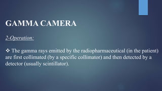
Nuclear Medicine
W. P. Rivindu H. Wickramanayake
Group no. 04a
3rd Year 2nd Semester – 2017 December
Tbilisi State Medical University, Georgia
 This is a branch of medicine that uses radiation from
radioactive tracers to provide information about the function
of specific organs.
 In some cases, radioactivity can be used to treat certain
conditions such as an overactive thyroid.
 Nuclear medicine studies use ionizing radiation, as do x-ray
studies.
 Radioactive tracers or radiopharmaceuticals commonly used
are quickly eliminated from the body through its natural
functions.
 In addition, the tracers used rapidly lose their radioactivity.
Radioactivity;
The ability of radioactive (unstable) atoms to spontaneously decay
via - Fission & Emission of energy to become stable atoms
which are; 1) Alpha
2) Beta
3) Gamma Particles/Photons
 In most cases, the dose of radiation necessary for a scan is very
small.
 For example, a patient having a lung scan is exposed to the same
dose of radiation they would receive from eight return air flights
between Sydney and London.
1) Alpha Particles
- Positive Charge, Heavier (the nucleus)
- Low penetration (paper width)
2) Beta Particles
- Negative Charge (but also has positive ions)
- May penetrate tissue (10mm-12mm)
3) Gamma Particles
- No charge
- Travel straight/radically (short wave length)
- Independently travel straight in air
- High penetration ability
What is a half-life
 Nuclear medicines used for diagnosis or treatment generally have
short half lives.
 A half-life is the time it takes for the level of radioactivity to drop
to half the starting level.
 Nuclear medicines typically have a half-life of several hours or
days.
 This means they rapidly lose their radioactivity level within the
predetermined half-life.
What is Pharmaceutical?
 The radioactive materials administered to patients are known as
radiopharmaceuticals.
 These consist of :
 Chemical molecule which determines the behavior of the
radiopharmaceutical in the body a radionuclide.
 The radiation emitted by the radionuclide may be detected from
outside the body by a radionuclide imaging device (a gamma
camera) or may be detected.
 In a sample of a body fluid (e.g. plasma or urine)
Radiopharmaceuticals
 Diagnostic radiopharmaceuticals must deliver the minimum
possible radiation dose to the patient while still obtaining the
required diagnostic information.
 Therapy radiopharmaceuticals must deliver the maximum
radiation dose to the diseased organ or tumor, while
minimizing the radiation dose to non target tissues such as
the bone marrow.
 Ensure minimal irradiation of other parts.
GAMMA CAMERA
 1-Principle:
 The Gamma or Scintillation Camera is an imaging device that is
most commonly used in nuclear medicine. It is also called the
Anger Camera.
 Gamma Cameras detect radiation from the entire field of view
simultaneously and therefore are capable of recording dynamic
as well as static images of the area of interest in the patient.
 The gamma cameras usually consists of several components: a
detector, a collimator, PM tube, a preamplifier, an amplifier, a
pulsed-height analyzer (PHA), an X-, Y-positioning circuit, and
display or recording device.
GAMMA CAMERA
2-Operation:
 The gamma rays emitted by the radiopharmaceutical (in the patient)
are first collimated (by a specific collimator) and then detected by a
detector (usually scintillator).
Single Photon Emission Computed
Tomography (SPECT)
 Tomograms: a series of views (profiles) are acquired at
different angles around
 Filters: Fourier transform "Sampling and correction
(SPECT)
 SPECT is also widely used and the process of injecting a
radioactive tracer is the same as the PLANAR technique.
 Instead of being stationary, the gamma camera moves around the
body providing a series of images. This takes about 20-30 minutes.
 SPECT and PLANAR imaging are highly convenient technologies
as they use radiopharmaceuticals, which can be easily distributed,
stored and mixed ready for use at nuclear medicine clinics and
hospitals.
Collimator
 A collimator is a device that narrows a beam of particles or waves.
To "narrow" can mean either to cause the directions of motion to
become more aligned in a specific direction (i.s., collimated or
parallel) or to cause the spatial cross section of the beam to become
smaller.
 Gamma rays are emitted isotropically (in all directions)
 Using only a detector would not result in an image because there
will be no relationship between the position at which the gamma
rays hit the detector and the origin of the gamma rays (in the
patient)
Detector
 Detector, by which increase the thickness of a detector increase
probability of Complete absorption of gamma ray.
 Detector are used in gamma camera, but this decrease the
sensitivity Of the camera, because many gamma rays () may
escape from the detector without interaction.
Detector
1) Spatial resolution: the smallest separation required between two
small objects to be detected and distinguished as two separated
objects.
2) Energy resolution: bill width half maximum (FWHM)
3) Non-Uniformity: the slightly different response of different areas
of the detector of the camera to a uniform radioactive source.
4) Spatial distortion: random or systematic error of determining event
location.
5) Counting-rate: counts / unit of time. It is assessed as the Observed
count-rate (gamma camera) vs. True count-rate (source activity)
6) Sensitivity: count-rate / unit of radioactivity
References:
Books:
1) Nuclrear Medicine and PET/CT, sixth edition.
2) The Essential Physics of Medical Imaging, third edition.
3) Physics Radiobiology of Nuclear Medicine, third edition.
Others:
1) Nuclear Medicine.ppt
2) Introduction to Nuclear Medicine.ppt
3) Imaging with Radionuclides.ppt
Nuclear Medicine - Rivin

Nuclear Medicine - Rivin

  • 1.
    Nuclear Medicine W. P.Rivindu H. Wickramanayake Group no. 04a 3rd Year 2nd Semester – 2017 December Tbilisi State Medical University, Georgia
  • 2.
     This isa branch of medicine that uses radiation from radioactive tracers to provide information about the function of specific organs.  In some cases, radioactivity can be used to treat certain conditions such as an overactive thyroid.  Nuclear medicine studies use ionizing radiation, as do x-ray studies.  Radioactive tracers or radiopharmaceuticals commonly used are quickly eliminated from the body through its natural functions.  In addition, the tracers used rapidly lose their radioactivity.
  • 3.
    Radioactivity; The ability ofradioactive (unstable) atoms to spontaneously decay via - Fission & Emission of energy to become stable atoms which are; 1) Alpha 2) Beta 3) Gamma Particles/Photons  In most cases, the dose of radiation necessary for a scan is very small.  For example, a patient having a lung scan is exposed to the same dose of radiation they would receive from eight return air flights between Sydney and London.
  • 4.
    1) Alpha Particles -Positive Charge, Heavier (the nucleus) - Low penetration (paper width) 2) Beta Particles - Negative Charge (but also has positive ions) - May penetrate tissue (10mm-12mm) 3) Gamma Particles - No charge - Travel straight/radically (short wave length) - Independently travel straight in air - High penetration ability
  • 5.
    What is ahalf-life  Nuclear medicines used for diagnosis or treatment generally have short half lives.  A half-life is the time it takes for the level of radioactivity to drop to half the starting level.  Nuclear medicines typically have a half-life of several hours or days.  This means they rapidly lose their radioactivity level within the predetermined half-life.
  • 6.
    What is Pharmaceutical? The radioactive materials administered to patients are known as radiopharmaceuticals.  These consist of :  Chemical molecule which determines the behavior of the radiopharmaceutical in the body a radionuclide.  The radiation emitted by the radionuclide may be detected from outside the body by a radionuclide imaging device (a gamma camera) or may be detected.  In a sample of a body fluid (e.g. plasma or urine)
  • 7.
    Radiopharmaceuticals  Diagnostic radiopharmaceuticalsmust deliver the minimum possible radiation dose to the patient while still obtaining the required diagnostic information.  Therapy radiopharmaceuticals must deliver the maximum radiation dose to the diseased organ or tumor, while minimizing the radiation dose to non target tissues such as the bone marrow.  Ensure minimal irradiation of other parts.
  • 8.
    GAMMA CAMERA  1-Principle: The Gamma or Scintillation Camera is an imaging device that is most commonly used in nuclear medicine. It is also called the Anger Camera.  Gamma Cameras detect radiation from the entire field of view simultaneously and therefore are capable of recording dynamic as well as static images of the area of interest in the patient.  The gamma cameras usually consists of several components: a detector, a collimator, PM tube, a preamplifier, an amplifier, a pulsed-height analyzer (PHA), an X-, Y-positioning circuit, and display or recording device.
  • 12.
    GAMMA CAMERA 2-Operation:  Thegamma rays emitted by the radiopharmaceutical (in the patient) are first collimated (by a specific collimator) and then detected by a detector (usually scintillator).
  • 14.
    Single Photon EmissionComputed Tomography (SPECT)  Tomograms: a series of views (profiles) are acquired at different angles around  Filters: Fourier transform "Sampling and correction
  • 15.
    (SPECT)  SPECT isalso widely used and the process of injecting a radioactive tracer is the same as the PLANAR technique.  Instead of being stationary, the gamma camera moves around the body providing a series of images. This takes about 20-30 minutes.  SPECT and PLANAR imaging are highly convenient technologies as they use radiopharmaceuticals, which can be easily distributed, stored and mixed ready for use at nuclear medicine clinics and hospitals.
  • 17.
    Collimator  A collimatoris a device that narrows a beam of particles or waves. To "narrow" can mean either to cause the directions of motion to become more aligned in a specific direction (i.s., collimated or parallel) or to cause the spatial cross section of the beam to become smaller.  Gamma rays are emitted isotropically (in all directions)  Using only a detector would not result in an image because there will be no relationship between the position at which the gamma rays hit the detector and the origin of the gamma rays (in the patient)
  • 21.
    Detector  Detector, bywhich increase the thickness of a detector increase probability of Complete absorption of gamma ray.  Detector are used in gamma camera, but this decrease the sensitivity Of the camera, because many gamma rays () may escape from the detector without interaction.
  • 22.
    Detector 1) Spatial resolution:the smallest separation required between two small objects to be detected and distinguished as two separated objects. 2) Energy resolution: bill width half maximum (FWHM) 3) Non-Uniformity: the slightly different response of different areas of the detector of the camera to a uniform radioactive source. 4) Spatial distortion: random or systematic error of determining event location. 5) Counting-rate: counts / unit of time. It is assessed as the Observed count-rate (gamma camera) vs. True count-rate (source activity) 6) Sensitivity: count-rate / unit of radioactivity
  • 23.
    References: Books: 1) Nuclrear Medicineand PET/CT, sixth edition. 2) The Essential Physics of Medical Imaging, third edition. 3) Physics Radiobiology of Nuclear Medicine, third edition. Others: 1) Nuclear Medicine.ppt 2) Introduction to Nuclear Medicine.ppt 3) Imaging with Radionuclides.ppt