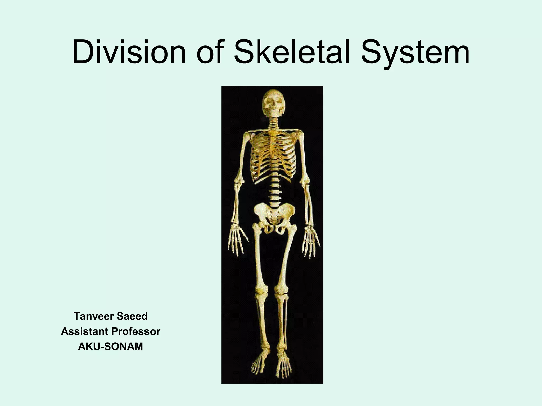
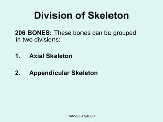
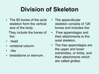
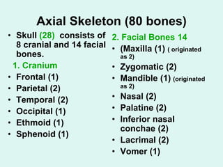
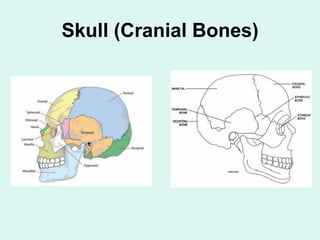
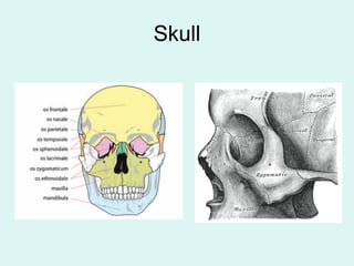
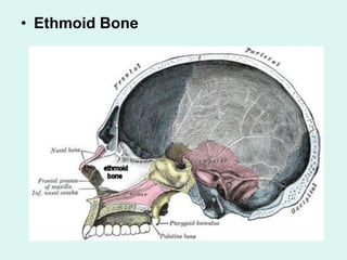
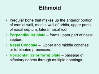
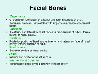
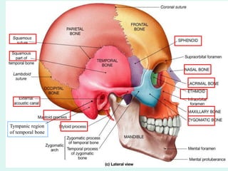
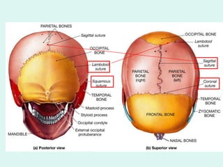
Division of Skeletal System
Tanveer Saeed
Assistant Professor
AKU-SONAM
TANVEER SAEED
Division of Skeleton
206 BONES: These bones can be grouped
in two divisions:
1. Axial Skeleton
2. Appendicular Skeleton
Division of Skeleton
• The 80 bones of the axial
skeleton form the vertical
axis of the body.
They include the bones of
the:
• head
• vertebral column
• ribs
• breastbone or sternum.
• The appendicular
skeleton consists of 126
bones and includes the:
• Free appendages and
their attachments to the
axial skeleton.
• The free appendages are
the upper and lower
extremities, or limbs, and
their attachments which
are called girdles.
Axial Skeleton (80 bones)
• Skull (28) consists of
8 cranial and 14 facial
bones.
1. Cranium
• Frontal (1)
• Parietal (2)
• Temporal (2)
• Occipital (1)
• Ethmoid (1)
• Sphenoid (1)
2. Facial Bones 14
• (Maxilla (1) ( originated
as 2)
• Zygomatic (2)
• Mandible (1) (originated
as 2)
• Nasal (2)
• Palatine (2)
• Inferior nasal
conchae (2)
• Lacrimal (2)
• Vomer (1)
Axial Skeleton Cont’d
• Auditory Ossicles (6 bones)
• Malleus (2)
• Incus (2)
• Stapes (2)
• Hyoid (1)
Total skull bones = 29
Hyoid bone
Skull (Cranial Bones)
Skull
Frontal and Parietal Bones
• Located anteriorly, forms part of orbital
roof, and anterior portion of cranial floor.
• Supraorbital margin – Arched ridge just
below eyebrows, forms superior edge of
orbits.
• Supraorbital foramina – in supraorbital
margin slightly medial to midpoint,
transmits nerve and blood vessels.
• Parietal Bones
• Superior lateral portion of cranium.
Frontal Bone
Occipital Bone
• Forms posterior wall and
floor of cranium.
• Foramen Magnum –
large passageway for
spinal cord exit from
cranium.
• Occipital Condyles –
located lateral to
foramen magnum
articulates with atlas
(C1).
• External Occipital
Protuberance –
prominent projection on
posterior surface.
Occipital Bone
Temporal Bone
Divided into four parts:
• Zygomatic process – projection that articulates
with zygomatic bone.
• Mastoid process – protuberance inferior and
posterior to ear, muscle attachment. Styloid
process – slender spike of bone extending
inferior and anteriorly.
• Squamous part - thin fan shaped part that
articulates with the parietal bone.
• Petrous portion- forms part of the base of the
skull and contains organ of hearing.
Temporal Bone
Sphenoid Bone
• Keystone of cranial floor.
• Resembles bat wings, lies deep and slightly
superior to nose and throat, forms parts of orbital
wall floor and posterior.
• Greater wings – lateral projection from body,
forms lateral wall of orbit.
• Lesser wings – superior portion of sphenoid
body, forms posterior orbital wall.
• Sella turcica –saddle shaped depression on
superior surface of body, contains Pituitary gland
(Hypophyseal gland).
Sphenoid Bone
• Ethmoid Bone
Ethmoid
• Irregular bone that makes up the anterior portion
of cranial wall, medial wall of orbits, upper parts
of nasal septum, lateral nasal roof.
• Perpendicular plate – forms upper part of nasal
septum.
• Nasal Conchae – Upper and middle conchae
or turbinated processes.
• Horizontal (cribriform) plate – passage of
olfactory nerves through multiple openings.
Facial Bones
Zygomatics
• Cheekbone, forms part of anterior and lateral surface of orbit.
• Temporal process – articulates with zygomatic process of temporal
bone.
Lacrimals
• Posterior and lateral to nasal bones in median wall of orbits, forms
lateral of nasal cavity.
Palatines
• Posterior portion of hard palate, inferior and lateral surface of nasal
cavity, inferior surface of orbit.
Nasal bones
• Superior portion of nasal cavity.
Vomer
• Inferior and posterior nasal septum.
Inferior Nasal Conchae
• Turbinated bones forms posterior of nasal cavity.
Tympanic region
of temporal bone
Maxillae or Upper Jaw
• Upper jaw bones, part of floor of orbit,
anterior portion of roof of mouth and floor
of nose and part of lateral walls of nasal
cavity.
• Alveolar ridge or process-- carries teeth.
• Maxillary Sinus--- on each side lined with
ciliated mucous membrane.
Mandible or Lower Jaw
• Movable bone of the skull.
• Rami (ramus)
• Condyloid process – part of
each ramus that articulates
with mandibular fossa of
temporal bone forming
temporomandibular joint.
• Coronoid process--- gives
attachment to muscles and
ligaments.
Paranasal Sinuses
• Spaces or cavities in bone those
associated with nasal cavity called
paranasal sinuses and includes:
• frontal, sphenoidal, ethmoidal, and
maxillary.
• Function – Humidify, warm and filter
inspired air through nasal cavity.
Paranasal Sinuses
Fontanels
• “Soft spots” where ossification is incomplete at
birth, allows some compression of cranium at
birth to pass through the birth canal.
• Frontal (anterior) –intersection of sagittal and
coronal sutures, diamond shaped largest
fontanel.
• When does it closes?
• Occipital (posterior) – intersection of sagittal
and lambdoidal sutures.
• When does it closes?
Fontanelles
Sutures
• Sutures are immovable joints between cranial
bones.
• Sagittal – between parietal bones.
• Squamous – between temporal and parietal
bones.
• Coronal (frontal) – between frontal and parietal
bones.
• Lambdoidal – between parietal and occipital.
Thank You
Practice

Division of skeletal system 2

  • 1.
    Division of SkeletalSystem Tanveer Saeed Assistant Professor AKU-SONAM
  • 2.
    TANVEER SAEED Division ofSkeleton 206 BONES: These bones can be grouped in two divisions: 1. Axial Skeleton 2. Appendicular Skeleton
  • 3.
    Division of Skeleton •The 80 bones of the axial skeleton form the vertical axis of the body. They include the bones of the: • head • vertebral column • ribs • breastbone or sternum. • The appendicular skeleton consists of 126 bones and includes the: • Free appendages and their attachments to the axial skeleton. • The free appendages are the upper and lower extremities, or limbs, and their attachments which are called girdles.
  • 4.
    Axial Skeleton (80bones) • Skull (28) consists of 8 cranial and 14 facial bones. 1. Cranium • Frontal (1) • Parietal (2) • Temporal (2) • Occipital (1) • Ethmoid (1) • Sphenoid (1) 2. Facial Bones 14 • (Maxilla (1) ( originated as 2) • Zygomatic (2) • Mandible (1) (originated as 2) • Nasal (2) • Palatine (2) • Inferior nasal conchae (2) • Lacrimal (2) • Vomer (1)
  • 5.
    Axial Skeleton Cont’d •Auditory Ossicles (6 bones) • Malleus (2) • Incus (2) • Stapes (2) • Hyoid (1) Total skull bones = 29 Hyoid bone
  • 6.
  • 7.
  • 8.
    Frontal and ParietalBones • Located anteriorly, forms part of orbital roof, and anterior portion of cranial floor. • Supraorbital margin – Arched ridge just below eyebrows, forms superior edge of orbits. • Supraorbital foramina – in supraorbital margin slightly medial to midpoint, transmits nerve and blood vessels. • Parietal Bones • Superior lateral portion of cranium.
  • 9.
  • 10.
    Occipital Bone • Formsposterior wall and floor of cranium. • Foramen Magnum – large passageway for spinal cord exit from cranium. • Occipital Condyles – located lateral to foramen magnum articulates with atlas (C1). • External Occipital Protuberance – prominent projection on posterior surface.
  • 11.
  • 12.
    Temporal Bone Divided intofour parts: • Zygomatic process – projection that articulates with zygomatic bone. • Mastoid process – protuberance inferior and posterior to ear, muscle attachment. Styloid process – slender spike of bone extending inferior and anteriorly. • Squamous part - thin fan shaped part that articulates with the parietal bone. • Petrous portion- forms part of the base of the skull and contains organ of hearing.
  • 13.
  • 14.
    Sphenoid Bone • Keystoneof cranial floor. • Resembles bat wings, lies deep and slightly superior to nose and throat, forms parts of orbital wall floor and posterior. • Greater wings – lateral projection from body, forms lateral wall of orbit. • Lesser wings – superior portion of sphenoid body, forms posterior orbital wall. • Sella turcica –saddle shaped depression on superior surface of body, contains Pituitary gland (Hypophyseal gland).
  • 15.
  • 16.
  • 17.
    Ethmoid • Irregular bonethat makes up the anterior portion of cranial wall, medial wall of orbits, upper parts of nasal septum, lateral nasal roof. • Perpendicular plate – forms upper part of nasal septum. • Nasal Conchae – Upper and middle conchae or turbinated processes. • Horizontal (cribriform) plate – passage of olfactory nerves through multiple openings.
  • 18.
    Facial Bones Zygomatics • Cheekbone,forms part of anterior and lateral surface of orbit. • Temporal process – articulates with zygomatic process of temporal bone. Lacrimals • Posterior and lateral to nasal bones in median wall of orbits, forms lateral of nasal cavity. Palatines • Posterior portion of hard palate, inferior and lateral surface of nasal cavity, inferior surface of orbit. Nasal bones • Superior portion of nasal cavity. Vomer • Inferior and posterior nasal septum. Inferior Nasal Conchae • Turbinated bones forms posterior of nasal cavity.
  • 19.
  • 20.
    Maxillae or UpperJaw • Upper jaw bones, part of floor of orbit, anterior portion of roof of mouth and floor of nose and part of lateral walls of nasal cavity. • Alveolar ridge or process-- carries teeth. • Maxillary Sinus--- on each side lined with ciliated mucous membrane.
  • 21.
    Mandible or LowerJaw • Movable bone of the skull. • Rami (ramus) • Condyloid process – part of each ramus that articulates with mandibular fossa of temporal bone forming temporomandibular joint. • Coronoid process--- gives attachment to muscles and ligaments.
  • 23.
    Paranasal Sinuses • Spacesor cavities in bone those associated with nasal cavity called paranasal sinuses and includes: • frontal, sphenoidal, ethmoidal, and maxillary. • Function – Humidify, warm and filter inspired air through nasal cavity.
  • 24.
  • 25.
    Fontanels • “Soft spots”where ossification is incomplete at birth, allows some compression of cranium at birth to pass through the birth canal. • Frontal (anterior) –intersection of sagittal and coronal sutures, diamond shaped largest fontanel. • When does it closes? • Occipital (posterior) – intersection of sagittal and lambdoidal sutures. • When does it closes?
  • 26.
  • 27.
    Sutures • Sutures areimmovable joints between cranial bones. • Sagittal – between parietal bones. • Squamous – between temporal and parietal bones. • Coronal (frontal) – between frontal and parietal bones. • Lambdoidal – between parietal and occipital.
  • 29.