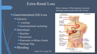
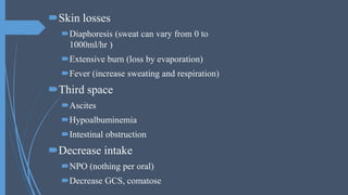
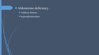
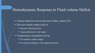
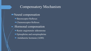
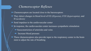
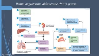
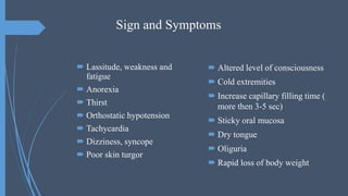
Isotonic Fluid Imbalance
Hypovolemia:
Mehmood ul hassan
Assistant Head Nurse SICU
Objectives
 Describe the isotonic volume deficit
 Discuss the pathophysiology of isotonic volume deficit
 Enlist the clinical manifestation of isotonic volume deficit
Isotonic Fluid Volume Deficit
 It result when water and electrolytes are lost in an isotonic fashion
 Sodium level remain normal in this imbalance
 It may occur alone or in combination with other imbalances
causes
Extra-Renal Loss Renal loss
Extra-Renal Loss
Gastrointestinal (GI) Loss
Gastric:
 vomiting
Gastrointestinal suctioning
Intestinal:
Diarrhea
Ileostomies
Pancreatic or Biliary fistula
Drainage bags
Bleeding
Daily volumes of fluid ingested, secreted,
absorbed, and excreted from the GI tract
Figure 24.21, page 958
Skin losses
Diaphoresis (sweat can vary from 0 to
1000ml/hr )
Extensive burn (loss by evaporation)
Fever (increase sweating and respiration)
Third space
Ascites
Hypoalbuminemia
Intestinal obstruction
Decrease intake
NPO (nothing per oral)
Decrease GCS, comatose
Renal loss
Cause intrinsic to kidney injury
Renal disease
Salt wasting nephritis
Diuretic phase of acute renal failure
Cause extrinsic to kidney injury
Diuretic excess
Osmotic diuresis
Diabetic glycosuria
Enteral or parenteral hyper-alimentation
Mannitol therapy
Aldosterone deficiency
Addison disease
hypoaldosteronism
Hemodynamic Response to Fluid volume Deficit
 Volume depletion result in decrease Cardiac output (CO)
 Decrease cardiac output result in
Decrease blood pressure
 hypo-perfusion to vital organ
 Compensatory mechanism activate
To maintain cardiac output
To maintain adequate vital organs perfusion
Compensatory Mechanism
Neural compensation
Baroreceptor Reflexes
Chemoreceptor Reflexes
 Hormonal compensation
Renin–angiotensin–aldosterone
Epinephrine and norepinephrine
 Antidiuretic hormone (ADH)
Neural compensation
Baroreceptor Reflexes
Pressure sensed receptor present in carotid artery and aorta
They sensed fall in blood pressure
Send signal to vasomotor center in brain
Brain activate sympathetic response that cause
Peripheral vasoconstriction
Increased heart rate
Increase cardiac contractility
Increase cardiac output
Increase blood pressure and perfusion to vital organ
Regulation of blood pressure
via baroreceptor reflexes
Figure 21.14, pg, 778
Chemoreceptor Reflexes
 Chemoreceptors are located close to the baroreceptors
 They detect changes in blood level of O2 (Hypoxia), CO2 (hypercapnia), and
H (acidosis)
 Send impulses to the cardiovascular center
 In response, the cardiovascular center increases sympathetic stimulation
Vasoconstriction of arterioles and veins
 Increase blood pressure
 These chemoreceptors also provide input to the respiratory center in the brain
stem to adjust the rate of breathing
Hormonal compensation
 Lowered blood pressure stimulates the kidneys cells to secrete the enzyme renin
 Renin converts angiotensinogen, into angiotensin I
 angiotensin-converting enzyme (ACE) converts angiotensin I into the hormone
angiotensin II, which raises blood pressure in two ways
First, angiotensin II is a potent vasoconstrictor; it raises blood pressure by
increasing systemic vascular resistance
Second, it stimulates secretion of aldosterone, which increases reabsorption
of sodium ions (Na) and water by the kidney
The water reabsorption increases total blood volume, which increases blood
pressure
Renin–angiotensin–aldosterone (RAA) system.
Renin–angiotensin–aldosterone (RAA) system
F.g 18.16 pg, 667
Epinephrine and norepinephrine
 In response to sympathetic stimulation, the adrenal medulla releases
epinephrine and norepinephrine
 These hormones increase cardiac output by:
Increasing the rate
Increase force of heart contractions
Vasoconstriction of arterioles and veins in the skin and
abdominal organs
Antidiuretic hormone (ADH)
 ADH is produced by the hypothalamus and released from the
posterior pituitary in response to dehydration or decreased blood
volume.
 ADH causes vasoconstriction, which increases blood pressure. For
this reason ADH is also called vasopressin
Regulation of secretion and actions of
antidiuretic hormone (ADH)
Figure 18.9 pg, 657
Sign and Symptoms
 Lassitude, weakness and
fatigue
 Anorexia
 Thirst
 Orthostatic hypotension
 Tachycardia
 Dizziness, syncope
 Poor skin turgor
 Altered level of consciousness
 Cold extremities
 Increase capillary filling time (
more then 3-5 sec)
 Sticky oral mucosa
 Dry tongue
 Oliguria
 Rapid loss of body weight
References:
 Tortora, G. J., Derrickson, B., Kuwaki, T., Kurosawa, M., & Takahashi, K. (2010). Tōtora jintai
no kōzō to kinō. Tōkyō: Maruzen.
 Porth, C., & Hannon, R. A. (2010). Porth pathophysiology: Concepts of altered health states.
Philadelphia, PA: Wolters Kluwer Health/Lippincott Williams & Wilkins.
 Scales, K. (2005). Vascular access: A guide to peripheral venous cannulation. Nursing
Standard, 19(49), 48-52.

Hypovolemia

  • 1.
    Isotonic Fluid Imbalance Hypovolemia: Mehmoodul hassan Assistant Head Nurse SICU
  • 2.
    Objectives  Describe theisotonic volume deficit  Discuss the pathophysiology of isotonic volume deficit  Enlist the clinical manifestation of isotonic volume deficit
  • 3.
    Isotonic Fluid VolumeDeficit  It result when water and electrolytes are lost in an isotonic fashion  Sodium level remain normal in this imbalance  It may occur alone or in combination with other imbalances
  • 4.
  • 5.
    Extra-Renal Loss Gastrointestinal (GI)Loss Gastric:  vomiting Gastrointestinal suctioning Intestinal: Diarrhea Ileostomies Pancreatic or Biliary fistula Drainage bags Bleeding Daily volumes of fluid ingested, secreted, absorbed, and excreted from the GI tract Figure 24.21, page 958
  • 6.
    Skin losses Diaphoresis (sweatcan vary from 0 to 1000ml/hr ) Extensive burn (loss by evaporation) Fever (increase sweating and respiration) Third space Ascites Hypoalbuminemia Intestinal obstruction Decrease intake NPO (nothing per oral) Decrease GCS, comatose
  • 7.
    Renal loss Cause intrinsicto kidney injury Renal disease Salt wasting nephritis Diuretic phase of acute renal failure Cause extrinsic to kidney injury Diuretic excess Osmotic diuresis Diabetic glycosuria Enteral or parenteral hyper-alimentation Mannitol therapy
  • 8.
  • 9.
    Hemodynamic Response toFluid volume Deficit  Volume depletion result in decrease Cardiac output (CO)  Decrease cardiac output result in Decrease blood pressure  hypo-perfusion to vital organ  Compensatory mechanism activate To maintain cardiac output To maintain adequate vital organs perfusion
  • 10.
    Compensatory Mechanism Neural compensation BaroreceptorReflexes Chemoreceptor Reflexes  Hormonal compensation Renin–angiotensin–aldosterone Epinephrine and norepinephrine  Antidiuretic hormone (ADH)
  • 11.
    Neural compensation Baroreceptor Reflexes Pressuresensed receptor present in carotid artery and aorta They sensed fall in blood pressure Send signal to vasomotor center in brain Brain activate sympathetic response that cause Peripheral vasoconstriction Increased heart rate Increase cardiac contractility Increase cardiac output Increase blood pressure and perfusion to vital organ
  • 12.
    Regulation of bloodpressure via baroreceptor reflexes Figure 21.14, pg, 778
  • 13.
    Chemoreceptor Reflexes  Chemoreceptorsare located close to the baroreceptors  They detect changes in blood level of O2 (Hypoxia), CO2 (hypercapnia), and H (acidosis)  Send impulses to the cardiovascular center  In response, the cardiovascular center increases sympathetic stimulation Vasoconstriction of arterioles and veins  Increase blood pressure  These chemoreceptors also provide input to the respiratory center in the brain stem to adjust the rate of breathing
  • 14.
    Hormonal compensation  Loweredblood pressure stimulates the kidneys cells to secrete the enzyme renin  Renin converts angiotensinogen, into angiotensin I  angiotensin-converting enzyme (ACE) converts angiotensin I into the hormone angiotensin II, which raises blood pressure in two ways First, angiotensin II is a potent vasoconstrictor; it raises blood pressure by increasing systemic vascular resistance Second, it stimulates secretion of aldosterone, which increases reabsorption of sodium ions (Na) and water by the kidney The water reabsorption increases total blood volume, which increases blood pressure Renin–angiotensin–aldosterone (RAA) system.
  • 15.
  • 16.
    Epinephrine and norepinephrine In response to sympathetic stimulation, the adrenal medulla releases epinephrine and norepinephrine  These hormones increase cardiac output by: Increasing the rate Increase force of heart contractions Vasoconstriction of arterioles and veins in the skin and abdominal organs
  • 17.
    Antidiuretic hormone (ADH) ADH is produced by the hypothalamus and released from the posterior pituitary in response to dehydration or decreased blood volume.  ADH causes vasoconstriction, which increases blood pressure. For this reason ADH is also called vasopressin
  • 18.
    Regulation of secretionand actions of antidiuretic hormone (ADH) Figure 18.9 pg, 657
  • 19.
    Sign and Symptoms Lassitude, weakness and fatigue  Anorexia  Thirst  Orthostatic hypotension  Tachycardia  Dizziness, syncope  Poor skin turgor  Altered level of consciousness  Cold extremities  Increase capillary filling time ( more then 3-5 sec)  Sticky oral mucosa  Dry tongue  Oliguria  Rapid loss of body weight
  • 20.
    References:  Tortora, G.J., Derrickson, B., Kuwaki, T., Kurosawa, M., & Takahashi, K. (2010). Tōtora jintai no kōzō to kinō. Tōkyō: Maruzen.  Porth, C., & Hannon, R. A. (2010). Porth pathophysiology: Concepts of altered health states. Philadelphia, PA: Wolters Kluwer Health/Lippincott Williams & Wilkins.  Scales, K. (2005). Vascular access: A guide to peripheral venous cannulation. Nursing Standard, 19(49), 48-52.