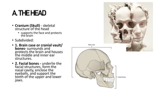
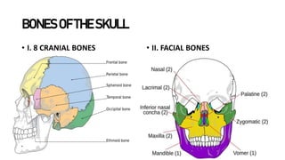
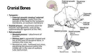
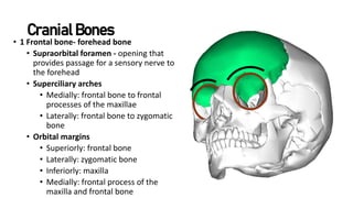
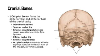
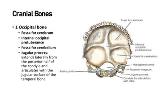
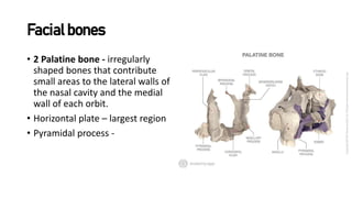
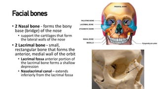
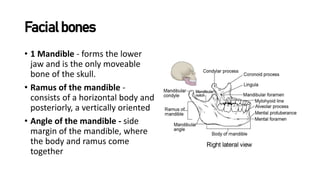
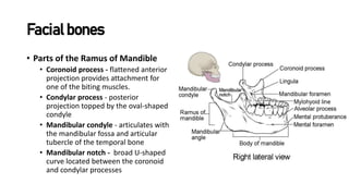
The document provides an overview of the bones that make up the skull and face. It describes the cranial bones that form the brain case and protect the brain, including the frontal, parietal, temporal, occipital, sphenoid, and ethmoid bones. It also details the facial bones that underlie the facial structures and form parts of the nose, orbits, and jaws. This includes the maxilla, palatine, zygomatic, nasal, lacrimal, inferior nasal conchae, vomer, and mandible bones. Finally, it discusses the joints between cranial bones and the foramina and fossae located on the interior base of the skull.
The skull is formed by cranial and facial bones. The cranial bones form the cranium which encloses and protects the brain, while the facial bones form the framework of the face. There are 22 bones that make up the skull which are divided into cranial bones and facial bones. The eight large cranial bones are the paired parietal and temporal bones along with the unpaired frontal, occipital, sphenoid, and ethmoid bones. These bones form the cranial vault which encloses the brain and has openings for nerves and blood vessels. The facial bones form the structures of the face and openings for senses.
The document describes the three cranial fossae (anterior, middle, posterior) and their boundaries. The anterior fossa is formed by the frontal bone, ethmoid, and lesser sphenoid wing and contains the frontal lobes. The middle fossa is formed by the sphenoid, temporal, and parietal bones and contains the temporal lobes. The posterior fossa is formed mainly by the occipital bone and contains the cerebellum and brainstem. It also discusses the orbital complex, nasal complex, infratemporal fossa, and muscles of mastication.
The document provides an overview of basic skull anatomy, including:
- The skull is composed of 22 bones that form the cranium and face.
- It describes the various views of the skull and identifies the bones that make up the different regions.
- The interior of the skull is divided into three cranial fossae that house different parts of the brain.
- Key anatomical structures like foramina, canals, and sinuses that transmit nerves and vessels are outlined.
head old lecture assignment presentation 123AmanuelIbrahim
The document discusses the osteology of the head and neck, specifically focusing on the bones that make up the skull. It describes the two main parts of the skull - the neurocranium which forms the brain case, and the viscerocranium which forms the facial bones. It provides details on the individual bones, their anatomical features, and articulations that comprise these two regions.
The document discusses the bones that make up the human skull. It is divided into two parts: the axial skeleton and appendicular skeleton. The axial skeleton includes the skull bones, which are divided into the cranium and mandible. The cranium contains eight bones, while the mandible is a single bone. The document further describes several important skull bones in more detail, including the maxilla, mandible, nasal bones, ethmoid bone, and sphenoid bone. It outlines the structures and features of each bone.
The document discusses the anatomy of the skull and facial bones. It describes the four types of fontanels present at birth, located between cranial bones. It then details the bones that make up the skull, including the cranial and facial bones. For each bone, it outlines the key features and structures, such as foramina and sutures, as well as their locations and functions.
The skull base forms the floor of the cranial cavity and separates the brain from facial structures. It is composed of five bones: ethmoid, sphenoid, occipital, and paired temporal and frontal bones. The anterior skull base is formed by the orbital plates of the frontal bone laterally, the cribriform plate of the ethmoid bone in the midline, and the lesser sphenoid wing posteriorly. The middle skull base is formed mainly by the sphenoid bone and the temporal bone anterior to the petrous ridge. The posterior skull base is formed by the occipital bone and parts of the temporal and sphenoid bones. Infections and tumors originating from the deep facial spaces can
This document provides an overview of head and neck anatomy, focusing on the bones of the skull and face. It describes the various bones that make up the cranium and face, including their names, locations, landmarks, and articulation points. Key bones discussed include the frontal, temporal, parietal, occipital, sphenoid, ethmoid, maxilla, zygomatic, lacrimal, palatine, nasal, vomer and mandible. The document also covers bones of the middle ear, temporomandibular joint, hyoid bone, and fetal skull sutures.
This is an educational presentation that describes methods of studying skull. Various Normas has been explained with diagrams. The presentation is the continuation of previously uploaded matter wherein major bones of the skull was explained. link to previous ppt is https://www.slideshare.net/AyshahHashimi/skull-copy
The document provides an overview of osteology and classification of bones in the facial skeleton. It begins with an introduction to bone structure and function. It then classifies bones based on shape and development. The document outlines the bones that make up the skull, including the calvarium (paired and unpaired bones), and provides details on the norma views (verticalis, occipitalis, lateralis, frontalis, basalis). It describes each individual bone, its landmarks and clinical relevance. In summary, the document categorizes and describes in detail the bones that make up the skull and facial skeleton.
The document provides an overview of the osteology (bone structure) of the human skull. It describes the various bones that make up the skull and how they articulate. It discusses the skull from 5 anatomical views (normas): superior (norma verticalis), anterior (norma frontalis), lateral (norma lateralis), posterior (norma occipitalis), and inferior (norma basalis). Each view identifies the bones visible from that perspective and important anatomical landmarks. The document also details the individual bones that comprise the cranial and facial skeleton.
It contains following subheadings:
-maxilla and mandible anatomy
-TMJ(Temporo mandibular joint)
-Muscles of mastication
By:
Dr. Syed Irfan Qadeer
Prof. and HOD Department of Anatomy
SPIDMS,Lucknow
The axial skeleton includes the bones of the skull, vertebral column, ribs, and sternum. The skull has 22 bones that form the cranium and face. The cranium contains the brain and sense organs and is formed by the occipital, parietal, frontal, temporal, sphenoid, and ethmoid bones. These bones have markings that indicate muscle attachments and openings for blood vessels and cranial nerves. The face contains 14 bones, including the paired maxillae, palatine, nasal, zygomatic, lacrimal, and conchae bones, as well as the single vomer and mandible bones.
This document provides an overview of the anatomy of the skull and cranial cavity. It describes the bones that make up the skull, including the cranium and facial bones. It details the cranial fossae and structures within them, such as foramina for cranial nerves and blood vessels. It also summarizes the meninges and dural projections, cranial venous channels, and some types of skull fractures and hemorrhages that can result from head trauma.
The temporal bone is formed from four fused elements and houses important structures of the inner ear, middle ear, and cranial nerves. The petrous part is the hardest bone in the body, containing the inner ear structures including the cochlea which is adult size at birth. Many cranial nerves pass through foramina in the temporal bone, making the skull base susceptible to fractures and infiltration. Clinical signs of cranial nerve palsies or pulsatile tinnitus could indicate underlying pathology visualized by imaging tests. Examination of cranial nerve function is important for diagnosing conditions affecting the temporal bone.
The skull consists of several bones joined by sutures. It is composed of an outer and inner table of compact bone separated by diploe. The skull bones are divided into those of the cranium (vault and base) and face. The cranium contains the frontal, parietal, occipital, temporal, sphenoid and ethmoid bones. The face contains zygomatic, maxilla, nasal, lacrimal, vomer and palatine bones. The dura mater of the brain has an endosteal layer attached to the inner skull surface and a meningeal layer which is the dura mater proper covering the brain.
The document provides information about the bones that make up the human skull. It discusses 28 individual bones and where they are located. Some of the key bones mentioned include the frontal bone, which forms the forehead; the parietal bones, which make up much of the skull vault; the occipital bone, which forms the back of the skull; and the sphenoid bone, which contributes to parts of the middle cranial fossa, orbits, nasal cavity, and infratemporal fossa. Sutures and joints between the skull bones are also described. The document provides detailed information on features and articulation points of each bone.
The skull consists of 22 bones that form the cerebral and facial cranium. The cerebral cranium contains 8 bones that protect the brain, while the facial cranium contains 14 bones that make up the face. Key bones of the cerebral cranium include the frontal, parietal, occipital, sphenoid, ethmoid, and temporal bones. The facial cranium contains bones like the maxilla, zygomatic, and mandible. Sutures join these bones together. Other structures discussed include the hyoid bone, middle ear ossicles (malleus, incus, stapes), and openings in the skull.
تلخيصات بسيطه تخص طلاب اسنان في ماده الاناتومي
تابعونا علي الصفحه
https://www.facebook.com/dentology7/?ref=bookmarks
او
https://www.facebook.com/Doctor.Hossam.A
The document provides an overview of the osteology of the human skull. It describes the external features of the skull from the anterior, lateral, posterior, superior and inferior views. It also describes the internal features of the cranial cavity including the anterior, middle and posterior sections. Key bones are discussed such as the mandible, frontal bone, parietal bone, occipital bone and temporal bone. The neurocranium and viscerocranium are defined. Several craniometric points are also defined that are used for anatomical measurements and landmarks. Buttresses that transmit forces across the skull are also noted.
The document summarizes the division of the skeletal system into the axial and appendicular skeleton. The axial skeleton consists of 80 bones including the skull, vertebral column, ribs, and sternum. It forms the vertical axis of the body. The appendicular skeleton has 126 bones and includes the limbs and their attachments via girdles. The skull is made up of 28 cranial bones including the frontal, parietal, temporal, occipital, ethmoid, and sphenoid bones, as well as 14 facial bones such as the maxilla, zygomatic, and mandible.
This is an educational presentation on skull anatomy. It features the structure of various bones of the skull, their exact location their characteristics features and various muscles attached to it.
The skull base forms the floor of the cranial cavity and separates the brain from facial structures. It is composed of five bones. The anterior skull base is formed by the frontal bone laterally, cribriform plate of ethmoid bone in the midline, and lesser sphenoid wing posteriorly. The middle skull base is formed mainly by the sphenoid bone and temporal bone anterior to the petrous ridge. It contains several openings such as the foramen ovale, rotundum, spinosum and cavernous sinus. The posterior skull base is formed by the occipital bone and parts of the temporal and sphenoid bones. It contains openings like the jugular foramen, hypogloss
The cranial cavity contains the brain and its meninges, cranial nerves, arteries, veins, and venous sinuses
The bones that take part in formation of cranial cavity are frontal, parietal, temporal, occipital and ethmoid
1-Vault of the Skull
2-Base of the Skull
This document provides an overview of the anatomy of the maxilla and mandible bones. It describes the main parts and features of each bone, including their surfaces, processes, sinuses, foramina, fossae and other anatomical structures. It also discusses the muscles, arteries, veins and nerves associated with each bone. Applied clinical implications are presented, such as how injuries to specific muscles or nerves during surgical procedures can impact surrounding structures or functions.
QUICK guide to Skeletal System- Dr Gurjant Singh (PT)Dr. Gurjant Singh
So you have read well about human skeletal system and now just want to revise or go through it quickly? Here is the perfect one for you
Dont forget to like and follow for more presentations
ANAMOLOUS SECONDARY GROWTH IN DICOT ROOTS.pptxRASHMI M G
Abnormal or anomalous secondary growth in plants. It defines secondary growth as an increase in plant girth due to vascular cambium or cork cambium. Anomalous secondary growth does not follow the normal pattern of a single vascular cambium producing xylem internally and phloem externally.
This presentation explores a brief idea about the structural and functional attributes of nucleotides, the structure and function of genetic materials along with the impact of UV rays and pH upon them.
Unlocking the mysteries of reproduction: Exploring fecundity and gonadosomati...AbdullaAlAsif1
The pygmy halfbeak Dermogenys colletei, is known for its viviparous nature, this presents an intriguing case of relatively low fecundity, raising questions about potential compensatory reproductive strategies employed by this species. Our study delves into the examination of fecundity and the Gonadosomatic Index (GSI) in the Pygmy Halfbeak, D. colletei (Meisner, 2001), an intriguing viviparous fish indigenous to Sarawak, Borneo. We hypothesize that the Pygmy halfbeak, D. colletei, may exhibit unique reproductive adaptations to offset its low fecundity, thus enhancing its survival and fitness. To address this, we conducted a comprehensive study utilizing 28 mature female specimens of D. colletei, carefully measuring fecundity and GSI to shed light on the reproductive adaptations of this species. Our findings reveal that D. colletei indeed exhibits low fecundity, with a mean of 16.76 ± 2.01, and a mean GSI of 12.83 ± 1.27, providing crucial insights into the reproductive mechanisms at play in this species. These results underscore the existence of unique reproductive strategies in D. colletei, enabling its adaptation and persistence in Borneo's diverse aquatic ecosystems, and call for further ecological research to elucidate these mechanisms. This study lends to a better understanding of viviparous fish in Borneo and contributes to the broader field of aquatic ecology, enhancing our knowledge of species adaptations to unique ecological challenges.
Travis Hills' Endeavors in Minnesota: Fostering Environmental and Economic Pr...Travis Hills MN
Travis Hills of Minnesota developed a method to convert waste into high-value dry fertilizer, significantly enriching soil quality. By providing farmers with a valuable resource derived from waste, Travis Hills helps enhance farm profitability while promoting environmental stewardship. Travis Hills' sustainable practices lead to cost savings and increased revenue for farmers by improving resource efficiency and reducing waste.
EWOCS-I: The catalog of X-ray sources in Westerlund 1 from the Extended Weste...Sérgio Sacani
Context. With a mass exceeding several 104 M⊙ and a rich and dense population of massive stars, supermassive young star clusters
represent the most massive star-forming environment that is dominated by the feedback from massive stars and gravitational interactions
among stars.
Aims. In this paper we present the Extended Westerlund 1 and 2 Open Clusters Survey (EWOCS) project, which aims to investigate
the influence of the starburst environment on the formation of stars and planets, and on the evolution of both low and high mass stars.
The primary targets of this project are Westerlund 1 and 2, the closest supermassive star clusters to the Sun.
Methods. The project is based primarily on recent observations conducted with the Chandra and JWST observatories. Specifically,
the Chandra survey of Westerlund 1 consists of 36 new ACIS-I observations, nearly co-pointed, for a total exposure time of 1 Msec.
Additionally, we included 8 archival Chandra/ACIS-S observations. This paper presents the resulting catalog of X-ray sources within
and around Westerlund 1. Sources were detected by combining various existing methods, and photon extraction and source validation
were carried out using the ACIS-Extract software.
Results. The EWOCS X-ray catalog comprises 5963 validated sources out of the 9420 initially provided to ACIS-Extract, reaching a
photon flux threshold of approximately 2 × 10⁻⁸ photons cm−2
s
−1
. The X-ray sources exhibit a highly concentrated spatial distribution,
with 1075 sources located within the central 1 arcmin. We have successfully detected X-ray emissions from 126 out of the 166 known
massive stars of the cluster, and we have collected over 71 000 photons from the magnetar CXO J164710.20-455217.
What is greenhouse gasses and how many gasses are there to affect the Earth.moosaasad1975
What are greenhouse gasses how they affect the earth and its environment what is the future of the environment and earth how the weather and the climate effects.
The debris of the ‘last major merger’ is dynamically youngSérgio Sacani
The Milky Way’s (MW) inner stellar halo contains an [Fe/H]-rich component with highly eccentric orbits, often referred to as the
‘last major merger.’ Hypotheses for the origin of this component include Gaia-Sausage/Enceladus (GSE), where the progenitor
collided with the MW proto-disc 8–11 Gyr ago, and the Virgo Radial Merger (VRM), where the progenitor collided with the
MW disc within the last 3 Gyr. These two scenarios make different predictions about observable structure in local phase space,
because the morphology of debris depends on how long it has had to phase mix. The recently identified phase-space folds in Gaia
DR3 have positive caustic velocities, making them fundamentally different than the phase-mixed chevrons found in simulations
at late times. Roughly 20 per cent of the stars in the prograde local stellar halo are associated with the observed caustics. Based
on a simple phase-mixing model, the observed number of caustics are consistent with a merger that occurred 1–2 Gyr ago.
We also compare the observed phase-space distribution to FIRE-2 Latte simulations of GSE-like mergers, using a quantitative
measurement of phase mixing (2D causticality). The observed local phase-space distribution best matches the simulated data
1–2 Gyr after collision, and certainly not later than 3 Gyr. This is further evidence that the progenitor of the ‘last major merger’
did not collide with the MW proto-disc at early times, as is thought for the GSE, but instead collided with the MW disc within
the last few Gyr, consistent with the body of work surrounding the VRM.
Or: Beyond linear.
Abstract: Equivariant neural networks are neural networks that incorporate symmetries. The nonlinear activation functions in these networks result in interesting nonlinear equivariant maps between simple representations, and motivate the key player of this talk: piecewise linear representation theory.
Disclaimer: No one is perfect, so please mind that there might be mistakes and typos.
dtubbenhauer@gmail.com
Corrected slides: dtubbenhauer.com/talks.html
When I was asked to give a companion lecture in support of ‘The Philosophy of Science’ (https://shorturl.at/4pUXz) I decided not to walk through the detail of the many methodologies in order of use. Instead, I chose to employ a long standing, and ongoing, scientific development as an exemplar. And so, I chose the ever evolving story of Thermodynamics as a scientific investigation at its best.
Conducted over a period of >200 years, Thermodynamics R&D, and application, benefitted from the highest levels of professionalism, collaboration, and technical thoroughness. New layers of application, methodology, and practice were made possible by the progressive advance of technology. In turn, this has seen measurement and modelling accuracy continually improved at a micro and macro level.
Perhaps most importantly, Thermodynamics rapidly became a primary tool in the advance of applied science/engineering/technology, spanning micro-tech, to aerospace and cosmology. I can think of no better a story to illustrate the breadth of scientific methodologies and applications at their best.
1. INTRODUCTION OF THE
HEAD AND NECK
• Bonesof theSkull
• Cranialbones
• Facialbones
• Base andForamenof theskull
2. A. THE HEAD
• Cranium (Skull) - skeletal
structure of the head
• supports the face and protects
the brain
• Subdivided:
• 1. Brain case or cranial vault/
bones- surrounds and
protects the brain and houses
the middle and inner ear
structures.
• 2. Facial bones - underlie the
facial structures, form the
nasal cavity, enclose the
eyeballs, and support the
teeth of the upper and lower
jaws.
4. Cranial bones
• Cranial Cavity – interior space
that is almost completely
occupied by the brain
• Boundaries:
• Superiorly, Lateral and
Posterior: Calvaria (skullcap)
• Floor/ Base:
• Anterior Cranial Fossa
• Middle Cranial Fossa
• Posterior Cranial Fossa
5. • 2 Parietal Bone – upper
lateral side of the skull
• Parietal foramen -
inconstant foramina
• transmit the emissary
veins, draining to the
superior sagittal
sinus, and
occasionally a branch
of the occipital artery
Cranial Bones
6. • 2 Temporal bone – lower lateral side
• Squamous
• AKA squama temporalis, flattened largest part of
the temporal bone, forming part of the temporal
fossa.
• Zygomatic Process - lower part of the squama
temporalis, forms the posterior portion of the
zygomatic arch.
• Mandibular fossa – articulates the head of the
mandible. it allows the mouth to be closed and
opened, meaning it exists to perform mastication.
• Articular tubercle - contribute to the
temporomandibular joint
Cranial Bones
7. • Tympanic:
• External acoustic meatus/ external
acoustic meatus - leads from the
outside of the head to the tympanic
membrane, or eardrum membrane
• Styloid process - attachment point for
muscles and ligaments, such as the
stylomandibular ligament of the TMJ.
• Petromastoid
• Mastoid process – attachment of
muscles
• Petrous part is pyramidal shaped and
lies at the base of temporal bone. It
contains the inner ear.
• Mastoid air cells - hollowed out areas,
equalizing the pressure within the
middle ear in the case of auditory
tube dysfunction
Cranial Bones
8. • 1 Frontal bone- forehead bone
• Supraorbital foramen - opening that
provides passage for a sensory nerve to
the forehead
• Superciliary arches
• Medially: frontal bone to frontal
processes of the maxillae
• Laterally: frontal bone to zygomatic
bone
• Orbital margins
• Superiorly: frontal bone
• Laterally: zygomatic bone
• Inferiorly: maxilla
• Medially: frontal process of the
maxilla and frontal bone
Cranial Bones
9. • 1 Occipital bone - forms the
posterior skull and posterior base
of the cranial cavity
• Supreme nuchal line
• Superior nuchal line
• External occipital protuberance -
serves as an attachment site for a
ligament
• Inferior nuchal line
• External occipital crest
• Occipital condyle - articulate with the
superior aspect of the lateral mass of
the first cervical vertebra,(atlas).
Cranial Bones
10. • 1 Occipital bone
• Fossa for cerebrum
• Internal occipital
protuberance
• Fossa for cerebellum
• Jugular process-
extends laterally from
the posterior half of
the condyle and
articulates with the
jugular surface of the
temporal bone.
Cranial Bones
11. • 1 Sphenoid bone- AKA “wasp bone,”
• located in the middle and toward the front
of the skull, just in front of the occipital
bone
• Body – center cubical shaped
• Greater wing – acts as floor of the middle
cranial fossa, lateral wall of the skull,
posterolateral wall of the orbit
• Lesser Wing - separates the anterior
cranial fossa from the middle cranial fossa.
• Medial pterygoid plate- supports post
opening of the nasal cavity
• Lateral pterygoid plate- origin of
pterygoid muscles
Cranial Bones
12. • 1 Ethmoid bone
• Separates the nasal
cavity from the brain. it
is located at the roof of
the nose, between the
two orbits
Cranial Bones
13. • Crista Galli (rooster’s comb/
crest) - small upward bony
projection located at the
midline, functions as an anterior
attachment point for one of the
covering layers of the brain.
• Cribriform plate - small,
flattened area with numerous
small openings (olfactory
foramina).
• Superior and Middle nasal
concha - shelves of bone that
project into the nasal cavity
• Perpendicular plate - forms the
upper portion of the nasal
septum.
Cranial Bones
14. Joints of the Cranial bones
Sutures- Immobile Joints
• united bones that composed the skull
Sutural ligaments - dense, fibrous
connective tissue between the bones
A. Frontal Suture/ Coronal Suture- parietal and
frontal
B. Squamosal Suture- parietal and temporal
C. Lambdoidal Suture- parietal and occipital
D. Sagittal Suture- parietal and parietal
NOTE: TEMPOROMANDIBULAR JOINT- UNITES
THE SKULL AND MANDIBLE
15. • Forms the upper and lower
jaws, the nose, nasal cavity
and nasal septum, and the
orbit.
• Paired bones are the maxilla,
palatine, zygomatic, nasal,
lacrimal, and inferior nasal
conchae bones.
• Unpaired bones are the vomer
and mandible bones.
Facial bones
16. • 2 Maxillary Bone/ Maxilla – one of a
pair that together form the upper jaw,
much of the hard palate, the medial
floor of the orbit, and the lateral base
of the nose
• Alveolar process of the maxilla-
curved, inferior margin of the
maxillary bone that forms the upper
jaw and contains the upper teeth
• Alveolus - deep socket each tooth is
anchored
• Infraorbital foramen - exit for a
sensory nerve that supplies the nose,
upper lip, and anterior cheek.
Facial bones
17. • Palatine process - from each maxillary
bone can be seen joining together at
the midline to form the anterior
three-quarters of the hard palate
• Hard palate - bony plate that forms
the roof of the mouth and floor of the
nasal cavity, separating the oral and
nasal cavities.
• Incisive foramen – AKA anterior
palatine foramen, or nasopalatine
foramen
• funnel-shaped opening in the
bone of the oral hard palate
immediately behind the incisor
teeth
Facial bones
18. • 2 Palatine bone - irregularly
shaped bones that contribute
small areas to the lateral walls of
the nasal cavity and the medial
wall of each orbit.
• Horizontal plate – largest region
• Pyramidal process -
Facial bones
19. • 2 Zygomatic bone (cheekbone).
• forms much of the lateral wall of
the orbit and the lateral-inferior
margins of the anterior orbital
opening.
• Temporal process - projects
posteriorly, where it forms the
anterior portion of the zygomatic
arch
• Zygomaticofacial foramen -
transmits zygomatic nerve and
vessels to temporal fossa and
cheek
Facial bones
20. Facial bones
• 2 Nasal bone - forms the bony
base (bridge) of the nose
• support the cartilages that form
the lateral walls of the nose
• 2 Lacrimal bone - small,
rectangular bone that forms the
anterior, medial wall of the orbit
• Lacrimal fossa anterior portion of
the lacrimal bone forms a shallow
depression
• Nasolacrimal canal – extends
inferiorly from the lacrimal fossa
21. • 2 Inferior nasal
conchae - form a
curved bony plate
that projects into the
nasal cavity space
from the lower lateral
wall
• 1 Vomer - triangular-
shaped and forms the
posterior-inferior part
of the nasal septum
Facial bones
22. • 1 Mandible - forms the lower
jaw and is the only moveable
bone of the skull.
• Ramus of the mandible -
consists of a horizontal body and
posteriorly, a vertically oriented
• Angle of the mandible - side
margin of the mandible, where
the body and ramus come
together
Facial bones
23. • Parts of the Ramus of Mandible
• Coronoid process - flattened anterior
projection provides attachment for
one of the biting muscles.
• Condylar process - posterior
projection topped by the oval-shaped
condyle
• Mandibular condyle - articulates with
the mandibular fossa and articular
tubercle of the temporal bone
• Mandibular notch - broad U-shaped
curve located between the coronoid
and condylar processes
Facial bones
24. • Mylohyoid line— bony ridge extends along
the inner aspect of the mandibular body,
attachment of the muscle that forms the floor
of the oral cavity
• Mandibular foramen— opening is located on
the medial side of the ramus of the mandible.
• leads into a tunnel that runs down the
length of the mandibular body.
• sensory nerve and blood vessels that
supply the lower teeth enter the
mandibular foramen and then follow this
tunnel.
• Lingula—small flap of bone located
immediately next to the mandibular foramen,
on the medial side of the ramus.
• ligament that anchors the mandible
during opening and closing of the mouth
extends down from the base of the skull
and attaches to the lingula.
Facial bones
25. • Body of the Mandible
• Alveolar process of the
mandible— upper border of the
mandibular body and serves to
anchor the lower teeth.
• Mental protuberance— forward
projection from the inferior margin
of the anterior mandible that
forms the chin
• Mental foramen— opening
located on each side of the
anterior-lateral mandible, exit site
for a sensory nerve that supplies
the chin.
Facial bones
26. • Anterior cranial fossa - most
anterior and the shallowest of
the three cranial fossae.
• Boundaries:
• Anterior: Frontal bone - forms
mainly the floor for this space.
• Posterior: Lesser wings of the
sphenoid bone - form the
prominent ledge that marks the
boundary.
• Midline: Crista galli and
Cribriform plates.
Floor/Base of Cranial Cavity
27. • Middle cranial fossa - deeper
and situated posterior to the
anterior fossa
• Boundaries:
• Anteriorly: Lesser wings of the
sphenoid bone
• Posteriorly: Petrous ridges
(petrous portion of the temporal
bones).
• Midline: Sella turcica
Floor/Base of Cranial Cavity
28. • Optic canal— opening is located at the
anterior lateral corner of the Sella turcica ,
provides for passage of the optic nerve into
the orbit.
• Superior orbital fissure— large, irregular
opening in the lesser wing of sphenoid into
the posterior orbit, provides passage to the
nerves to the eyeball and associated
muscles, and sensory nerves to the
forehead.
• Foramen rotundum— rounded opening
located in the greater wing of sphenoid, just
inferior to the superior orbital fissure, exit
point for a major sensory nerve that supplies
the cheek, nose, and upper teeth.
• Foramen ovale — large, oval-shaped
opening in the base of greater wing of
sphenoid, provides passage for a major
sensory nerve to the lateral head, cheek,
chin, and lower teeth.
Floor/Base of Cranial Cavity
29. • Foramen spinosum— small opening, located
posteromedial part of greater wing of
sphenoid bone posterolateral to foramen
ovale, entry point for an important artery that
supplies the covering layers surrounding the
brain
• Carotid canal— located on the inferior aspect
of the skull, anteromedial to the styloid
process, passageway through which a major
artery to the brain enters the skull,
• Foramen lacerum— irregular opening located
in the between the sphenoid bone, apex of
petrous temporal and basilar part of occipital,
immediately inferior to the exit of the carotid
canal
• artifact of the dry skull-filled with
cartilage, nothing passes this foramen
Floor/Base of Cranial Cavity
30. • Posterior cranial fossa - most
posterior and deepest portion of
the cranial cavity
• contains the cerebellum of the brain.
• Boundaries:
• Anterior: Petrous ridges of Temporal
bone
• Floor: Occipital bone
• Posterior: Foramen magnum (“great
aperture”) the opening that provides
for passage of the spinal cord.
Floor/Base of Cranial Cavity
31. • Internal acoustic meatus - Located on the
medial wall of the petrous ridge in the
posterior cranial fossa, provides for
passage of the nerve from the hearing
and equilibrium organs of the inner ear,
and the nerve that supplies the muscles
of the face
• Hypoglossal canal – located in the
inferior aspect of the skull at the base of
the occipital condyle, provide passage for
an important nerve to the tongue.
• Jugular foramen – large irregularly
shaped located inferior to the internal
acoustic meatus, provides opening for the
exit of several cranial nerves from the
brain and exit point through the base of
the skull for all the venous return blood
leaving the brain.
Floor/Base of Cranial Cavity