Download to read offline

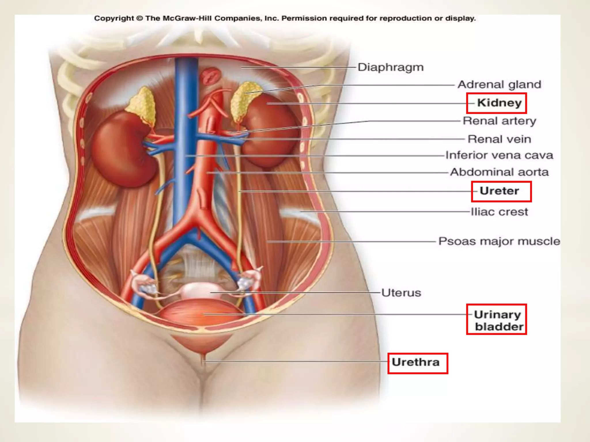

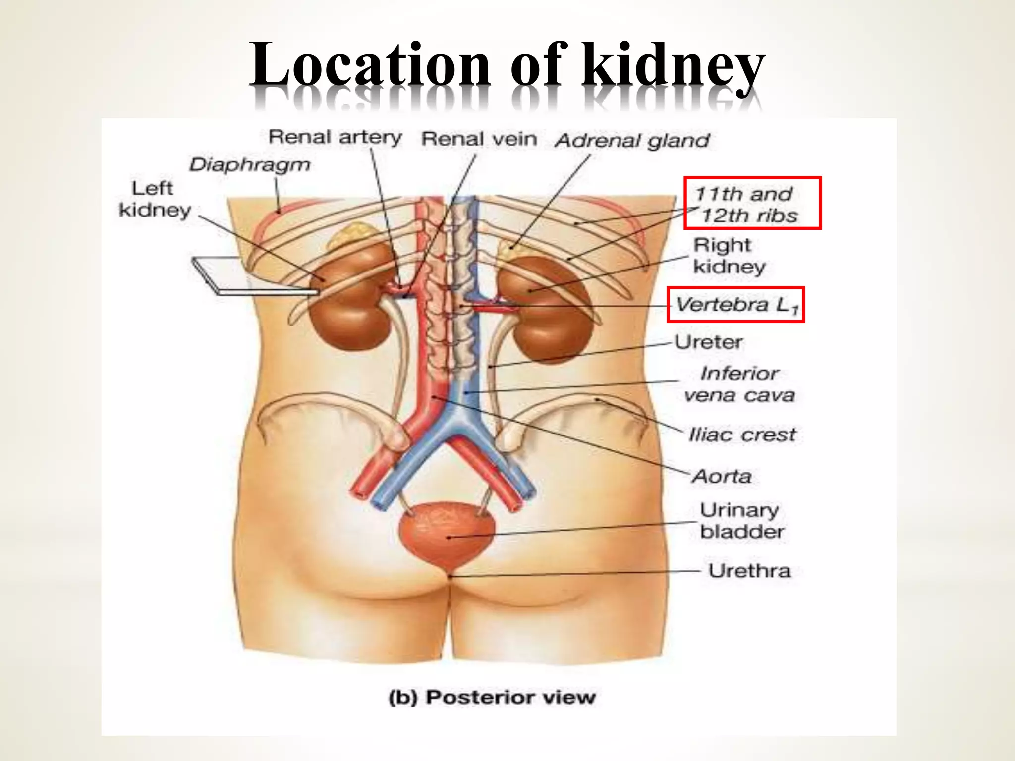
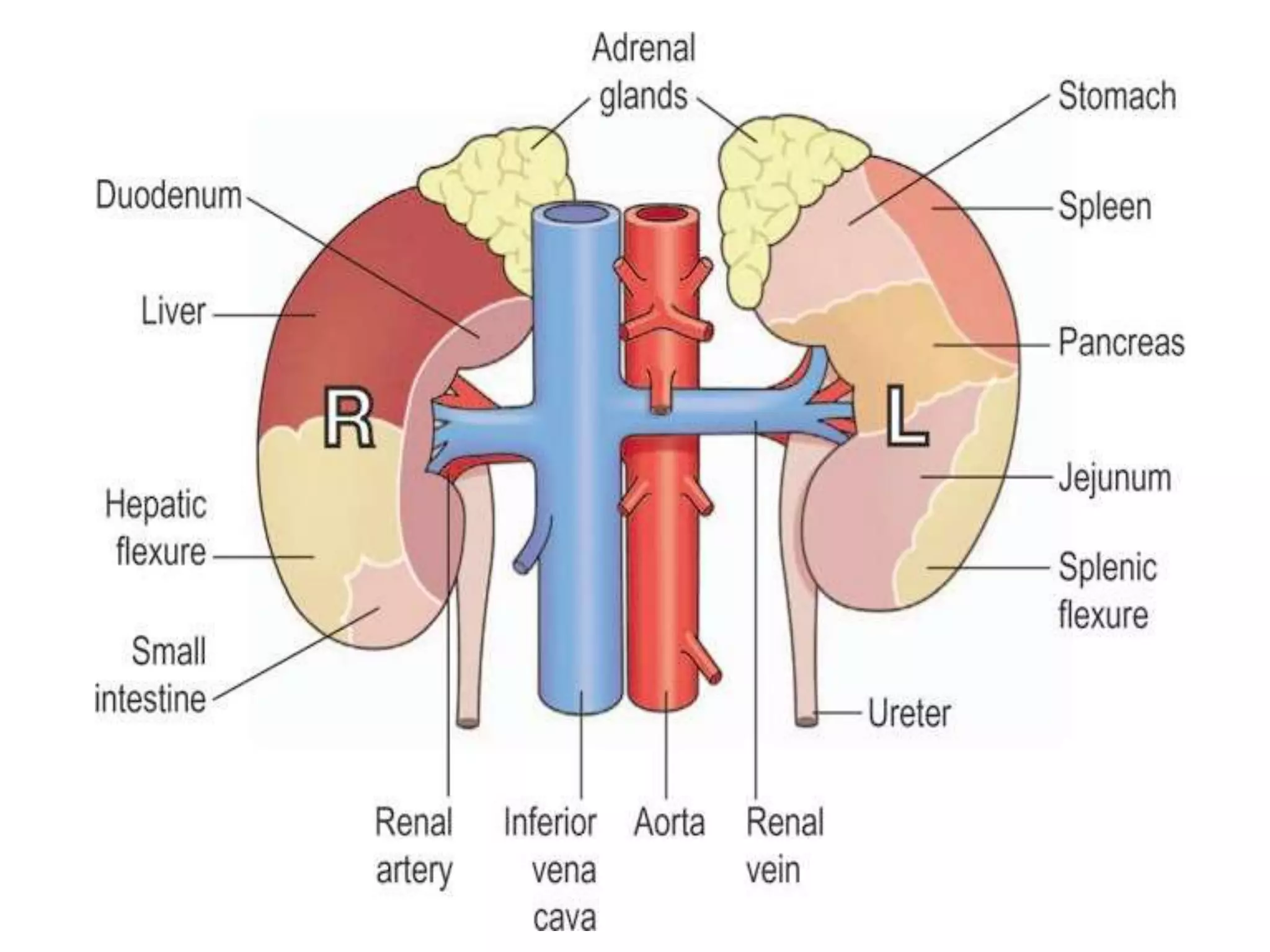
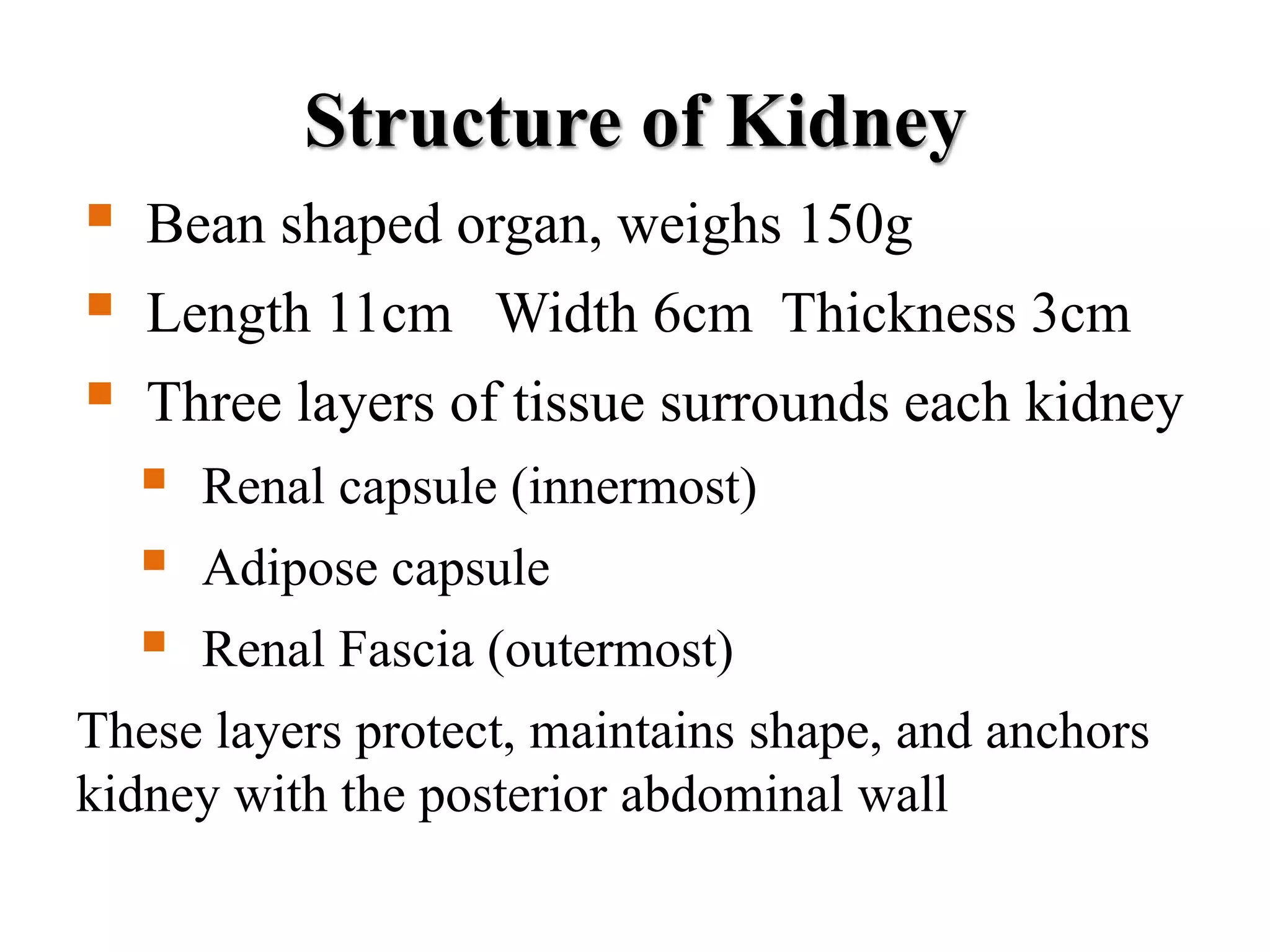
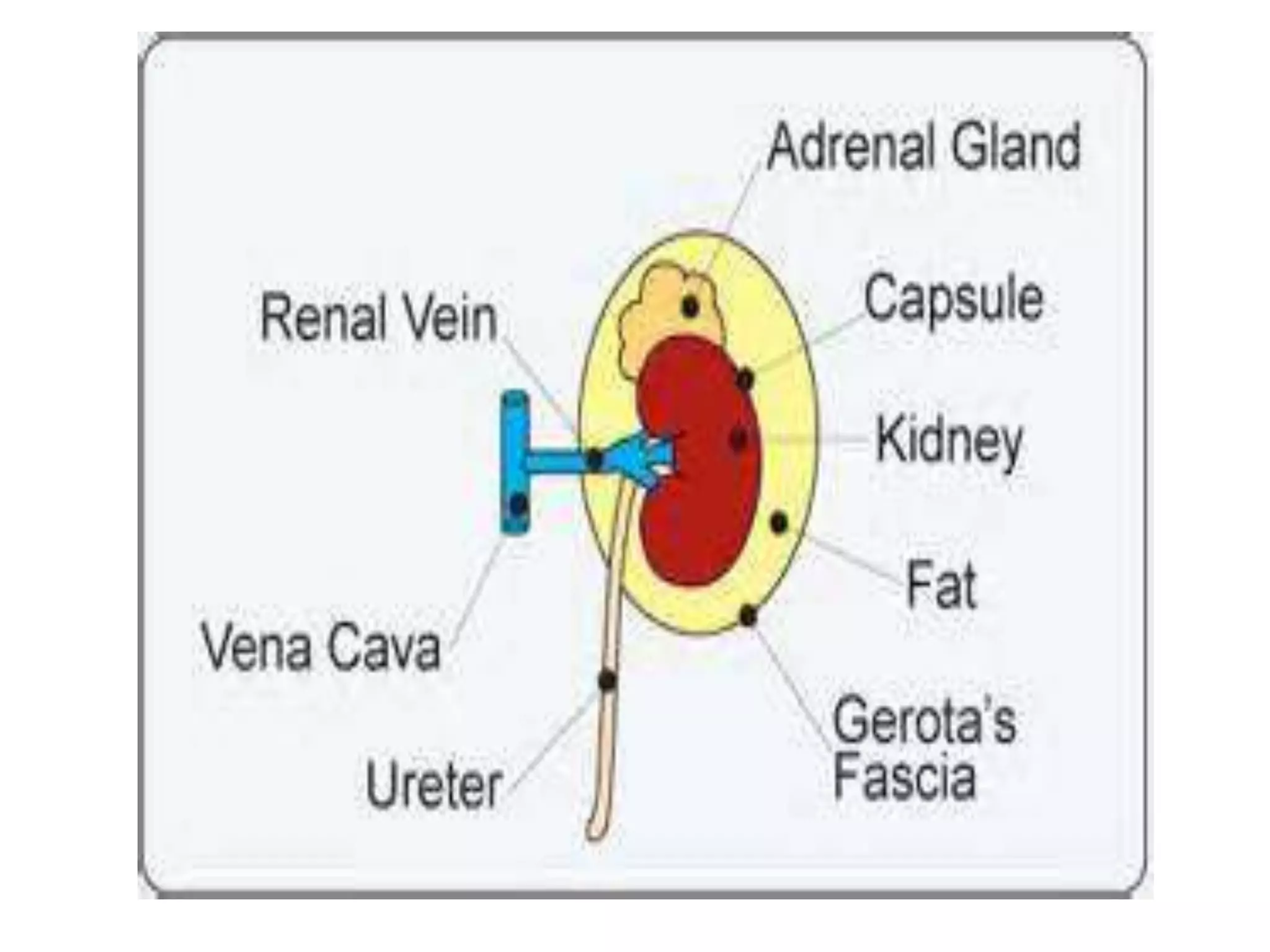
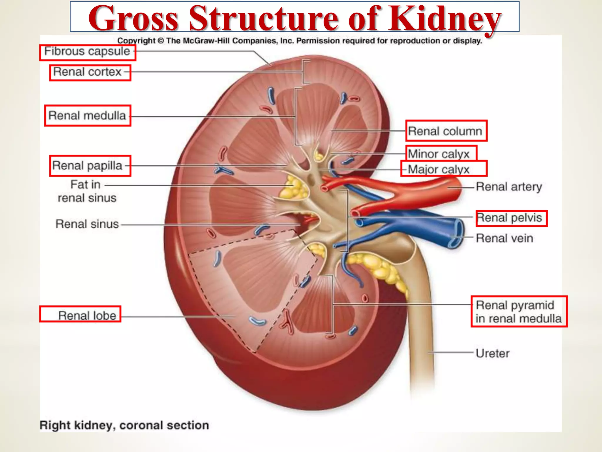
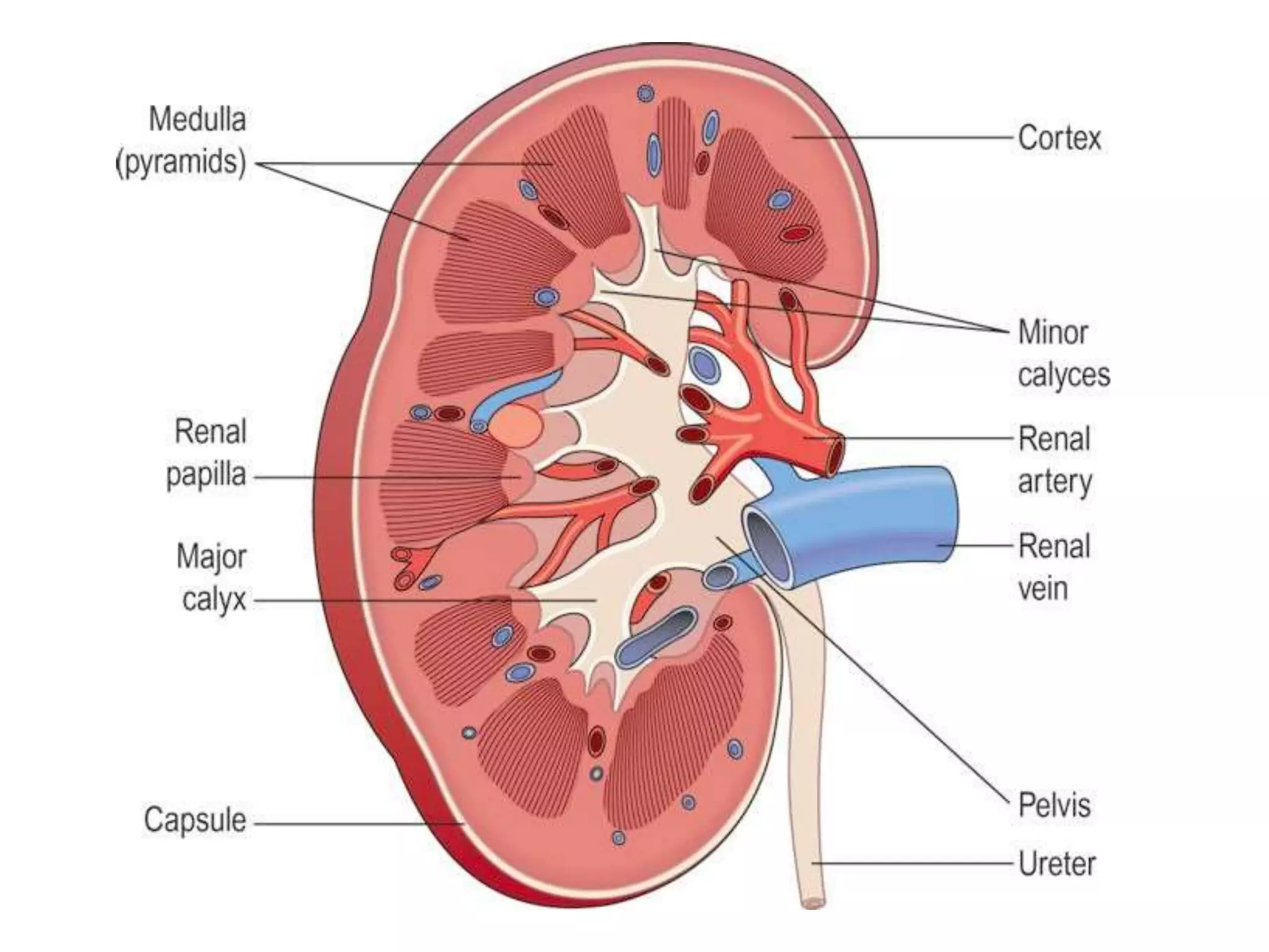
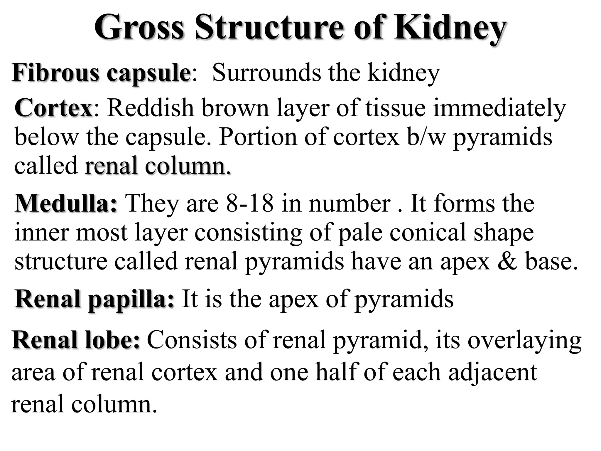
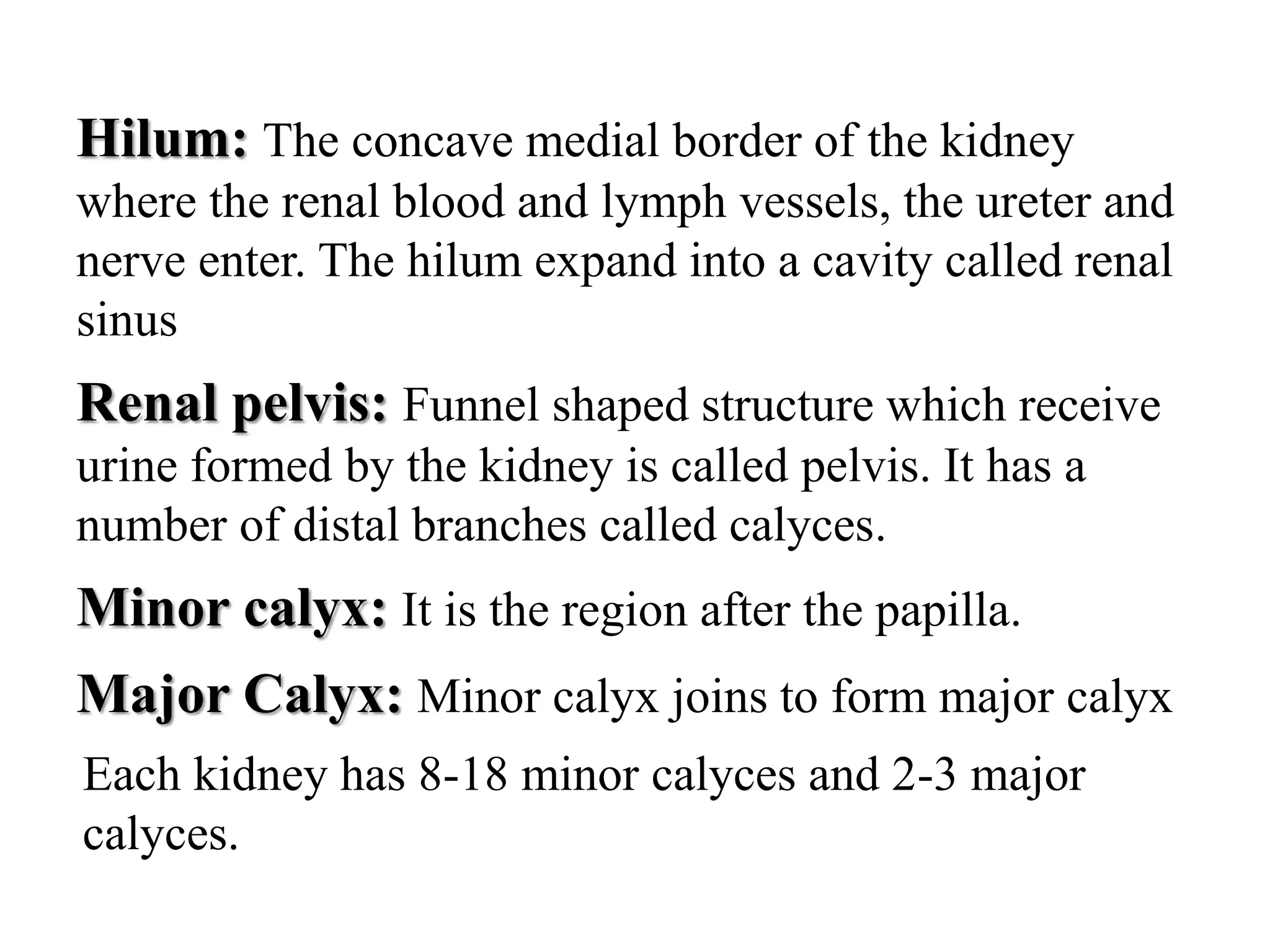

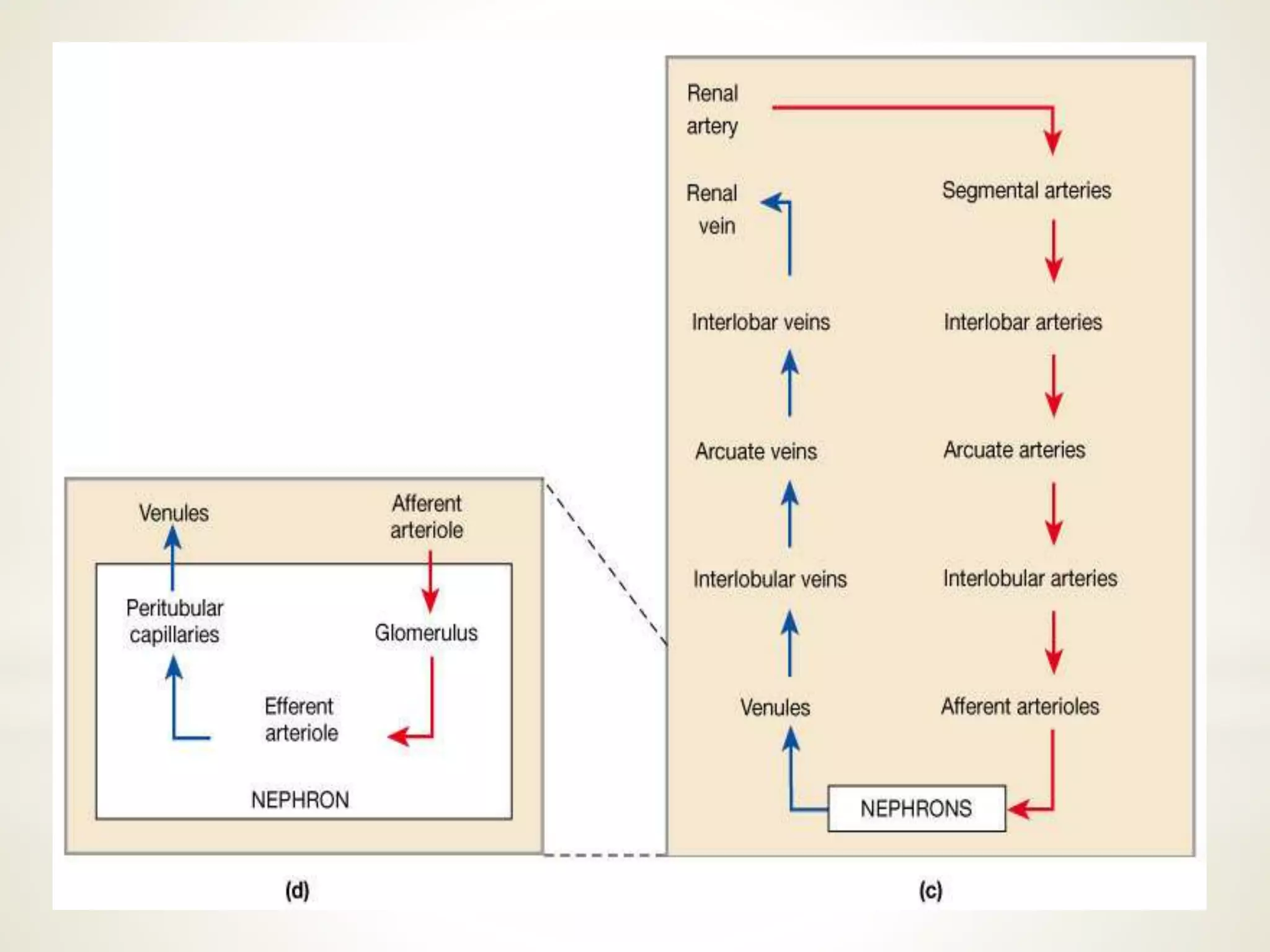

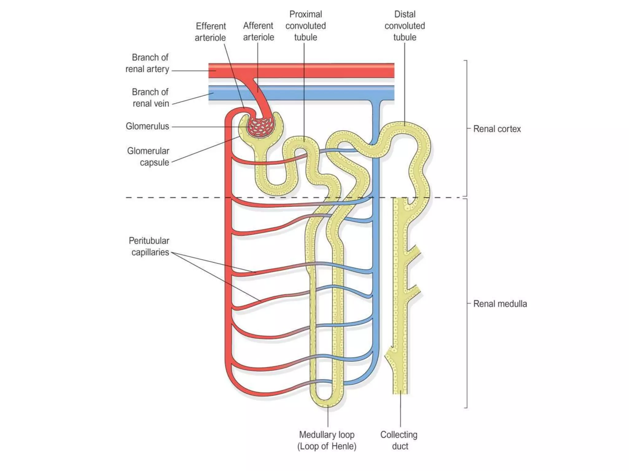
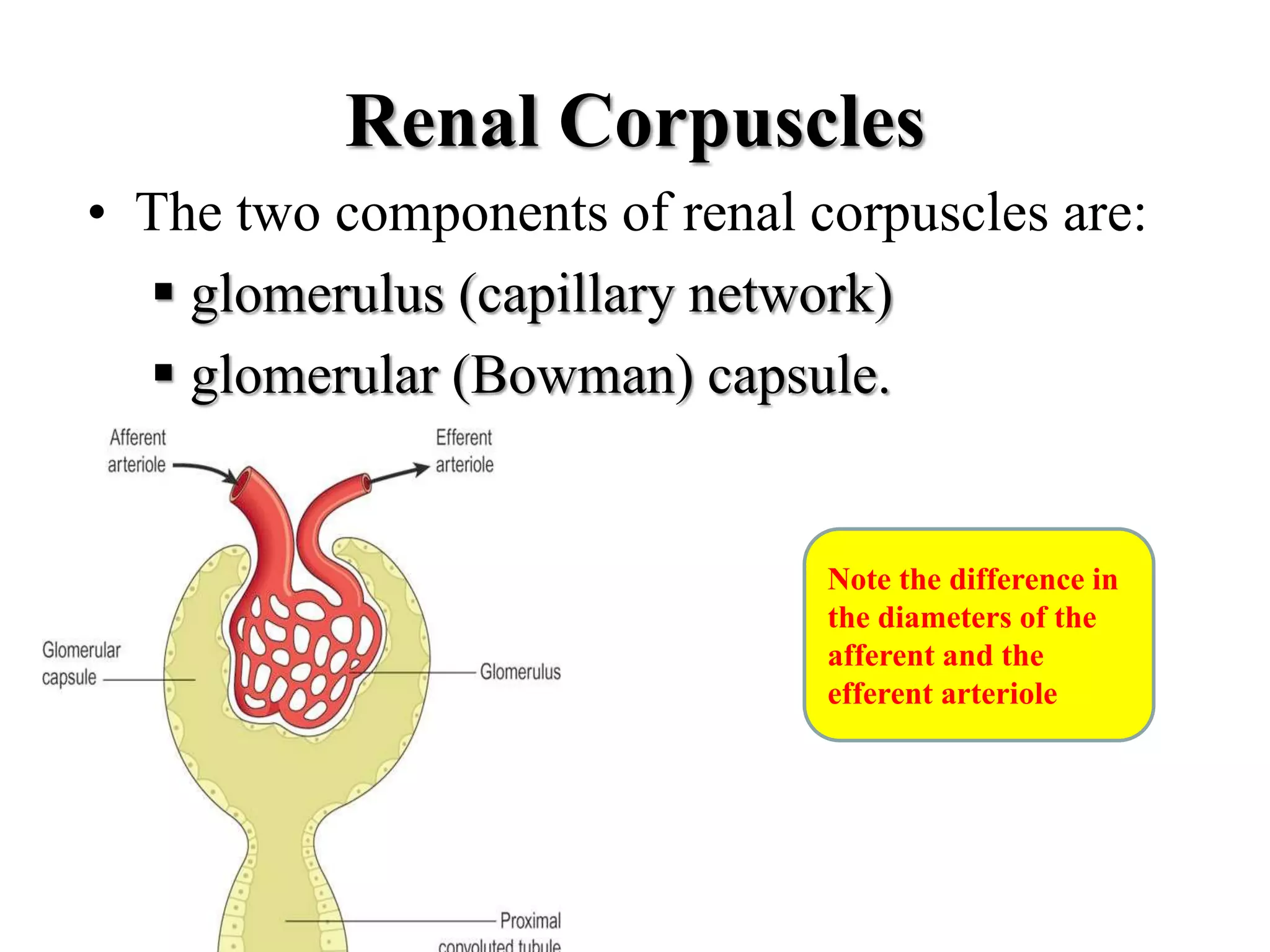
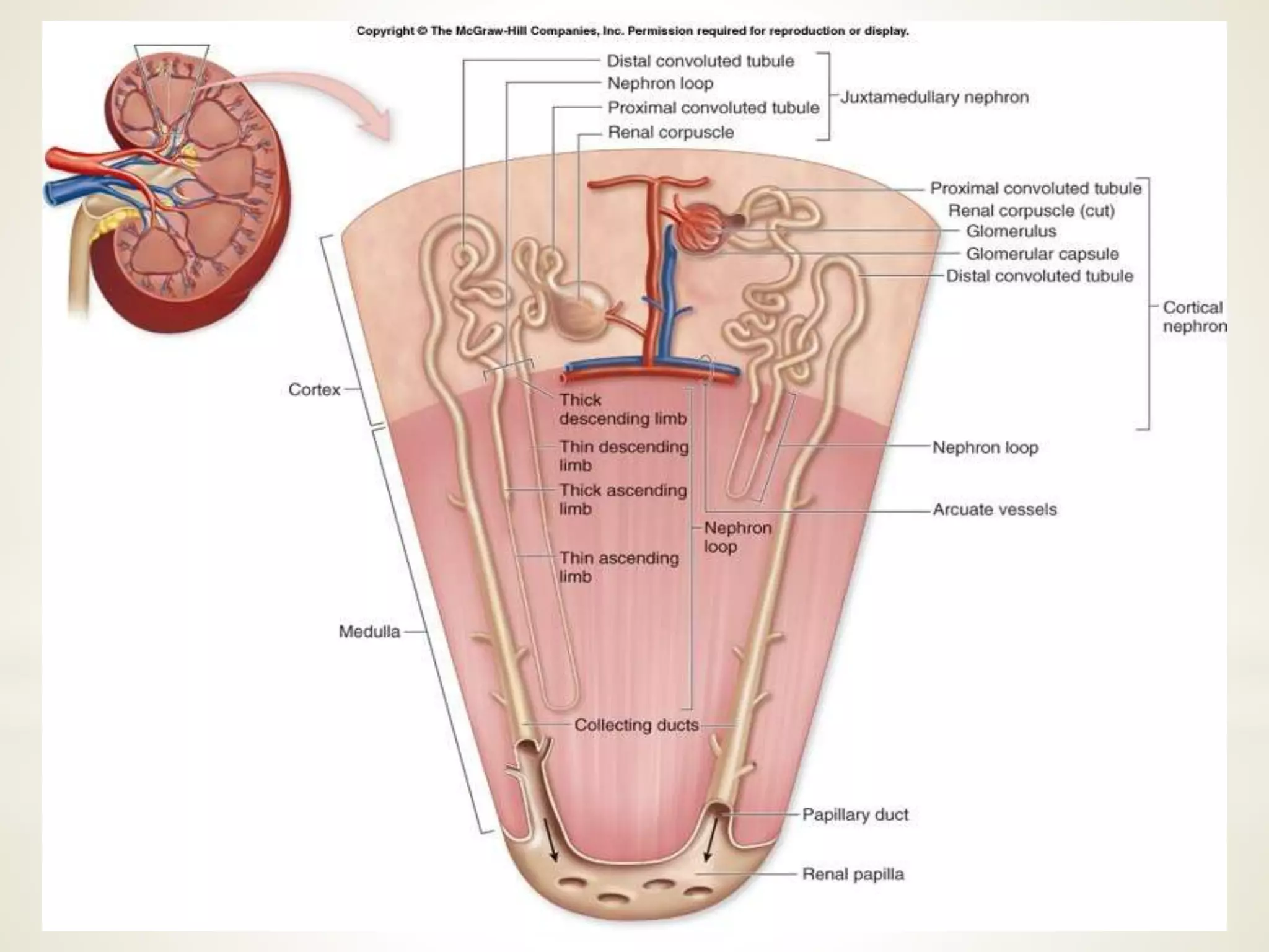
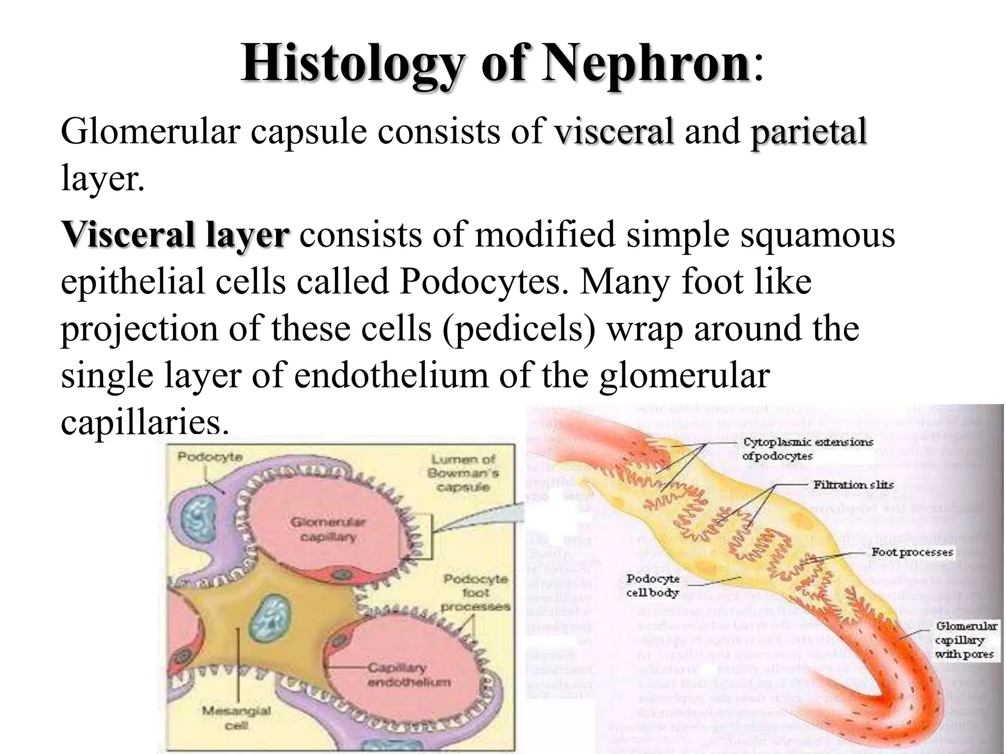
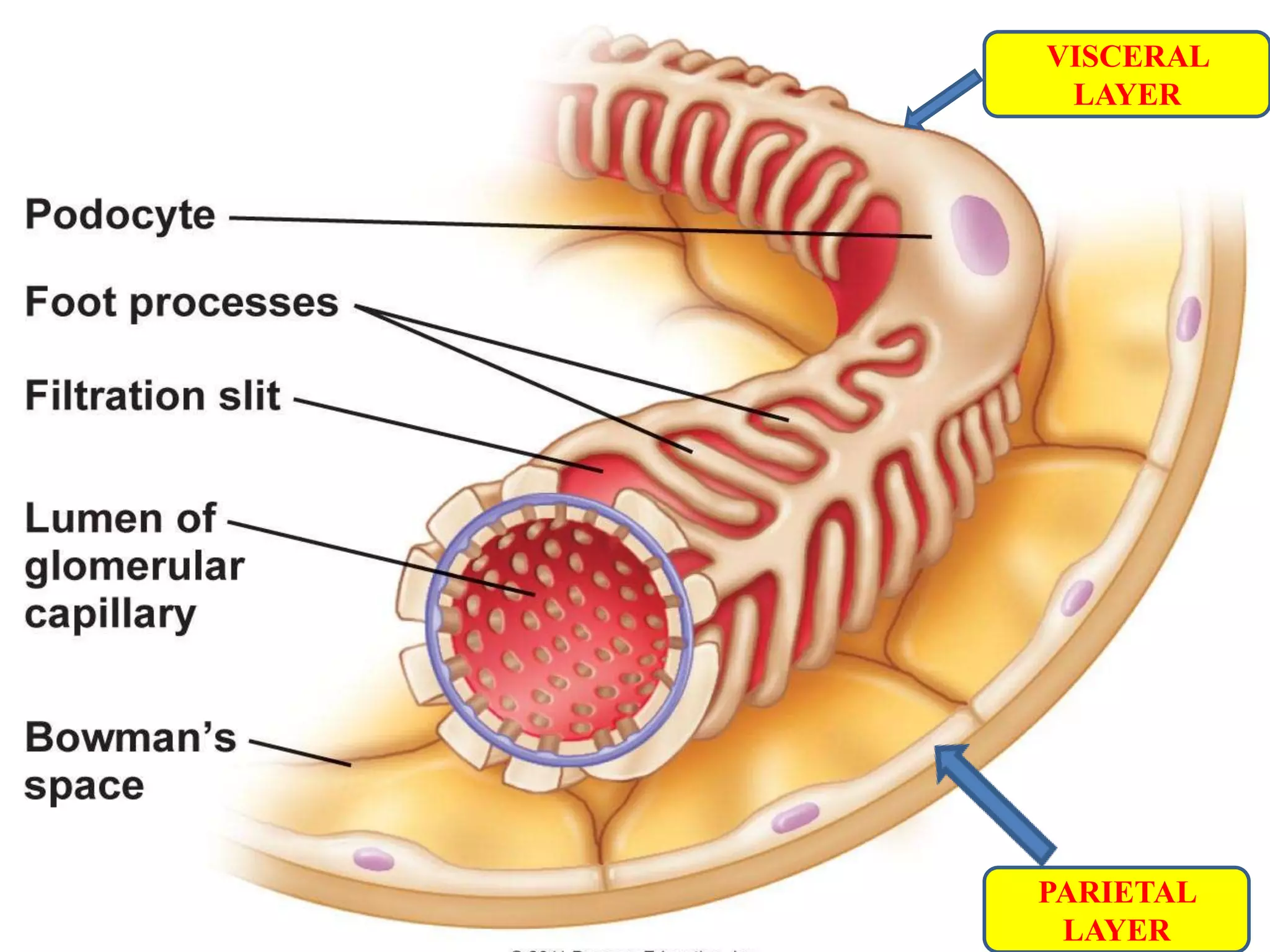
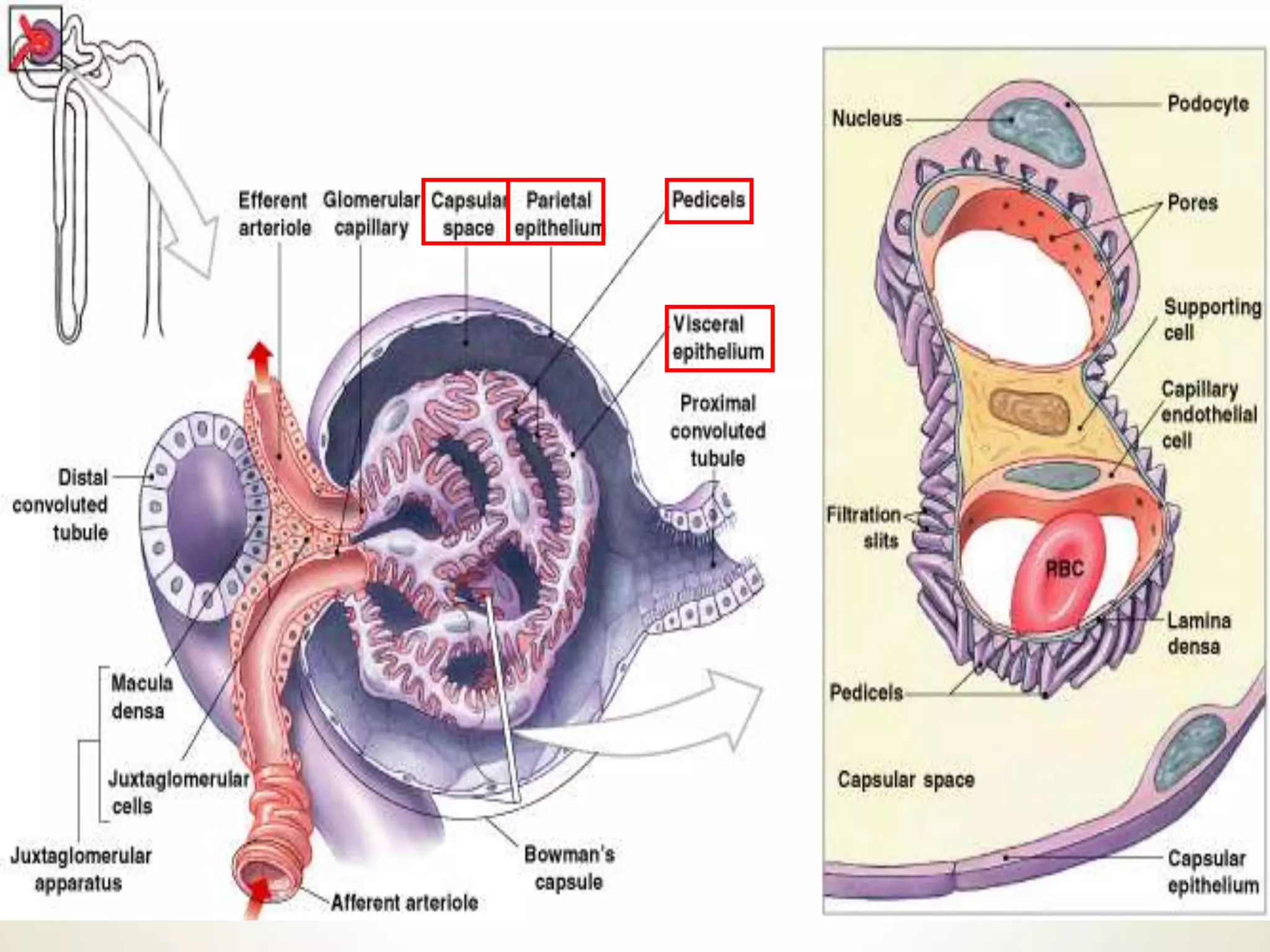
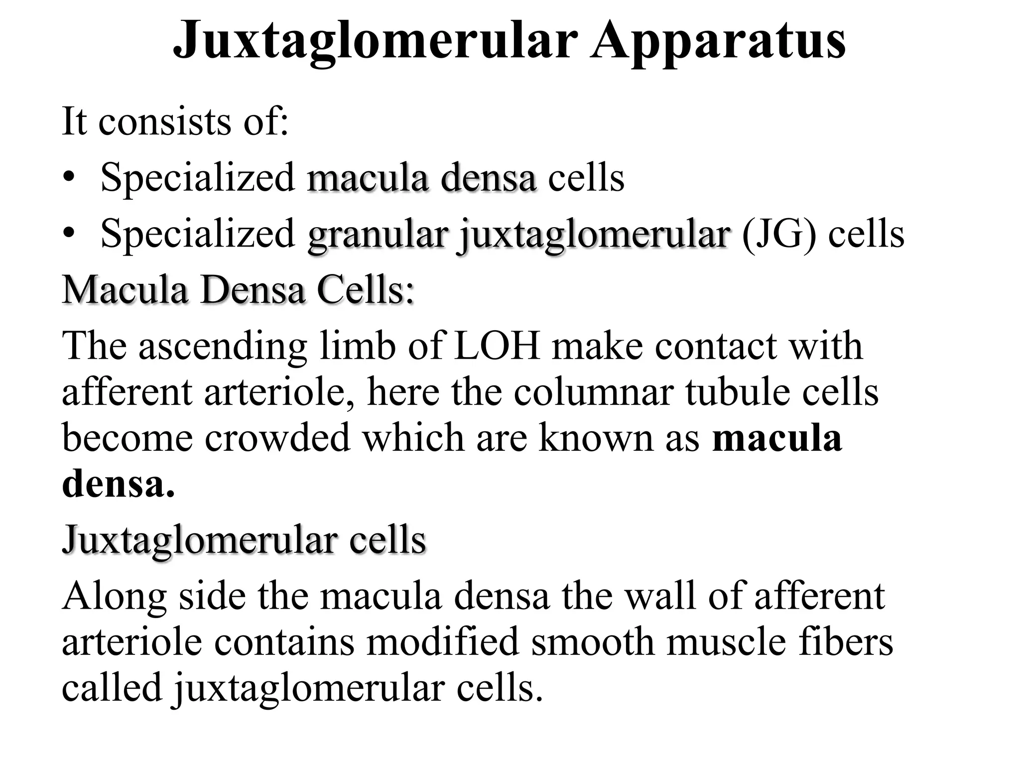


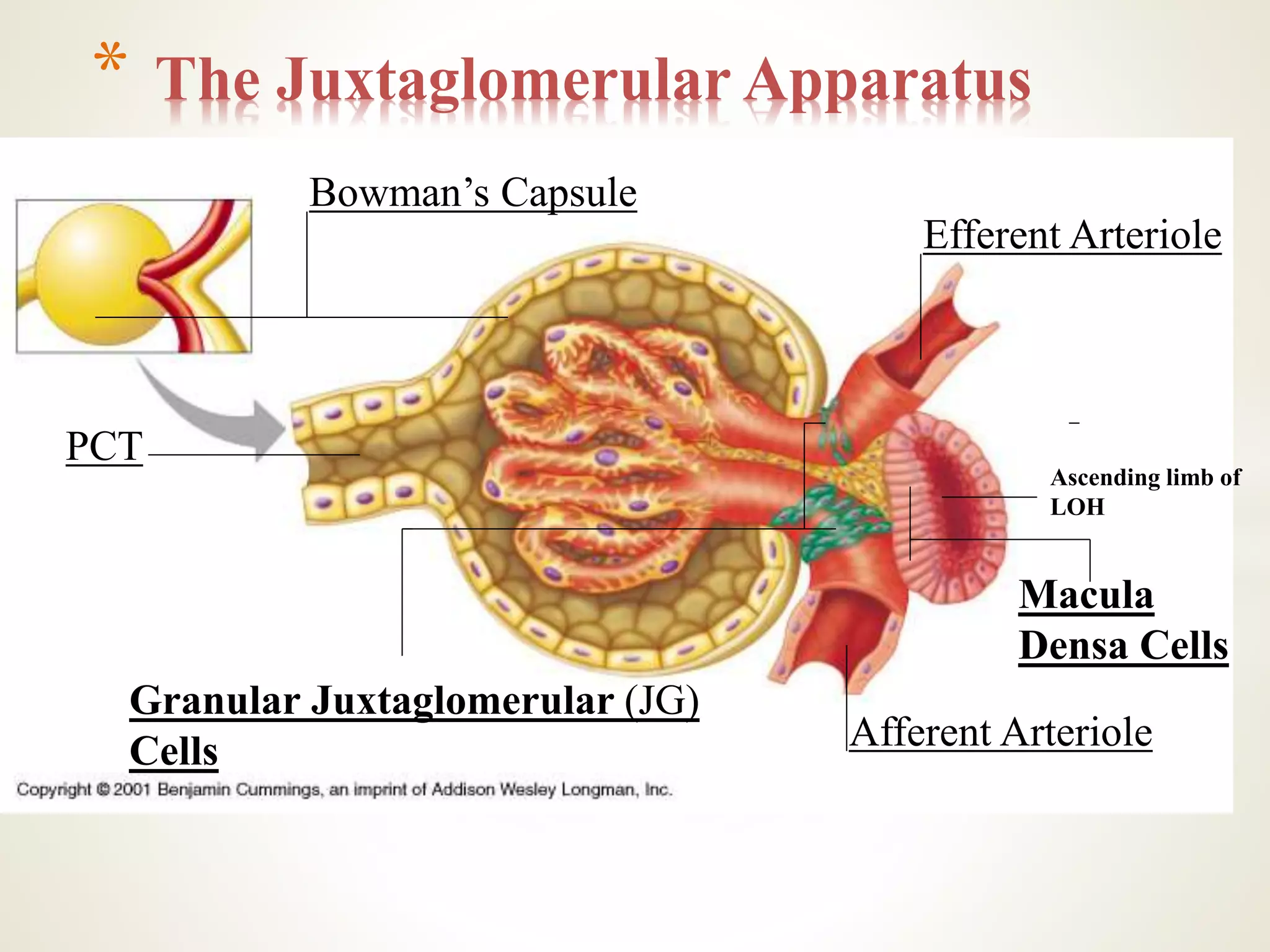

This document discusses the labeling exercise for the micturition reflex and provides an overview of the urinary system. It defines the urinary system and lists its main organs as the kidneys, ureters, urinary bladder, and urethra. It describes the location of the kidneys and provides diagrams of the gross and microscopic kidney structure, labeling components such as the cortex, medulla, nephrons, renal corpuscles, and tubules. It also briefly explains the roles of filtration, reabsorption, and secretion in urine formation and discusses homeostasis functions of the kidneys.