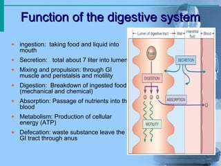
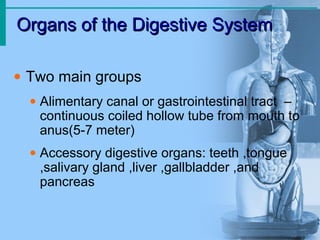
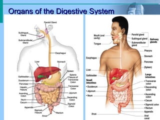
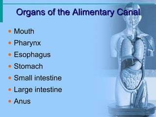
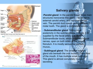
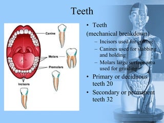
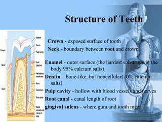
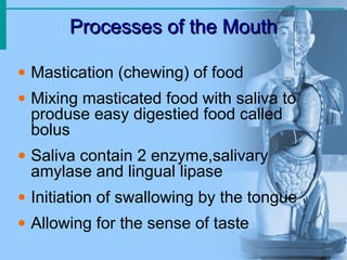
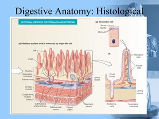
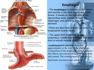
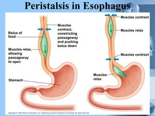
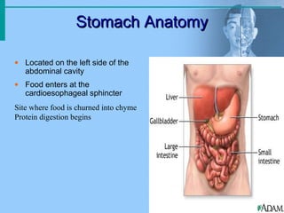
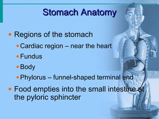
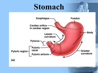
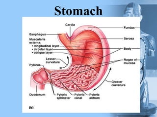
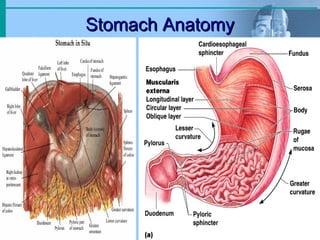
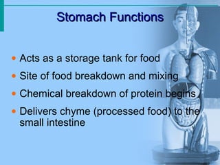
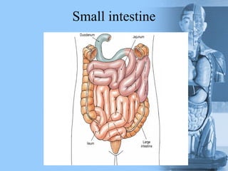
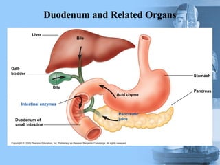
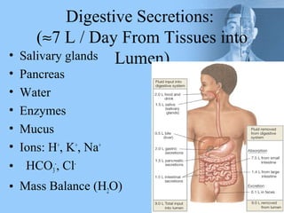
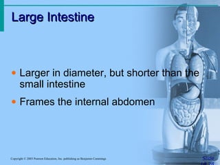
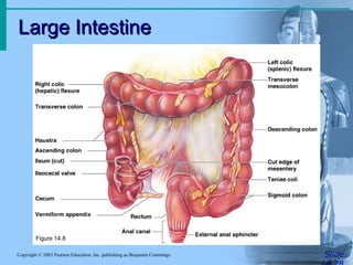
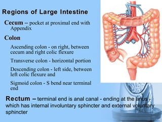
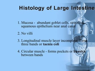
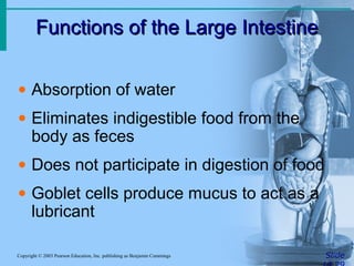
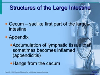
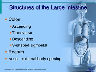
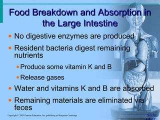
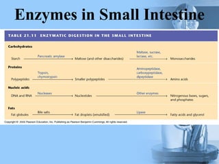
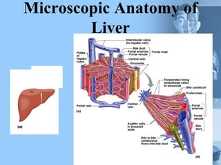
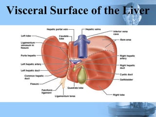
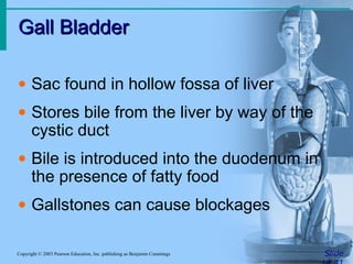
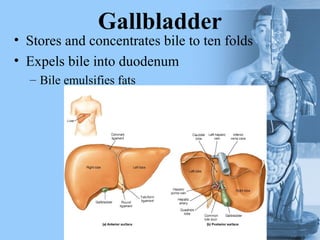
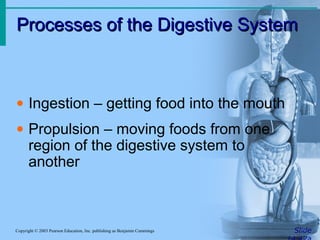
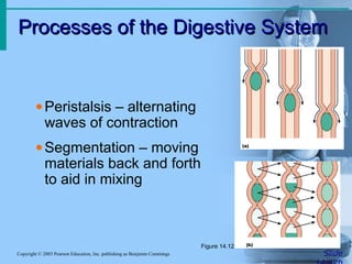
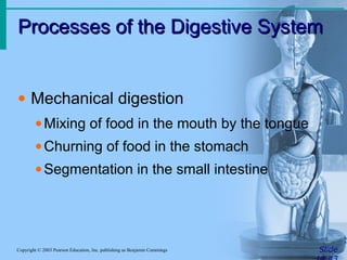
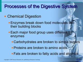
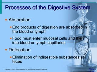
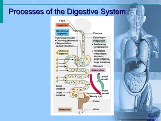
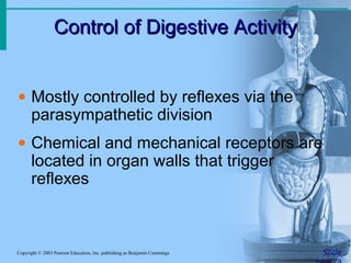
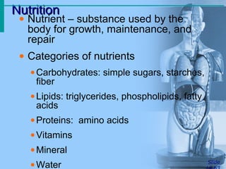
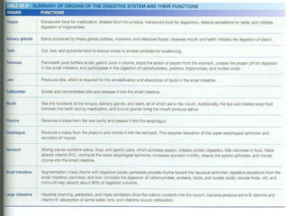
The digestive system breaks down ingested food into nutrients that can be absorbed and used by the body. It consists of the alimentary canal and accessory organs. The alimentary canal includes the mouth, esophagus, stomach, small intestine, large intestine, and anus. Accessory organs include the teeth, tongue, salivary glands, liver, gallbladder and pancreas. Food is ingested, digested, absorbed, and waste is eliminated in a multi-step process involving both mechanical and chemical breakdown as well as nutrient absorption throughout the alimentary canal.
The urinary system, also known as the renal system or urinary tract, consists of the kidneys, ureters, bladder, and the urethra. The purpose of the urinary system is to eliminate waste from the body, regulate blood volume and blood pressure, control levels of electrolytes and metabolites, and regulate blood pH.
In humans, the respiratory tract is the part of the anatomy of the respiratory system involved with the process of respiration. Air is breathed in through the nose or the mouth. In the nasal cavity, a layer of mucous membrane acts as a filter and traps pollutants and other harmful substances found in the air.
Digestion is the breakdown of large insoluble food molecules into small water-soluble food molecules so that they can be absorbed into the watery blood plasma. In certain organisms, these smaller substances are absorbed through the small intestine into the blood stream.
This PowerPoint presentation details out the anatomy of the human digestive system. Their are general terminologies that involves the topic but over-all this work focuses on how digestion takes place in the human body. The details coming from this presentation are combined from four different and liable sources/references including Biology (Thomson Asian Edition). I can say that this presentation is brief and well-organized so I hope this could help you in your class or seminars. Thanks.
An overview of the GIT with detailed study of the organs, along with their anatomy and physiology. It will find u easier to go through this complex function within our body.
Announcement about my previous presentations - Thank youAreej Abu Hanieh
ANNOUNCEMENT Thank you for all of you, my followers who sent me messages with a lot of love and appreciations, I finally graduated after 6 years of studying in Birzeit University , In doctor of Pharmacy department I hope all of you benefited from all the presentations posted before Thank you a new PharmD GraduatedAreej ^^
Observation of Io’s Resurfacing via Plume Deposition Using Ground-based Adapt...Sérgio Sacani
Since volcanic activity was first discovered on Io from Voyager images in 1979, changes
on Io’s surface have been monitored from both spacecraft and ground-based telescopes.
Here, we present the highest spatial resolution images of Io ever obtained from a groundbased telescope. These images, acquired by the SHARK-VIS instrument on the Large
Binocular Telescope, show evidence of a major resurfacing event on Io’s trailing hemisphere. When compared to the most recent spacecraft images, the SHARK-VIS images
show that a plume deposit from a powerful eruption at Pillan Patera has covered part
of the long-lived Pele plume deposit. Although this type of resurfacing event may be common on Io, few have been detected due to the rarity of spacecraft visits and the previously low spatial resolution available from Earth-based telescopes. The SHARK-VIS instrument ushers in a new era of high resolution imaging of Io’s surface using adaptive
optics at visible wavelengths.
Comparing Evolved Extractive Text Summary Scores of Bidirectional Encoder Rep...University of Maribor
Slides from:
11th International Conference on Electrical, Electronics and Computer Engineering (IcETRAN), Niš, 3-6 June 2024
Track: Artificial Intelligence
https://www.etran.rs/2024/en/home-english/
Richard's entangled aventures in wonderlandRichard Gill
Since the loophole-free Bell experiments of 2020 and the Nobel prizes in physics of 2022, critics of Bell's work have retreated to the fortress of super-determinism. Now, super-determinism is a derogatory word - it just means "determinism". Palmer, Hance and Hossenfelder argue that quantum mechanics and determinism are not incompatible, using a sophisticated mathematical construction based on a subtle thinning of allowed states and measurements in quantum mechanics, such that what is left appears to make Bell's argument fail, without altering the empirical predictions of quantum mechanics. I think however that it is a smoke screen, and the slogan "lost in math" comes to my mind. I will discuss some other recent disproofs of Bell's theorem using the language of causality based on causal graphs. Causal thinking is also central to law and justice. I will mention surprising connections to my work on serial killer nurse cases, in particular the Dutch case of Lucia de Berk and the current UK case of Lucy Letby.
Slide 1: Title Slide
Extrachromosomal Inheritance
Slide 2: Introduction to Extrachromosomal Inheritance
Definition: Extrachromosomal inheritance refers to the transmission of genetic material that is not found within the nucleus.
Key Components: Involves genes located in mitochondria, chloroplasts, and plasmids.
Slide 3: Mitochondrial Inheritance
Mitochondria: Organelles responsible for energy production.
Mitochondrial DNA (mtDNA): Circular DNA molecule found in mitochondria.
Inheritance Pattern: Maternally inherited, meaning it is passed from mothers to all their offspring.
Diseases: Examples include Leber’s hereditary optic neuropathy (LHON) and mitochondrial myopathy.
Slide 4: Chloroplast Inheritance
Chloroplasts: Organelles responsible for photosynthesis in plants.
Chloroplast DNA (cpDNA): Circular DNA molecule found in chloroplasts.
Inheritance Pattern: Often maternally inherited in most plants, but can vary in some species.
Examples: Variegation in plants, where leaf color patterns are determined by chloroplast DNA.
Slide 5: Plasmid Inheritance
Plasmids: Small, circular DNA molecules found in bacteria and some eukaryotes.
Features: Can carry antibiotic resistance genes and can be transferred between cells through processes like conjugation.
Significance: Important in biotechnology for gene cloning and genetic engineering.
Slide 6: Mechanisms of Extrachromosomal Inheritance
Non-Mendelian Patterns: Do not follow Mendel’s laws of inheritance.
Cytoplasmic Segregation: During cell division, organelles like mitochondria and chloroplasts are randomly distributed to daughter cells.
Heteroplasmy: Presence of more than one type of organellar genome within a cell, leading to variation in expression.
Slide 7: Examples of Extrachromosomal Inheritance
Four O’clock Plant (Mirabilis jalapa): Shows variegated leaves due to different cpDNA in leaf cells.
Petite Mutants in Yeast: Result from mutations in mitochondrial DNA affecting respiration.
Slide 8: Importance of Extrachromosomal Inheritance
Evolution: Provides insight into the evolution of eukaryotic cells.
Medicine: Understanding mitochondrial inheritance helps in diagnosing and treating mitochondrial diseases.
Agriculture: Chloroplast inheritance can be used in plant breeding and genetic modification.
Slide 9: Recent Research and Advances
Gene Editing: Techniques like CRISPR-Cas9 are being used to edit mitochondrial and chloroplast DNA.
Therapies: Development of mitochondrial replacement therapy (MRT) for preventing mitochondrial diseases.
Slide 10: Conclusion
Summary: Extrachromosomal inheritance involves the transmission of genetic material outside the nucleus and plays a crucial role in genetics, medicine, and biotechnology.
Future Directions: Continued research and technological advancements hold promise for new treatments and applications.
Slide 11: Questions and Discussion
Invite Audience: Open the floor for any questions or further discussion on the topic.
Introduction:
RNA interference (RNAi) or Post-Transcriptional Gene Silencing (PTGS) is an important biological process for modulating eukaryotic gene expression.
It is highly conserved process of posttranscriptional gene silencing by which double stranded RNA (dsRNA) causes sequence-specific degradation of mRNA sequences.
dsRNA-induced gene silencing (RNAi) is reported in a wide range of eukaryotes ranging from worms, insects, mammals and plants.
This process mediates resistance to both endogenous parasitic and exogenous pathogenic nucleic acids, and regulates the expression of protein-coding genes.
What are small ncRNAs?
micro RNA (miRNA)
short interfering RNA (siRNA)
Properties of small non-coding RNA:
Involved in silencing mRNA transcripts.
Called “small” because they are usually only about 21-24 nucleotides long.
Synthesized by first cutting up longer precursor sequences (like the 61nt one that Lee discovered).
Silence an mRNA by base pairing with some sequence on the mRNA.
Discovery of siRNA?
The first small RNA:
In 1993 Rosalind Lee (Victor Ambros lab) was studying a non- coding gene in C. elegans, lin-4, that was involved in silencing of another gene, lin-14, at the appropriate time in the
development of the worm C. elegans.
Two small transcripts of lin-4 (22nt and 61nt) were found to be complementary to a sequence in the 3' UTR of lin-14.
Because lin-4 encoded no protein, she deduced that it must be these transcripts that are causing the silencing by RNA-RNA interactions.
Types of RNAi ( non coding RNA)
MiRNA
Length (23-25 nt)
Trans acting
Binds with target MRNA in mismatch
Translation inhibition
Si RNA
Length 21 nt.
Cis acting
Bind with target Mrna in perfect complementary sequence
Piwi-RNA
Length ; 25 to 36 nt.
Expressed in Germ Cells
Regulates trnasposomes activity
MECHANISM OF RNAI:
First the double-stranded RNA teams up with a protein complex named Dicer, which cuts the long RNA into short pieces.
Then another protein complex called RISC (RNA-induced silencing complex) discards one of the two RNA strands.
The RISC-docked, single-stranded RNA then pairs with the homologous mRNA and destroys it.
THE RISC COMPLEX:
RISC is large(>500kD) RNA multi- protein Binding complex which triggers MRNA degradation in response to MRNA
Unwinding of double stranded Si RNA by ATP independent Helicase
Active component of RISC is Ago proteins( ENDONUCLEASE) which cleave target MRNA.
DICER: endonuclease (RNase Family III)
Argonaute: Central Component of the RNA-Induced Silencing Complex (RISC)
One strand of the dsRNA produced by Dicer is retained in the RISC complex in association with Argonaute
ARGONAUTE PROTEIN :
1.PAZ(PIWI/Argonaute/ Zwille)- Recognition of target MRNA
2.PIWI (p-element induced wimpy Testis)- breaks Phosphodiester bond of mRNA.)RNAse H activity.
MiRNA:
The Double-stranded RNAs are naturally produced in eukaryotic cells during development, and they have a key role in regulating gene expression .
This presentation explores a brief idea about the structural and functional attributes of nucleotides, the structure and function of genetic materials along with the impact of UV rays and pH upon them.
THE IMPORTANCE OF MARTIAN ATMOSPHERE SAMPLE RETURN.Sérgio Sacani
The return of a sample of near-surface atmosphere from Mars would facilitate answers to several first-order science questions surrounding the formation and evolution of the planet. One of the important aspects of terrestrial planet formation in general is the role that primary atmospheres played in influencing the chemistry and structure of the planets and their antecedents. Studies of the martian atmosphere can be used to investigate the role of a primary atmosphere in its history. Atmosphere samples would also inform our understanding of the near-surface chemistry of the planet, and ultimately the prospects for life. High-precision isotopic analyses of constituent gases are needed to address these questions, requiring that the analyses are made on returned samples rather than in situ.
2. Function of the digestive systemFunction of the digestive system
• ingestion: taking food and liquid into
mouth
• Secretion: total about 7 liter into lumen
• Mixing and propulsion: through GI
muscle and peristalsis and motility
• Digestion: Breakdown of ingested food
(mechanical and chemical)
• Absorption: Passage of nutrients into the
blood
• Metabolism: Production of cellular
energy (ATP)
• Defecation: waste substance leave the
GI tract through anus
3. Organs of the Digestive SystemOrgans of the Digestive System
• Two main groups
• Alimentary canal or gastrointestinal tract –
continuous coiled hollow tube from mouth to
anus(5-7 meter)
• Accessory digestive organs: teeth ,tongue
,salivary gland ,liver ,gallbladder ,and
pancreas
4. Organs of the Digestive SystemOrgans of the Digestive System
5. Organs of the Alimentary CanalOrgans of the Alimentary Canal
• Mouth
• Pharynx
• Esophagus
• Stomach
• Small intestine
• Large intestine
• Anus
6. Mouth (Oral Cavity) AnatomyMouth (Oral Cavity) Anatomy
• Lips (labia) – protect
the anterior opening
• Cheeks – form the
lateral walls
• Hard palate – forms
the anterior roof
• Soft palate – forms
the posterior roof
• Uvula – fleshy
projection of the
soft palate
7. Mouth (Oral Cavity) AnatomyMouth (Oral Cavity) Anatomy
• Vestibule – space
between lips
externally and teeth
and gums internally
• Oral cavity – area
contained by the
teeth
• Tongue – attached
at hyoid bone and
styloid processes of
the skull, and by the
lingual frenulum
8. TongueTongue
• Dorsum (upper part of
tongue covered with papillae
taste receptor and buds)
• filiform papillae
• fungiform papillae
• circumvallate papillae
• Paltine tonsil and
lingual tonsil
9. Salivary glands
-Parotid gland: In the parotid fossa, three main
structures transverse this gland – facial nerve,
external carotid artery, and retromandibular
vein. The parotid duct opens near the upper 2nd
molar tooth. The gland is completely serous.
- Submandibular gland: Sitting most
posteriorly in the submandibular triangle, it is
supplied by the facial artery and vein.
Submandibular ducts, which cross the lingual
nerves, open on both sides of the tongue
frenulum. It is mostly serous but partially
mucus,.
- Sublingual gland: The smallest salivary
gland sits beneath the oral mucosa in the floor
of the mouth. It has multiple small openings.
This gland is almost completely mucus-
secreting.
10. Teeth
• Teeth
(mechanical breakdown)
– Incisors used for cutting
– Canines used for stabbing
and holding
– Molars large surface area
used for grinding
• Primary or deciduous
teeth 20
• Secondary or permanent
teeth 32
11. Structure of Teeth
Crown - exposed surface of tooth
Neck - boundary between root and crown
Enamel - outer surface (the hardest substance in the
body 95% calcium salts)
Dentin – bone-like, but noncellular(70% calcium
salts)
Pulp cavity - hollow with blood vessels and nerves
Root canal - canal length of root
gingival sulcus - where gum and tooth meet
12. Processes of the MouthProcesses of the Mouth
• Mastication (chewing) of food
• Mixing masticated food with saliva to
produse easy digestied food called
bolus
• Saliva contain 2 enzyme,salivary
amylase and lingual lipase
• Initiation of swallowing by the tongue
• Allowing for the sense of taste
13. Layers of Alimentary Canal OrgansLayers of Alimentary Canal Organs
• Submucosa
•Just beneath the mucosa
•Soft connective tissue with blood vessels,
nerve endings, and lymphatics also contain
submucosal plexus
14. Layers of Alimentary Canal OrgansLayers of Alimentary Canal Organs
• Mucosa
• Innermost layer
• Moist membrane
1. Surface epithelium : secretion and
absorbtion,renew every 5-7 days also
contain enteroendocrine cells
2. Small amount of connective tissue
(lamina propria): contain blood and
lymphatic vessele also contain MALT
3. Small smooth muscle layer
15. Layers of Alimentary Canal OrgansLayers of Alimentary Canal Organs
• Muscularis externa – smooth muscle
1. Inner circular layer
2. Outer longitudinal layer
Between them is myenteric plexus
• Serosa
• Outermost layer – visceral peritoneum
• Layer of serous fluid-producing cells
(mesothelium)
18. Pharynx AnatomyPharynx Anatomy
• Nasopharynx –
not part of the
digestive system
• Oropharynx –
posterior to oral
cavity
• Laryngopharynx –
below the oropharynx
and connected to
the esophagus
19. Pharynx FunctionPharynx Function
• Serves as a passageway for air and
food
• Food is propelled to the esophagus by
two muscle layers
•Longitudinal inner layer
•Circular outer layer
• Food movement is by alternating
contractions of the muscle layers
(peristalsis)
20. EsophagusEsophagus
• Runs from pharynx to stomach through
the diaphragm( 25 cm)
• Conducts food by peristalsis
(slow rhythmic squeezing): contraction
of circular layer above the food and
contraction of longitudinal below the
food
• Passageway for food only (respiratory
system branches off after the pharynx)
21. Esophagus
- The esophagus is posterior to the larynx
and trachea in the neck region and upper
thorax. It travels on the right side of the
descending aorta, passes through the
diaphragm, and connects with the
stomach.
-There are also inner circular and outer
longitudinal muscle layers.
- The upper third is skeletal muscle
(voluntary), middle third is mixed, and lower
third is smooth muscle (involuntary).
-esophagogastric junction is located
approximately at the level of the diaphragm.
Contractions of the diaphragm create sphincter-
like effects, preventing reflux of stomach acids
and content. The esophagogastric junction is a
functional, not anatomical, sphincter.
22. Peristalsis in Esophagus
Bolus of
food
Muscles relax,
allowing
passageway
to open
Stomach
Muscles
contract,
constricting
passageway
and pushing
bolus down
Muscles
relax
Muscles contract
Muscles relax
Muscles contract
23. Stomach AnatomyStomach Anatomy
• Located on the left side of the
abdominal cavity
• Food enters at the
cardioesophageal sphincter
Site where food is churned into chyme
Protein digestion begins
24. Stomach AnatomyStomach Anatomy
• Regions of the stomach
•Cardiac region – near the heart
•Fundus
•Body
•Phylorus – funnel-shaped terminal end
• Food empties into the small intestine at
the pyloric sphincter
28. Stomach AnatomyStomach Anatomy
• Layers of peritoneum attached to the
stomach
•Lesser omentum – attaches the liver to the
lesser curvature
•Greater omentum – attaches the greater
curvature to the transverse colon which
Contains fat to insulate, cushion, and
protect abdominal organs
31. Stomach FunctionsStomach Functions
• Acts as a storage tank for food
• Site of food breakdown and mixing
• Chemical breakdown of protein begins
• Delivers chyme (processed food) to the
small intestine
32. Specialized Mucosa of theSpecialized Mucosa of the
StomachStomach
• Simple columnar epithelium
• Mucous neck cells – produce a sticky
alkaline mucus
• Gastric glands – secrete gastric juice
• Chief cells – produce protein-digesting
enzymes (pepsinogens)
• Parietal cells – produce hydrochloric acid
and Intrinsic factor(B12 absorption)
• Endocrine cells (G cell) – produce gastrin
which stimulates both parietal and chief cells)
33. Structure of the Stomach MucosaStructure of the Stomach Mucosa
• Gastric pits
formed by
folded mucosa
• Glands and
specialized
cells are in the
gastric gland
region
35. Peritoneum
• Is the largest serous membrane of the body consist of
mesothelium
• Divide into
1. Parietal peritoneum: lines the wall of abdominopelvic
cavity internally
2. Visceral peritoneum: cover some oh the organs in the
cavity
3. The space between them contain fluid and called
peritoneal cavity this cavity may be accumulated by
several liters of fluid state called ascites
36. MembranesMembranes
Mesenteries - double sheets of peritoneum, surrounding and
suspending portions of the digestive
organs
Peritoneal folds
1. falciform ligament:- attach the liver to anterior abdominal
wall and diaphragm
2. Greater omentum - "fatty apron", hangs anteriorly from
stomach, double layer encloses fat
3. Lesser omentum - between stomach and liver
4. Mesentery proper - suspends and wraps the small intestine
5. Mesocolon - suspends and wraps the colon, parts are
i. transverse mesocolon
ii. sigmoid mesocolon
• Ascending and descending ,pancreas, first 2 parts of the
duodenum and kidneys are Retroperitoneal structure
40. Small IntestineSmall Intestine
• The body’s major digestive organ
• Site of nutrient absorption into the blood
• Muscular tube extending form the
pyloric sphincter to the ileocecal valve
• Suspended from the posterior
abdominal wall by the mesentery
41. Subdivisions of the Small IntestineSubdivisions of the Small Intestine
• Duodenum(25cm)
•Attached to the stomach
•Curves around the head of the pancreas
•Fixed retroperitoneal structure
• Jejunum (2.5m)
•Attaches anteriorly to the duodenum
• Ileum (3.5m)
•Extends from jejunum to large intestine
44. Duodenum and Related Organs
Liver
Bile
Gall-
bladder
Bile
Duodenum of
small intestine
Acid chyme
Pancreatic
juice
Intestinal enzymes
Stomach
Pancreas
58. CecumCecum –– pocket at proximal end with
Appendix
ColonColon
Ascending colon - on right, between
cecum and right colic flexure
Transverse colon - horizontal portion
Descending colon - left side, between
left colic flexure and
Sigmoid colon - S bend near terminal
end
Regions of Large IntestineRegions of Large Intestine
RectumRectum –– terminal end is anal canal - ending at the anus -
which has internal involuntary sphincter and external voluntary
sphincter
59. 1. Mucosa - abundant goblet cells, stratified
squamous epithelium near anal canal
2. No villi
3. Longitudinal muscle layer incomplete, forms
three bands or taenia coli
4. Circular muscle - forms pockets or haustra
between bands
Histology of Large IntestineHistology of Large Intestine
68. PancreasPancreas
Slide
• Produces a wide spectrum of digestive
enzymes that break down all categories of food
• Enzymes are secreted into the duodenum
• Alkaline fluid introduced with enzymes
neutralizes acidic chyme
• Endocrine products of pancreas (langerhans
island)
•Insulin
•Glucagons
•Somatostatin
71. Composition and Function of
Pancreatic Juice
• Examples include
• Trypsinogen is activated to trypsin
• Procarboxypeptidase is activated to
carboxypeptidase
• Active enzymes secreted
• Amylase, lipases, and nucleases
• These enzymes require ions or bile for
optimal activity
72. • Retroperitoneal :compose
of head, body and tail
• Endocrine and exocrine
gland
• Common bile duct and
major pancreatic duct lead
to ampulla of vater then to
second part of duodenum
through sphincter of oddi
PancreasPancreas
74. Liver
On right under diaphragm,
largest organ made up of 4
lobes (left and right, caudate,
and quadrate)
Hilus (porta hepatis) –
underside "entry" point
Gall bladder
Microscopic anatomy: Liver lobules and triads
89. NutritionNutrition
Slide
• Nutrient – substance used by the
body for growth, maintenance, and
repair
• Categories of nutrients
•Carbohydrates: simple sugars, starches,
fiber
•Lipids: triglycerides, phospholipids, fatty
acids
•Proteins: amino acids
•Vitamins
•Mineral
•Water
Editor's Notes
Masseter muscle zygomatic arch, cranial ridge = carnivore
Herbivore – diastema plate for incisors to press against
Epiglotis prevents food going down the wrong way
Enamel: Calcium phosphates. Hardest biological material produced.
makes bile, stores excess nutrients, makes plasma proteins, detoxifies harmful materials, stores iron & fat soluble vitamins