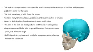
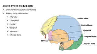
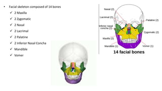
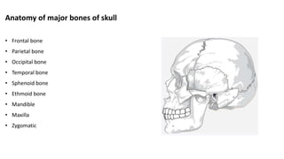
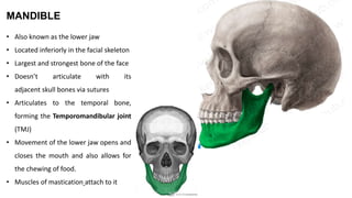
SlideShare a Scribd company logo
SKULL
BY DR AYSHAH HASHIMI
• The skull is a bony structure that forms the head. It supports the structures of the face and provides a
protective cavity for the brain
• The skull is made up of a 22 fused flat bones
• Contains many foramina, fossae, processes, and several cavities or sinuses
• Bones in skull develops from intramembranous ossification
• The joint in the skull are mostly sutures and few are 1° cartilaginous
• Only temporomandibular joint is synovial in nature that permits us to
speak, eat, drink and laugh
• Skull lodges brain, cochlear and vestibular apparatus, retina, olfactory
mucosa and taste buds
Skull is divided into two parts
• Cranium/Braincase/Calvaria/skullcap
• 8 bones forms the cranium
 2 Parietal
 2 Temporal
 Frontal
 Occipital
 Sphenoid
 Ethmoid Bone
Frontal Bone
Parietal Bone
Temporal Bone
Sphenoid
Occipital Bone
Ethmoid
• Facial skeleton composed of 14 bones
 2 Maxilla
 2 Zygomatic
 2 Nasal
 2 Lacrimal
 2 Palatine
 2 Inferior Nasal Concha
 Mandible
 Vomer
Anatomy of major bones of skull
• Frontal bone
• Parietal bone
• Occipital bone
• Temporal bone
• Sphenoid bone
• Ethmoid bone
• Mandible
• Maxilla
• Zygomatic
Frontal Bone
• Creates the smooth curvature of the forehead
• Protects the frontal lobe of the brain
• Involved in the three regions of the head, such are
the squamous part(forehead), the orbital part and
the nasal part
Squamous Part forms the major portion of the bone
and forms the forehead
Orbital part forms the roof of the orbit and ethmoidal
sinus
Nasal part forms the stem of the nose
Squamous Part comprises of
• Frontal sinuses present superior and medially to orbit
• Glabella (elevated surface above the nasal root)
• Zygomatic processes of frontal bone
• Supraorbital foramen
• Supraorbital notch
Frontal sinus
Glabella
Zygomatic processes
Supraorbital foramen
Supraorbital notch
Orbital part includes anterior and posterior ethmoidal foramen
Nasal part comprises of nasal process that’s ends below in a sharp spine
Anterior Ethmoidal Foramen
Posterior Ethmoidal Foramen
Surface Landmarks In Frontal Bone
Externally frontal bone includes- Frontal eminence, superciliary arch, glabella, supraorbital margin(frontal notch,
supraorbital notch), zygomatic process, temporal line, temporal surface
Parietal Bone
• Located on each side of the skull behind the frontal bone
• Irregular quadrilateral in shape- four angles, four margins, and two surfaces
• Angles - frontal angle, sphenoidal angle, occipital angle, mastoid angle,
• Margins/Border- frontal, occipital, squamous, sagittal
• External surface features- parietal tuber, superior temporal line, inferior temporal line, parietal foramen
• Internal surface has grooves for sinuses
Occipital Bone
• The occipital bone is an unpaired bone which covers the back of the head
• Convex externally and concave internally
• Protects cerebellum
• It has four parts: the basilar part, two condylar parts and the squamous part
• All four are arranged around a large opening, the foramen magnum
SQUAMOUS
PART
CONDYLAR
PART
BASILAR
PART
THE BASILAR PART
• Anterior to the foramen magnum
• Anteriorly it fuses with the sphenoid bone to form the clivus
• Pharyngeal tubercle found on the inferior surface of the basilar part
CLIVUS
Condylar Parts
• Located lateral to the foramen magnum
• kidney shaped prominences
• Articulate with the first cervical vertebra (atlanto-occipital joint)
• Posterior to condyles are the condylar canals where the
condylar emissary veins passes
Condylar Canals
Squamous Part
• External occipital protuberance in the midline
• Highest nuchal line
• Superior nuchal line
• Inferior nuchal line
Temporal Bone
• Pair of bilateral
• Many number of openings and canals
• Houses the structures forming the middle and inner ear
• It has 4 parts- squamous, petrous , mastoid and tympanic part
Squamous Part
• Antero-superior portion of the temporal bone
• Zygomatic process- anterior projection from the squamous part of temporal bone (articulate with the
temporal process of the zygomatic bone to form the zygomatic arch)
• Below the process is a mandibular fossa that articulate with the mandible to form temporomandibular joint
• Articular tubercle is an elevation in front of the mandibular fossa
External Acoustic
Meatus
Zygomatic Arch
Zygomatic Process
Mandibular Fossa
Temporomandibular Joint
Articular Tubercle
Mastoid Part
• Most posterior part of the temporal bone
• Downward conical projection called the Mastoid Process
• Mastoid process consists of mastoid air cells (reservoir of air, equalize pressure within the middle ear) and
mastoid antrum
• Various muscles are attached to it (SCM etc. )
• Mastoid notch is present on inferior surface of the mastoid process
Petrous Part
• Pyramid shaped mass of bone located between the sphenoid and occipital bones within the cranial cavity
• It has a base, an apex and three surfaces (anterior, posterior, inferior), three borders (anterior, superior,
posterior)
• The acoustic labyrinth is located within the petrous part (houses the inner ear)
• Trigeminal impression on anterior margin
• Tegmen Tympani forms the roof of the middle ear
Petrous Part
Tympanic Part
• Curved plate immediately below the origin of the zygomatic process
• Forms the anterior wall, floor and part of the posterior wall of the external acoustic meatus
• The styloid process is a narrow, pointed projection that extends downwards and anteriorly from the inferior
surface of the temporal bone
Sphenoid Bone
• Butterfly-shaped
• It consists of a body, paired greater wings and lesser wings, and two pterygoid processes
• Various foramen found- F. Rotundum, F. Ovale, F. Spinosum. Optic Canal and Superior orbital fissure
Body
Greater Wing
Lesser Wing
Pterygoid Processes
Body of Sphenoid Bone
• Six surfaces: superior, posterior, anterior, inferior, and two lateral surfaces
• Superior surface- comprises of Sella turcica that lodge pituitary gland
Lesser wings
• Forms lateral wall of Optic canal (transmit 2nd optic nerve and
ophthalmic artery)
• superior orbital fissure-slit-like’ gap between the lesser and greater
wings (transmit ophthalmic vein and 5thnerve, 6th abducent nerve, 3rd
oculomotor nerve, 4th trochlear nerve)
Greater wings
• Foramen rotundum, which transmits the 5th maxillary nerve
• Foramen ovale, which allows the passage of the 5th
mandibular nerve, accessory meningeal artery, lesser
petrosal nerve and emissary vein (mnemonic "MALE")
• Foramen spinosum
Pterygoid Processes
• Extensions of the basal surface of the sphenoid body
• Processes contain two canals known as the pterygoid canal and pharyngeal canal
• Pterygoid canal transmit major petrosal nerve, deep petrosal nerve and vessels
• Pharyngeal canal transmit pharyngeal nerve
Pterygoid Canal
Ethmoid Bone
• Single porous bone
• The ethmoid bone has four parts:
 Cribriform plate
 Perpendicular plate
 Ethmoidal labyrinth (2)
Ethmoidal labyrinths
Features consists of -
• Ethmoidal Air Cells (Anterior, Middle,
Posterior)
• Orbital Plate
• Superior Nasal Concha
• Middle Nasal Concha
Perpendicular plate
• Forms the nasal septum
Cribriform Plate of Ethmoid
• Forms the roof of the nasal cavity
• Has small openings which transmit the fibers of the olfactory nerves (CN I) from the olfactory epithelium
(nasal cavity) to the brain (cranial cavity)
• A small vertical protrusion on top of the plate is called as Crista galli
MANDIBLE
• Also known as the lower jaw
• Located inferiorly in the facial skeleton
• Largest and strongest bone of the face
• Doesn’t articulate with its
adjacent skull bones via sutures
• Articulates to the temporal bone,
forming the Temporomandibular joint
(TMJ)
• Movement of the lower jaw opens and
closes the mouth and also allows for
the chewing of food.
• Muscles of mastication attach to it
Features
Consist of
• Body
• Ramus
• Foramina
Ramus
• Extends cranially from the angle of the mandible, away from the body at an angle of 110 degrees
• Each ramus contains the following bony landmarks
 Angle of mandible
 Coronoid process
 Mandibular notch
 Condylar process
 Head
 Neck
 Pterygoid fovea
• 2 surfaces
 External - masseteric tuberosity
 Internal- mandibular foramen, mandibular canal, mylohyoid groove, pterygoid tuberosity
Mandibular Notch
Rami
Condylar process
Coronoid process
Body
Alveolar process: holds the teeth
Base
External surface
• Mandibular symphysis: small ridge in the midline
• Mental protuberance: forms the shape of the chin
• Mental tubercle
• Mental foramen (located below the second
premolar)
Internal surface
• Mental spine (attachment of genioglossus and
geniohyoid muscles.)
• Digastric fossa (attachment of digastric muscle)
• Mylohyoid line (mylohyoid muscle)
• Sublingual fovea (contains sublingual gland)
• Submandibular fovea (submandibular gland)
Maxilla Bone
• Upper jaw bone
• Paired bone
Features
• Body
• Four processes
 Frontal process
 Zygomatic process
 Palatine process
 Alveolar process
• Involved in the formation of the orbit,
nose and palate, holds the upper teeth
• Plays an important role for mastication
and communication
Processes
• Frontal process
• Zygomatic process
• Palatine process
• Alveolar process
Frontal process Zygomatic process
Palatine process
Alveolar process
Body
• Central portion of the maxilla
• Four surfaces
 Anterior
 Nasal notch
 Infra-orbital margin
 Infra-orbital foramen
 Canine fossa
 Orbital (floor of the orbit)
 Nasal (lateral wall of the nasal cavity)
 Maxillary sinus
 Infratemporal
 Maxillary tuberosity
 Alveolar foramina
 Alveolar canals
Nasal notch
Infra-orbital margin
Infra-orbital foramen
Canine fossa
Maxillary tuberosity
Alveolar foramina
Maxillary sinus
Zygomatic Bone
• Also called as cheek bone, malar bone
• Articulates with four bones
 Maxilla
 Temporal bone
 Sphenoid
 Frontal bone
Surfaces
• Orbital
• Temporal
• Lateral
(zygomaticofacial
foramen)
Processes
• frontal process
• temporal process
Skull   copy

More Related Content

What's hot

Histology of muscle pdf lecture notes by Dr.N.Mugunthan
Histology of muscle   pdf lecture notes by Dr.N.MugunthanHistology of muscle   pdf lecture notes by Dr.N.Mugunthan
Histology of muscle pdf lecture notes by Dr.N.Mugunthan
MUGUNTHAN Dr.Mugunthan
 
Cervical vertebrae
Cervical vertebraeCervical vertebrae
Cervical vertebrae
Dr. sana yaseen
 
General osteology
General osteologyGeneral osteology
General osteology
Jay Patel
 
Dural folds and cavernous sinus
Dural  folds and cavernous sinusDural  folds and cavernous sinus
Dural folds and cavernous sinusAravind Murugesan
 
Muscles of Head & Neck
Muscles of Head & NeckMuscles of Head & Neck
Muscles of Head & Neck
Dr Chirag Ananth
 
Osteology of facial skeleton
Osteology of facial skeletonOsteology of facial skeleton
Osteology of facial skeleton
K BHATTACHARJEE
 
Atlas(C1) & Axis(C2) Vertebrae.pptx
Atlas(C1) & Axis(C2) Vertebrae.pptxAtlas(C1) & Axis(C2) Vertebrae.pptx
Atlas(C1) & Axis(C2) Vertebrae.pptx
Rahul Sharma
 
Atlanto occipital and atlanto axial joint
Atlanto occipital and atlanto axial jointAtlanto occipital and atlanto axial joint
Atlanto occipital and atlanto axial joint
Shubham Singh
 
Cranial cavity
Cranial cavityCranial cavity
Cranial cavity
mehreen roohi
 
Histology ospee
Histology ospeeHistology ospee
Histology ospee
Muhammad Ramzan Ul Rehman
 
Deep cervical fascia of neck
Deep cervical fascia of neckDeep cervical fascia of neck
Deep cervical fascia of neck
SupriyaDhoot
 
Nervous tissue
Nervous tissueNervous tissue
Nervous tissue
amanlalit00121
 
Superficial & deep fascia
Superficial & deep fascia Superficial & deep fascia
Superficial & deep fascia
Yapa
 
Bone (basic)
Bone (basic)Bone (basic)
Bone (basic)
Vibhuti Nautiyal
 
Vertebral Column And Contents Of The Vertebral Canal
Vertebral Column And Contents Of The Vertebral CanalVertebral Column And Contents Of The Vertebral Canal
Vertebral Column And Contents Of The Vertebral Canal
MD Specialclass
 
Histology practicals MBBS - slides to identify
Histology practicals MBBS - slides to identify Histology practicals MBBS - slides to identify
Histology practicals MBBS - slides to identify
VikramanAdhiyaman
 
Histology of bone
Histology of boneHistology of bone
Histology of bone
Najeebullah2016
 
03.18.09(c): Eye Histology
03.18.09(c): Eye Histology03.18.09(c): Eye Histology
03.18.09(c): Eye Histology
Open.Michigan
 

What's hot (20)

Histology of muscle pdf lecture notes by Dr.N.Mugunthan
Histology of muscle   pdf lecture notes by Dr.N.MugunthanHistology of muscle   pdf lecture notes by Dr.N.Mugunthan
Histology of muscle pdf lecture notes by Dr.N.Mugunthan
 
Cervical vertebrae
Cervical vertebraeCervical vertebrae
Cervical vertebrae
 
General osteology
General osteologyGeneral osteology
General osteology
 
Dural folds and cavernous sinus
Dural  folds and cavernous sinusDural  folds and cavernous sinus
Dural folds and cavernous sinus
 
Muscles of Head & Neck
Muscles of Head & NeckMuscles of Head & Neck
Muscles of Head & Neck
 
Osteology of facial skeleton
Osteology of facial skeletonOsteology of facial skeleton
Osteology of facial skeleton
 
Histo – bone
Histo – boneHisto – bone
Histo – bone
 
Atlas(C1) & Axis(C2) Vertebrae.pptx
Atlas(C1) & Axis(C2) Vertebrae.pptxAtlas(C1) & Axis(C2) Vertebrae.pptx
Atlas(C1) & Axis(C2) Vertebrae.pptx
 
Atlanto occipital and atlanto axial joint
Atlanto occipital and atlanto axial jointAtlanto occipital and atlanto axial joint
Atlanto occipital and atlanto axial joint
 
Cranial cavity
Cranial cavityCranial cavity
Cranial cavity
 
Histology ospee
Histology ospeeHistology ospee
Histology ospee
 
Deep cervical fascia of neck
Deep cervical fascia of neckDeep cervical fascia of neck
Deep cervical fascia of neck
 
Nervous tissue
Nervous tissueNervous tissue
Nervous tissue
 
Superficial & deep fascia
Superficial & deep fascia Superficial & deep fascia
Superficial & deep fascia
 
Bone (basic)
Bone (basic)Bone (basic)
Bone (basic)
 
Vertebral Column And Contents Of The Vertebral Canal
Vertebral Column And Contents Of The Vertebral CanalVertebral Column And Contents Of The Vertebral Canal
Vertebral Column And Contents Of The Vertebral Canal
 
Histology practicals MBBS - slides to identify
Histology practicals MBBS - slides to identify Histology practicals MBBS - slides to identify
Histology practicals MBBS - slides to identify
 
Scalp
ScalpScalp
Scalp
 
Histology of bone
Histology of boneHistology of bone
Histology of bone
 
03.18.09(c): Eye Histology
03.18.09(c): Eye Histology03.18.09(c): Eye Histology
03.18.09(c): Eye Histology
 

Similar to Skull copy

Normas of skull
Normas of skullNormas of skull
Normas of skull
Ayshah Hashimi
 
Gross anatomy hsjjsjjshshdhhhdhdhgffaghhshshs
Gross anatomy hsjjsjjshshdhhhdhdhgffaghhshshsGross anatomy hsjjsjjshshdhhhdhdhgffaghhshshs
Gross anatomy hsjjsjjshshdhhhdhdhgffaghhshshs
HaileyesusNatnael
 
Nose & nasal cavity i
Nose & nasal cavity iNose & nasal cavity i
Nose & nasal cavity i
Rohit Paswan
 
OVERVIEW OF THE HEAD REGION.pptx.pdf
OVERVIEW OF THE HEAD REGION.pptx.pdfOVERVIEW OF THE HEAD REGION.pptx.pdf
OVERVIEW OF THE HEAD REGION.pptx.pdf
AbooddSandoqaa
 
Module-1-Bones-of-the-Skull.pptx
Module-1-Bones-of-the-Skull.pptxModule-1-Bones-of-the-Skull.pptx
Module-1-Bones-of-the-Skull.pptx
hehehehe62
 
Human skull (Bones of Jaw)
Human skull (Bones of Jaw)Human skull (Bones of Jaw)
Human skull (Bones of Jaw)
Dr. Yumna
 
Skull masters
Skull mastersSkull masters
Skull masters
HosSam AbuOd
 
norma basalis.pptx
norma basalis.pptxnorma basalis.pptx
norma basalis.pptx
MohamedElgendy772702
 
head old lecture assignment presentation 123
head old lecture assignment presentation 123head old lecture assignment presentation 123
head old lecture assignment presentation 123
AmanuelIbrahim
 
Ear seminar Temporal Bone presentation.pptx
Ear seminar Temporal Bone presentation.pptxEar seminar Temporal Bone presentation.pptx
Ear seminar Temporal Bone presentation.pptx
Srikrishna177594
 
Cranial fossae,mastication muscles
Cranial fossae,mastication musclesCranial fossae,mastication muscles
Cranial fossae,mastication muscles
IrinaHreniuc
 
Basic Skull Anatomy
Basic Skull AnatomyBasic Skull Anatomy
Basic Skull Anatomy
Hadi Munib
 
Anatomy head
Anatomy headAnatomy head
Anatomy headJSlinkyNY
 
Bone of skull and what have inside D/Gamil
Bone of skull and what have inside D/GamilBone of skull and what have inside D/Gamil
Bone of skull and what have inside D/Gamil
hartl1619
 
Lec 5 skull
Lec 5 skullLec 5 skull
Lec 5 skull
Meinah Razak
 
anatomy 2.pptx
anatomy 2.pptxanatomy 2.pptx
anatomy 2.pptx
MaharijNoor
 
cranialcavity-170730175308.pptx
cranialcavity-170730175308.pptxcranialcavity-170730175308.pptx
cranialcavity-170730175308.pptx
shehlazaman1
 
Anatomy of maxilla and mandible
Anatomy of maxilla and mandibleAnatomy of maxilla and mandible
Anatomy of maxilla and mandible
Samidha Arora
 

Similar to Skull copy (20)

Normas of skull
Normas of skullNormas of skull
Normas of skull
 
Gross anatomy hsjjsjjshshdhhhdhdhgffaghhshshs
Gross anatomy hsjjsjjshshdhhhdhdhgffaghhshshsGross anatomy hsjjsjjshshdhhhdhdhgffaghhshshs
Gross anatomy hsjjsjjshshdhhhdhdhgffaghhshshs
 
Nose & nasal cavity i
Nose & nasal cavity iNose & nasal cavity i
Nose & nasal cavity i
 
Division of skeletal system 2
Division of skeletal system 2Division of skeletal system 2
Division of skeletal system 2
 
OVERVIEW OF THE HEAD REGION.pptx.pdf
OVERVIEW OF THE HEAD REGION.pptx.pdfOVERVIEW OF THE HEAD REGION.pptx.pdf
OVERVIEW OF THE HEAD REGION.pptx.pdf
 
Module-1-Bones-of-the-Skull.pptx
Module-1-Bones-of-the-Skull.pptxModule-1-Bones-of-the-Skull.pptx
Module-1-Bones-of-the-Skull.pptx
 
Human skull (Bones of Jaw)
Human skull (Bones of Jaw)Human skull (Bones of Jaw)
Human skull (Bones of Jaw)
 
Skull masters
Skull mastersSkull masters
Skull masters
 
norma basalis.pptx
norma basalis.pptxnorma basalis.pptx
norma basalis.pptx
 
head old lecture assignment presentation 123
head old lecture assignment presentation 123head old lecture assignment presentation 123
head old lecture assignment presentation 123
 
Ear seminar Temporal Bone presentation.pptx
Ear seminar Temporal Bone presentation.pptxEar seminar Temporal Bone presentation.pptx
Ear seminar Temporal Bone presentation.pptx
 
Cranial fossae,mastication muscles
Cranial fossae,mastication musclesCranial fossae,mastication muscles
Cranial fossae,mastication muscles
 
Basic Skull Anatomy
Basic Skull AnatomyBasic Skull Anatomy
Basic Skull Anatomy
 
Anatomy head
Anatomy headAnatomy head
Anatomy head
 
Bone of skull and what have inside D/Gamil
Bone of skull and what have inside D/GamilBone of skull and what have inside D/Gamil
Bone of skull and what have inside D/Gamil
 
Lec 5 skull
Lec 5 skullLec 5 skull
Lec 5 skull
 
anatomy 2.pptx
anatomy 2.pptxanatomy 2.pptx
anatomy 2.pptx
 
Head and neck rs
Head and neck  rsHead and neck  rs
Head and neck rs
 
cranialcavity-170730175308.pptx
cranialcavity-170730175308.pptxcranialcavity-170730175308.pptx
cranialcavity-170730175308.pptx
 
Anatomy of maxilla and mandible
Anatomy of maxilla and mandibleAnatomy of maxilla and mandible
Anatomy of maxilla and mandible
 

More from Ayshah Hashimi

Brachial plexus
Brachial plexusBrachial plexus
Brachial plexus
Ayshah Hashimi
 
Brain anatomy
Brain anatomyBrain anatomy
Brain anatomy
Ayshah Hashimi
 
Brain anatomy
Brain anatomyBrain anatomy
Brain anatomy
Ayshah Hashimi
 
Psoriasis
PsoriasisPsoriasis
Psoriasis
Ayshah Hashimi
 
Epidermolysis bullosa
Epidermolysis bullosaEpidermolysis bullosa
Epidermolysis bullosa
Ayshah Hashimi
 
Keratodermas
KeratodermasKeratodermas
Keratodermas
Ayshah Hashimi
 
Ichthyoses
IchthyosesIchthyoses
Ichthyoses
Ayshah Hashimi
 
Skin examination
Skin examinationSkin examination
Skin examination
Ayshah Hashimi
 
Joints and its type
Joints and its typeJoints and its type
Joints and its type
Ayshah Hashimi
 
Structure of Muscle and its Type
Structure of Muscle and its TypeStructure of Muscle and its Type
Structure of Muscle and its Type
Ayshah Hashimi
 
Bones
BonesBones
Anatomy & physiology of skin
Anatomy & physiology of skinAnatomy & physiology of skin
Anatomy & physiology of skin
Ayshah Hashimi
 
Covid 19
Covid 19Covid 19
Covid 19
Ayshah Hashimi
 

More from Ayshah Hashimi (13)

Brachial plexus
Brachial plexusBrachial plexus
Brachial plexus
 
Brain anatomy
Brain anatomyBrain anatomy
Brain anatomy
 
Brain anatomy
Brain anatomyBrain anatomy
Brain anatomy
 
Psoriasis
PsoriasisPsoriasis
Psoriasis
 
Epidermolysis bullosa
Epidermolysis bullosaEpidermolysis bullosa
Epidermolysis bullosa
 
Keratodermas
KeratodermasKeratodermas
Keratodermas
 
Ichthyoses
IchthyosesIchthyoses
Ichthyoses
 
Skin examination
Skin examinationSkin examination
Skin examination
 
Joints and its type
Joints and its typeJoints and its type
Joints and its type
 
Structure of Muscle and its Type
Structure of Muscle and its TypeStructure of Muscle and its Type
Structure of Muscle and its Type
 
Bones
BonesBones
Bones
 
Anatomy & physiology of skin
Anatomy & physiology of skinAnatomy & physiology of skin
Anatomy & physiology of skin
 
Covid 19
Covid 19Covid 19
Covid 19
 

Recently uploaded

micro teaching on communication m.sc nursing.pdf
micro teaching on communication m.sc nursing.pdfmicro teaching on communication m.sc nursing.pdf
micro teaching on communication m.sc nursing.pdf
Anurag Sharma
 
Triangles of Neck and Clinical Correlation by Dr. RIG.pptx
Triangles of Neck and Clinical Correlation by Dr. RIG.pptxTriangles of Neck and Clinical Correlation by Dr. RIG.pptx
Triangles of Neck and Clinical Correlation by Dr. RIG.pptx
Dr. Rabia Inam Gandapore
 
New Drug Discovery and Development .....
New Drug Discovery and Development .....New Drug Discovery and Development .....
New Drug Discovery and Development .....
NEHA GUPTA
 
Evaluation of antidepressant activity of clitoris ternatea in animals
Evaluation of antidepressant activity of clitoris ternatea in animalsEvaluation of antidepressant activity of clitoris ternatea in animals
Evaluation of antidepressant activity of clitoris ternatea in animals
Shweta
 
Novas diretrizes da OMS para os cuidados perinatais de mais qualidade
Novas diretrizes da OMS para os cuidados perinatais de mais qualidadeNovas diretrizes da OMS para os cuidados perinatais de mais qualidade
Novas diretrizes da OMS para os cuidados perinatais de mais qualidade
Prof. Marcus Renato de Carvalho
 
24 Upakrama.pptx class ppt useful in all
24 Upakrama.pptx class ppt useful in all24 Upakrama.pptx class ppt useful in all
24 Upakrama.pptx class ppt useful in all
DrSathishMS1
 
New Directions in Targeted Therapeutic Approaches for Older Adults With Mantl...
New Directions in Targeted Therapeutic Approaches for Older Adults With Mantl...New Directions in Targeted Therapeutic Approaches for Older Adults With Mantl...
New Directions in Targeted Therapeutic Approaches for Older Adults With Mantl...
i3 Health
 
NVBDCP.pptx Nation vector borne disease control program
NVBDCP.pptx Nation vector borne disease control programNVBDCP.pptx Nation vector borne disease control program
NVBDCP.pptx Nation vector borne disease control program
Sapna Thakur
 
TEST BANK for Operations Management, 14th Edition by William J. Stevenson, Ve...
TEST BANK for Operations Management, 14th Edition by William J. Stevenson, Ve...TEST BANK for Operations Management, 14th Edition by William J. Stevenson, Ve...
TEST BANK for Operations Management, 14th Edition by William J. Stevenson, Ve...
kevinkariuki227
 
Pharynx and Clinical Correlations BY Dr.Rabia Inam Gandapore.pptx
Pharynx and Clinical Correlations BY Dr.Rabia Inam Gandapore.pptxPharynx and Clinical Correlations BY Dr.Rabia Inam Gandapore.pptx
Pharynx and Clinical Correlations BY Dr.Rabia Inam Gandapore.pptx
Dr. Rabia Inam Gandapore
 
For Better Surat #ℂall #Girl Service ❤85270-49040❤ Surat #ℂall #Girls
For Better Surat #ℂall #Girl Service ❤85270-49040❤ Surat #ℂall #GirlsFor Better Surat #ℂall #Girl Service ❤85270-49040❤ Surat #ℂall #Girls
For Better Surat #ℂall #Girl Service ❤85270-49040❤ Surat #ℂall #Girls
Savita Shen $i11
 
Hemodialysis: Chapter 3, Dialysis Water Unit - Dr.Gawad
Hemodialysis: Chapter 3, Dialysis Water Unit - Dr.GawadHemodialysis: Chapter 3, Dialysis Water Unit - Dr.Gawad
Hemodialysis: Chapter 3, Dialysis Water Unit - Dr.Gawad
NephroTube - Dr.Gawad
 
KDIGO 2024 guidelines for diabetologists
KDIGO 2024 guidelines for diabetologistsKDIGO 2024 guidelines for diabetologists
KDIGO 2024 guidelines for diabetologists
د.محمود نجيب
 
BRACHYTHERAPY OVERVIEW AND APPLICATORS
BRACHYTHERAPY OVERVIEW  AND  APPLICATORSBRACHYTHERAPY OVERVIEW  AND  APPLICATORS
BRACHYTHERAPY OVERVIEW AND APPLICATORS
Krishan Murari
 
Charaka Samhita Sutra sthana Chapter 15 Upakalpaniyaadhyaya
Charaka Samhita Sutra sthana Chapter 15 UpakalpaniyaadhyayaCharaka Samhita Sutra sthana Chapter 15 Upakalpaniyaadhyaya
Charaka Samhita Sutra sthana Chapter 15 Upakalpaniyaadhyaya
Dr KHALID B.M
 
Prix Galien International 2024 Forum Program
Prix Galien International 2024 Forum ProgramPrix Galien International 2024 Forum Program
Prix Galien International 2024 Forum Program
Levi Shapiro
 
HOT NEW PRODUCT! BIG SALES FAST SHIPPING NOW FROM CHINA!! EU KU DB BK substit...
HOT NEW PRODUCT! BIG SALES FAST SHIPPING NOW FROM CHINA!! EU KU DB BK substit...HOT NEW PRODUCT! BIG SALES FAST SHIPPING NOW FROM CHINA!! EU KU DB BK substit...
HOT NEW PRODUCT! BIG SALES FAST SHIPPING NOW FROM CHINA!! EU KU DB BK substit...
GL Anaacs
 
ARTIFICIAL INTELLIGENCE IN HEALTHCARE.pdf
ARTIFICIAL INTELLIGENCE IN  HEALTHCARE.pdfARTIFICIAL INTELLIGENCE IN  HEALTHCARE.pdf
ARTIFICIAL INTELLIGENCE IN HEALTHCARE.pdf
Anujkumaranit
 
Ocular injury ppt Upendra pal optometrist upums saifai etawah
Ocular injury  ppt  Upendra pal  optometrist upums saifai etawahOcular injury  ppt  Upendra pal  optometrist upums saifai etawah
Ocular injury ppt Upendra pal optometrist upums saifai etawah
pal078100
 
Couples presenting to the infertility clinic- Do they really have infertility...
Couples presenting to the infertility clinic- Do they really have infertility...Couples presenting to the infertility clinic- Do they really have infertility...
Couples presenting to the infertility clinic- Do they really have infertility...
Sujoy Dasgupta
 

Recently uploaded (20)

micro teaching on communication m.sc nursing.pdf
micro teaching on communication m.sc nursing.pdfmicro teaching on communication m.sc nursing.pdf
micro teaching on communication m.sc nursing.pdf
 
Triangles of Neck and Clinical Correlation by Dr. RIG.pptx
Triangles of Neck and Clinical Correlation by Dr. RIG.pptxTriangles of Neck and Clinical Correlation by Dr. RIG.pptx
Triangles of Neck and Clinical Correlation by Dr. RIG.pptx
 
New Drug Discovery and Development .....
New Drug Discovery and Development .....New Drug Discovery and Development .....
New Drug Discovery and Development .....
 
Evaluation of antidepressant activity of clitoris ternatea in animals
Evaluation of antidepressant activity of clitoris ternatea in animalsEvaluation of antidepressant activity of clitoris ternatea in animals
Evaluation of antidepressant activity of clitoris ternatea in animals
 
Novas diretrizes da OMS para os cuidados perinatais de mais qualidade
Novas diretrizes da OMS para os cuidados perinatais de mais qualidadeNovas diretrizes da OMS para os cuidados perinatais de mais qualidade
Novas diretrizes da OMS para os cuidados perinatais de mais qualidade
 
24 Upakrama.pptx class ppt useful in all
24 Upakrama.pptx class ppt useful in all24 Upakrama.pptx class ppt useful in all
24 Upakrama.pptx class ppt useful in all
 
New Directions in Targeted Therapeutic Approaches for Older Adults With Mantl...
New Directions in Targeted Therapeutic Approaches for Older Adults With Mantl...New Directions in Targeted Therapeutic Approaches for Older Adults With Mantl...
New Directions in Targeted Therapeutic Approaches for Older Adults With Mantl...
 
NVBDCP.pptx Nation vector borne disease control program
NVBDCP.pptx Nation vector borne disease control programNVBDCP.pptx Nation vector borne disease control program
NVBDCP.pptx Nation vector borne disease control program
 
TEST BANK for Operations Management, 14th Edition by William J. Stevenson, Ve...
TEST BANK for Operations Management, 14th Edition by William J. Stevenson, Ve...TEST BANK for Operations Management, 14th Edition by William J. Stevenson, Ve...
TEST BANK for Operations Management, 14th Edition by William J. Stevenson, Ve...
 
Pharynx and Clinical Correlations BY Dr.Rabia Inam Gandapore.pptx
Pharynx and Clinical Correlations BY Dr.Rabia Inam Gandapore.pptxPharynx and Clinical Correlations BY Dr.Rabia Inam Gandapore.pptx
Pharynx and Clinical Correlations BY Dr.Rabia Inam Gandapore.pptx
 
For Better Surat #ℂall #Girl Service ❤85270-49040❤ Surat #ℂall #Girls
For Better Surat #ℂall #Girl Service ❤85270-49040❤ Surat #ℂall #GirlsFor Better Surat #ℂall #Girl Service ❤85270-49040❤ Surat #ℂall #Girls
For Better Surat #ℂall #Girl Service ❤85270-49040❤ Surat #ℂall #Girls
 
Hemodialysis: Chapter 3, Dialysis Water Unit - Dr.Gawad
Hemodialysis: Chapter 3, Dialysis Water Unit - Dr.GawadHemodialysis: Chapter 3, Dialysis Water Unit - Dr.Gawad
Hemodialysis: Chapter 3, Dialysis Water Unit - Dr.Gawad
 
KDIGO 2024 guidelines for diabetologists
KDIGO 2024 guidelines for diabetologistsKDIGO 2024 guidelines for diabetologists
KDIGO 2024 guidelines for diabetologists
 
BRACHYTHERAPY OVERVIEW AND APPLICATORS
BRACHYTHERAPY OVERVIEW  AND  APPLICATORSBRACHYTHERAPY OVERVIEW  AND  APPLICATORS
BRACHYTHERAPY OVERVIEW AND APPLICATORS
 
Charaka Samhita Sutra sthana Chapter 15 Upakalpaniyaadhyaya
Charaka Samhita Sutra sthana Chapter 15 UpakalpaniyaadhyayaCharaka Samhita Sutra sthana Chapter 15 Upakalpaniyaadhyaya
Charaka Samhita Sutra sthana Chapter 15 Upakalpaniyaadhyaya
 
Prix Galien International 2024 Forum Program
Prix Galien International 2024 Forum ProgramPrix Galien International 2024 Forum Program
Prix Galien International 2024 Forum Program
 
HOT NEW PRODUCT! BIG SALES FAST SHIPPING NOW FROM CHINA!! EU KU DB BK substit...
HOT NEW PRODUCT! BIG SALES FAST SHIPPING NOW FROM CHINA!! EU KU DB BK substit...HOT NEW PRODUCT! BIG SALES FAST SHIPPING NOW FROM CHINA!! EU KU DB BK substit...
HOT NEW PRODUCT! BIG SALES FAST SHIPPING NOW FROM CHINA!! EU KU DB BK substit...
 
ARTIFICIAL INTELLIGENCE IN HEALTHCARE.pdf
ARTIFICIAL INTELLIGENCE IN  HEALTHCARE.pdfARTIFICIAL INTELLIGENCE IN  HEALTHCARE.pdf
ARTIFICIAL INTELLIGENCE IN HEALTHCARE.pdf
 
Ocular injury ppt Upendra pal optometrist upums saifai etawah
Ocular injury  ppt  Upendra pal  optometrist upums saifai etawahOcular injury  ppt  Upendra pal  optometrist upums saifai etawah
Ocular injury ppt Upendra pal optometrist upums saifai etawah
 
Couples presenting to the infertility clinic- Do they really have infertility...
Couples presenting to the infertility clinic- Do they really have infertility...Couples presenting to the infertility clinic- Do they really have infertility...
Couples presenting to the infertility clinic- Do they really have infertility...
 

Skull copy

  • 2. • The skull is a bony structure that forms the head. It supports the structures of the face and provides a protective cavity for the brain • The skull is made up of a 22 fused flat bones • Contains many foramina, fossae, processes, and several cavities or sinuses • Bones in skull develops from intramembranous ossification • The joint in the skull are mostly sutures and few are 1° cartilaginous • Only temporomandibular joint is synovial in nature that permits us to speak, eat, drink and laugh • Skull lodges brain, cochlear and vestibular apparatus, retina, olfactory mucosa and taste buds
  • 3. Skull is divided into two parts • Cranium/Braincase/Calvaria/skullcap • 8 bones forms the cranium  2 Parietal  2 Temporal  Frontal  Occipital  Sphenoid  Ethmoid Bone Frontal Bone Parietal Bone Temporal Bone Sphenoid Occipital Bone Ethmoid
  • 4. • Facial skeleton composed of 14 bones  2 Maxilla  2 Zygomatic  2 Nasal  2 Lacrimal  2 Palatine  2 Inferior Nasal Concha  Mandible  Vomer
  • 5. Anatomy of major bones of skull • Frontal bone • Parietal bone • Occipital bone • Temporal bone • Sphenoid bone • Ethmoid bone • Mandible • Maxilla • Zygomatic
  • 6. Frontal Bone • Creates the smooth curvature of the forehead • Protects the frontal lobe of the brain • Involved in the three regions of the head, such are the squamous part(forehead), the orbital part and the nasal part Squamous Part forms the major portion of the bone and forms the forehead Orbital part forms the roof of the orbit and ethmoidal sinus Nasal part forms the stem of the nose
  • 7. Squamous Part comprises of • Frontal sinuses present superior and medially to orbit • Glabella (elevated surface above the nasal root) • Zygomatic processes of frontal bone • Supraorbital foramen • Supraorbital notch Frontal sinus Glabella Zygomatic processes Supraorbital foramen Supraorbital notch
  • 8. Orbital part includes anterior and posterior ethmoidal foramen Nasal part comprises of nasal process that’s ends below in a sharp spine Anterior Ethmoidal Foramen Posterior Ethmoidal Foramen
  • 9. Surface Landmarks In Frontal Bone Externally frontal bone includes- Frontal eminence, superciliary arch, glabella, supraorbital margin(frontal notch, supraorbital notch), zygomatic process, temporal line, temporal surface
  • 10. Parietal Bone • Located on each side of the skull behind the frontal bone • Irregular quadrilateral in shape- four angles, four margins, and two surfaces • Angles - frontal angle, sphenoidal angle, occipital angle, mastoid angle, • Margins/Border- frontal, occipital, squamous, sagittal • External surface features- parietal tuber, superior temporal line, inferior temporal line, parietal foramen • Internal surface has grooves for sinuses
  • 11. Occipital Bone • The occipital bone is an unpaired bone which covers the back of the head • Convex externally and concave internally • Protects cerebellum • It has four parts: the basilar part, two condylar parts and the squamous part • All four are arranged around a large opening, the foramen magnum SQUAMOUS PART CONDYLAR PART BASILAR PART
  • 12. THE BASILAR PART • Anterior to the foramen magnum • Anteriorly it fuses with the sphenoid bone to form the clivus • Pharyngeal tubercle found on the inferior surface of the basilar part CLIVUS
  • 13. Condylar Parts • Located lateral to the foramen magnum • kidney shaped prominences • Articulate with the first cervical vertebra (atlanto-occipital joint) • Posterior to condyles are the condylar canals where the condylar emissary veins passes Condylar Canals
  • 14. Squamous Part • External occipital protuberance in the midline • Highest nuchal line • Superior nuchal line • Inferior nuchal line
  • 15. Temporal Bone • Pair of bilateral • Many number of openings and canals • Houses the structures forming the middle and inner ear • It has 4 parts- squamous, petrous , mastoid and tympanic part
  • 16. Squamous Part • Antero-superior portion of the temporal bone • Zygomatic process- anterior projection from the squamous part of temporal bone (articulate with the temporal process of the zygomatic bone to form the zygomatic arch) • Below the process is a mandibular fossa that articulate with the mandible to form temporomandibular joint • Articular tubercle is an elevation in front of the mandibular fossa External Acoustic Meatus Zygomatic Arch Zygomatic Process Mandibular Fossa Temporomandibular Joint Articular Tubercle
  • 17. Mastoid Part • Most posterior part of the temporal bone • Downward conical projection called the Mastoid Process • Mastoid process consists of mastoid air cells (reservoir of air, equalize pressure within the middle ear) and mastoid antrum • Various muscles are attached to it (SCM etc. ) • Mastoid notch is present on inferior surface of the mastoid process
  • 18. Petrous Part • Pyramid shaped mass of bone located between the sphenoid and occipital bones within the cranial cavity • It has a base, an apex and three surfaces (anterior, posterior, inferior), three borders (anterior, superior, posterior) • The acoustic labyrinth is located within the petrous part (houses the inner ear) • Trigeminal impression on anterior margin • Tegmen Tympani forms the roof of the middle ear Petrous Part
  • 19. Tympanic Part • Curved plate immediately below the origin of the zygomatic process • Forms the anterior wall, floor and part of the posterior wall of the external acoustic meatus • The styloid process is a narrow, pointed projection that extends downwards and anteriorly from the inferior surface of the temporal bone
  • 20. Sphenoid Bone • Butterfly-shaped • It consists of a body, paired greater wings and lesser wings, and two pterygoid processes • Various foramen found- F. Rotundum, F. Ovale, F. Spinosum. Optic Canal and Superior orbital fissure Body Greater Wing Lesser Wing Pterygoid Processes
  • 21. Body of Sphenoid Bone • Six surfaces: superior, posterior, anterior, inferior, and two lateral surfaces • Superior surface- comprises of Sella turcica that lodge pituitary gland
  • 22. Lesser wings • Forms lateral wall of Optic canal (transmit 2nd optic nerve and ophthalmic artery) • superior orbital fissure-slit-like’ gap between the lesser and greater wings (transmit ophthalmic vein and 5thnerve, 6th abducent nerve, 3rd oculomotor nerve, 4th trochlear nerve) Greater wings • Foramen rotundum, which transmits the 5th maxillary nerve • Foramen ovale, which allows the passage of the 5th mandibular nerve, accessory meningeal artery, lesser petrosal nerve and emissary vein (mnemonic "MALE") • Foramen spinosum
  • 23. Pterygoid Processes • Extensions of the basal surface of the sphenoid body • Processes contain two canals known as the pterygoid canal and pharyngeal canal • Pterygoid canal transmit major petrosal nerve, deep petrosal nerve and vessels • Pharyngeal canal transmit pharyngeal nerve Pterygoid Canal
  • 24. Ethmoid Bone • Single porous bone • The ethmoid bone has four parts:  Cribriform plate  Perpendicular plate  Ethmoidal labyrinth (2)
  • 25. Ethmoidal labyrinths Features consists of - • Ethmoidal Air Cells (Anterior, Middle, Posterior) • Orbital Plate • Superior Nasal Concha • Middle Nasal Concha Perpendicular plate • Forms the nasal septum
  • 26. Cribriform Plate of Ethmoid • Forms the roof of the nasal cavity • Has small openings which transmit the fibers of the olfactory nerves (CN I) from the olfactory epithelium (nasal cavity) to the brain (cranial cavity) • A small vertical protrusion on top of the plate is called as Crista galli
  • 27. MANDIBLE • Also known as the lower jaw • Located inferiorly in the facial skeleton • Largest and strongest bone of the face • Doesn’t articulate with its adjacent skull bones via sutures • Articulates to the temporal bone, forming the Temporomandibular joint (TMJ) • Movement of the lower jaw opens and closes the mouth and also allows for the chewing of food. • Muscles of mastication attach to it
  • 28. Features Consist of • Body • Ramus • Foramina
  • 29. Ramus • Extends cranially from the angle of the mandible, away from the body at an angle of 110 degrees • Each ramus contains the following bony landmarks  Angle of mandible  Coronoid process  Mandibular notch  Condylar process  Head  Neck  Pterygoid fovea • 2 surfaces  External - masseteric tuberosity  Internal- mandibular foramen, mandibular canal, mylohyoid groove, pterygoid tuberosity Mandibular Notch
  • 31. Body Alveolar process: holds the teeth Base External surface • Mandibular symphysis: small ridge in the midline • Mental protuberance: forms the shape of the chin • Mental tubercle • Mental foramen (located below the second premolar) Internal surface • Mental spine (attachment of genioglossus and geniohyoid muscles.) • Digastric fossa (attachment of digastric muscle) • Mylohyoid line (mylohyoid muscle) • Sublingual fovea (contains sublingual gland) • Submandibular fovea (submandibular gland)
  • 32. Maxilla Bone • Upper jaw bone • Paired bone Features • Body • Four processes  Frontal process  Zygomatic process  Palatine process  Alveolar process • Involved in the formation of the orbit, nose and palate, holds the upper teeth • Plays an important role for mastication and communication
  • 33. Processes • Frontal process • Zygomatic process • Palatine process • Alveolar process Frontal process Zygomatic process Palatine process Alveolar process
  • 34. Body • Central portion of the maxilla • Four surfaces  Anterior  Nasal notch  Infra-orbital margin  Infra-orbital foramen  Canine fossa  Orbital (floor of the orbit)  Nasal (lateral wall of the nasal cavity)  Maxillary sinus  Infratemporal  Maxillary tuberosity  Alveolar foramina  Alveolar canals Nasal notch Infra-orbital margin Infra-orbital margin Infra-orbital foramen Infra-orbital foramen Canine fossa Maxillary tuberosity Maxillary tuberosity Alveolar foramina Maxillary sinus
  • 35. Zygomatic Bone • Also called as cheek bone, malar bone • Articulates with four bones  Maxilla  Temporal bone  Sphenoid  Frontal bone
  • 36. Surfaces • Orbital • Temporal • Lateral (zygomaticofacial foramen) Processes • frontal process • temporal process

Editor's Notes

  1. Structures that pass through this foramen magnum include: the brainstem (medulla oblongata), spinal branch of accessory nerve, anterior and posterior spinal arteries, vertebral artery, spinal vein
  2. Angle of mandible: angle forming between the body and ramus of the mandible Coronoid process: muscular process located anteriorly, temporalis muscle attaches to it Masseteric tuberosity is for masseter muscle attachment Mandibular canal is a bony passage that transmits the inferior alveolar artery and nerve Mylohyoid groove is passage for the mylohyoid nerve and the mylohyoid branch of the inferior alveolar artery Pterygoid tuberosity gives attachment to medial pterygoid muscle