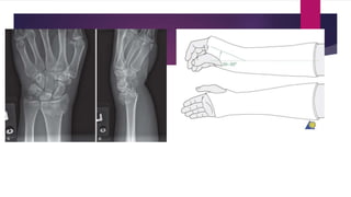
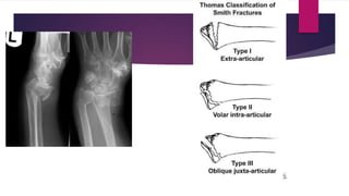
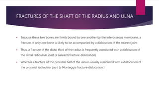
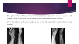
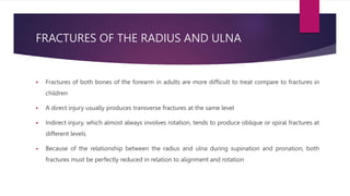
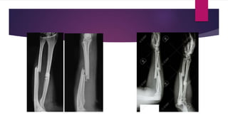
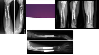
THE WRIST AND FOREARM
DISTAL END OF RADIUS
COLLES
 A fracture through the distal metaphysis of the radius, the Colles
 The patient either slips or trips, and in an attempt to break the fall , lands on open hand with
the forearm pronated, breaking the wrist
 The forces that break the distal end of the radius involve dorsoflexion and radial deviation as
well as supination, all of which responsible for the typical fracture deformity
 The fracture pattern is relatively constant, the main fracture line being transverse within the
distal 2 cm of the radius, there may be only two major fragments, but comminution of the thin
cortex is common
 The clinical deformity, frequently referred to as a "dinner fork deformity" is typical
 There is an obvious jog just proximal to the wrist due to the posterior displacement and
posterior tilt of the distal radial fragment
 The hand tends to be radially deviated and the wrist appears supinated in relation to the
forearm
 Two main types of Colles fracture can be differentiated radiographically
 In the stable type, there is one main transverse fracture line with little cortical comminution
 In the unstable type, there is gross comminution, particularly of the dorsal cortex, and also
significant crushing of the cancellous bone
 The intact periosteal hinge is on the dorsal aspect of the fracture in both types
 Undisplaced Colles (rare) fractures require only immobilization in a below-elbow cast for 4 weeks
 Displaced one can usually be well reduced by closed manipulation, but the major problem is maintenance
of reduction, particularly in the unstable type of Colles fracture
 In this type, with comminution of the dorsal cortex and crushing of the cancellous bone, the reduced
fracture tends to slip back toward the prereduction position of deformity
 The blood supply to bone at the distal end of the radius is excellent,so bony union is assured
 The main problem is not non union but malunion
 Closed reduction is obtained by using the principle of the intact periosteal hinge
 The fracture deformityis first increased to disimpact the fragments and to slacken the intact
periosteal hinge on the dorsal surface, after which the distal fragment is moved distally to
engage the proximal fragment.
 At this point, the dorsal displacement is corrected by pushing the distal fragment forward, the
angulation is straightened, the radial deviation is corrected by placing the hand in ulnar
deviation, and the supination deformity is corrected by placing the forearm in full pronation
 These maneuvers bring the distal radius out to length, tighten the intact periosteal hinge and
help to maintain the reduction
 The plaster cast then applied to hold the reduced position of the fracture, but the finger must left to
move freely
 The radiologic image must took after 1 and 2 weeks reduction, to make sure the fracture didn’t slip
 The immobilization then continued for 6 weeks, because most Colles' fratures are well united in an
acceptable position within 6 weeks
 The complications, which are often preventable, include finger stiffness, shoulder stiffness, malunion
with deformity and residual subluxation of the distal radioulnar joint
SMITH
 This fracture is a pronation injury, caused by a fall or blow on the back of the flexed wrist
 Reduction requires strong supination of the wrist but open reduction and internal fixation are
frequently necessary
 An above elbow cast is usually required during the 6 week period of immobilization to
maintain the position of supination
FRACTURES OF THE SHAFT OF THE RADIUS AND ULNA
 Because these two bones are firmly bound to one another by the interosseous membrane, a
fracture of only one bone is likely to be accompanied by a dislocation of the nearest joint
 Thus, a fracture of the distal third of the radius is frequently associated with a dislocation of
the distal radioulnar joint (a Galeazzi fracture-dislocation)
 Whereas a fracture of the proximal half of the ulna is usually associated with a dislocation of
the proximal radioulnar joint (a Monteggia fracture-dislocation )
FRACTURE OF THE RADIAL SHAFT AND DISLOCATION OF THE DISTAL
RADIOULNAR JOINT (GALEAZZI FRACTURE-DISLOCATION)
 Displaced fractures of the distal third of the radial shaft are not common, but when they do
occur, they are associated with complete disruption and dislocation of the distal radioulnar
joint
 In this injury, which is usually sustained by young adults, the distal fragment of the radius is
tilted posteriorly (anterior angulation at the fracture site)
 The carpus and hand are displaced with the radius and the resultant clinical deformity is
striking
 Radiographically, the nature of the fracture-dislocation is most apparent in the lateral
projection
 The optimum form of treatment for the Galeazzi fracture-dislocation is open reduction and
internal fixation of the radius, with either a plate and screws or an intramedullary nail
 When the radius is perfectly reduced , so also is the dislocation of the distal radioulnar joint
reduced
ISOLATED FRACTURE OF THE PROXIMAL 2/3 OF THE RADIAL SHAFT
 When the radial shaft is fractured in its upper two thirds, the fragments tend to override and
rotate
 As a result of the shortening of the radius there is, of course, some degree of subluxation at
the distal radioulnar joint
 Isolated fractures of the radial shaft are difficultt to reduce by dosed means and reduction, if
obtained, is difficult ro maintain
 The most suitable treatment is open reduction of the radius and internal fixation with either an
AO compression plate and screws or an intermedullary nail
 The most significant complication is malunion that affect the ROM (loss of pronation)
FRACTURES OF THE RADIUS AND ULNA
 Fractures of both bones of the forearm in adults are more difficult to treat compare to fractures in
children
 A direct injury usually produces transverse fractures at the same level
 Indirect injury, which almost always involves rotation, tends to produce oblique or spiral fractures at
different levels
 Because of the relationship between the radius and ulna during supination and pronation, both
fractures must be perfectly reduced in relation to alignment and rotation
 Closed reduction of both fractures maybe possible using traction and varying degrees of pronation
or supination depending on the deformity
 Fractures of the distal third are most stable in pronation, those in the middle third are most stable
in the midposition, and those in the proximal third are most stable in supination
 Even if accurate closed reduction can be obtained, fractures of both bones of the forearm are
unstable and tend to redisplace despite a carefully molded above-elbow cast
 Nevertheless, Sarmiento recommends treating fractures of both bones of the forearm by functional
fracture-bracing (after 3 to 5 weeks in an above-elbow cast), the position of supination is
satisfactory regardless of the level of the fractures
 Open reduction is usually required for fractures of both bones of the forearm in adults, either as
primary or secondary treatment after failure of closed reduction
 The radius and ulna should be approached through separate incisions to minimize the risk of cross-
union between the two bones
 The most effective form of intemal fixation for these fractures is an AO compression plate and
screws
 The most often complication is malunion or even non union
FRACTURE OF THE ULNAR SHAFT AND DISLOCATION OF THE PROXIMAL
RADIOULNAR JOINT (MONTEGGIA FRACTURE-DISLOCATION)
 An angulated fracture of the proximal half of the ulna is invariably accompanied by a
dislocation of the proximal radioulnar joint
 Thus, radiographic examination for fractures in the forearm should always include both the
wrist and elbow joints to avoid overlooking a fracture-dislocation
 In the common (extension) type of Monteggia fracture-dislocation, a hyperextension and
pronation injury produces a fracture of the proximal half of the ulna with anterior angulation
and anterior dislocation of the proximal radioulnar joint. This injury can also be produced by a
direct blow over the ulnar border of the forearm
 A rare variation of Monteggia fracture-dislocation is the flexion type, which is caused by a
flexion injury and characterized by posterior angulation of the fractured ulna and posterior
dislocation of the proximal radioulnar joint
 This type of injury is treated using the same principles as the extension type of Monteggia
fracture -dislocation
 Monteggia fracture-dislocations in adults are best treated by open reduction of the ulna so that its length
and alignment may be perfectly restored
 Internal fixation of the fracture should be obtained by means of either a compression plate and screws or
an intramedullary nail
 Correction of the ulnar deformity usually results in a closed reduction of the radial head, in which case it
is unnecessary to perform an open reduction of the dislocated proximal radioulnar joint or to repair the
ruptured annular ligament
 The limb should be immobilized in an above-elbow cast with the forearm in supination for approximately
3 months
THANK YOU

The Wrist and Forearm.pptx

  • 1.
  • 2.
  • 3.
    COLLES  A fracturethrough the distal metaphysis of the radius, the Colles  The patient either slips or trips, and in an attempt to break the fall , lands on open hand with the forearm pronated, breaking the wrist  The forces that break the distal end of the radius involve dorsoflexion and radial deviation as well as supination, all of which responsible for the typical fracture deformity  The fracture pattern is relatively constant, the main fracture line being transverse within the distal 2 cm of the radius, there may be only two major fragments, but comminution of the thin cortex is common
  • 4.
     The clinicaldeformity, frequently referred to as a "dinner fork deformity" is typical  There is an obvious jog just proximal to the wrist due to the posterior displacement and posterior tilt of the distal radial fragment  The hand tends to be radially deviated and the wrist appears supinated in relation to the forearm
  • 5.
     Two maintypes of Colles fracture can be differentiated radiographically  In the stable type, there is one main transverse fracture line with little cortical comminution  In the unstable type, there is gross comminution, particularly of the dorsal cortex, and also significant crushing of the cancellous bone  The intact periosteal hinge is on the dorsal aspect of the fracture in both types
  • 6.
     Undisplaced Colles(rare) fractures require only immobilization in a below-elbow cast for 4 weeks  Displaced one can usually be well reduced by closed manipulation, but the major problem is maintenance of reduction, particularly in the unstable type of Colles fracture  In this type, with comminution of the dorsal cortex and crushing of the cancellous bone, the reduced fracture tends to slip back toward the prereduction position of deformity  The blood supply to bone at the distal end of the radius is excellent,so bony union is assured  The main problem is not non union but malunion
  • 7.
     Closed reductionis obtained by using the principle of the intact periosteal hinge  The fracture deformityis first increased to disimpact the fragments and to slacken the intact periosteal hinge on the dorsal surface, after which the distal fragment is moved distally to engage the proximal fragment.  At this point, the dorsal displacement is corrected by pushing the distal fragment forward, the angulation is straightened, the radial deviation is corrected by placing the hand in ulnar deviation, and the supination deformity is corrected by placing the forearm in full pronation  These maneuvers bring the distal radius out to length, tighten the intact periosteal hinge and help to maintain the reduction
  • 8.
     The plastercast then applied to hold the reduced position of the fracture, but the finger must left to move freely  The radiologic image must took after 1 and 2 weeks reduction, to make sure the fracture didn’t slip  The immobilization then continued for 6 weeks, because most Colles' fratures are well united in an acceptable position within 6 weeks  The complications, which are often preventable, include finger stiffness, shoulder stiffness, malunion with deformity and residual subluxation of the distal radioulnar joint
  • 10.
    SMITH  This fractureis a pronation injury, caused by a fall or blow on the back of the flexed wrist  Reduction requires strong supination of the wrist but open reduction and internal fixation are frequently necessary  An above elbow cast is usually required during the 6 week period of immobilization to maintain the position of supination
  • 12.
    FRACTURES OF THESHAFT OF THE RADIUS AND ULNA  Because these two bones are firmly bound to one another by the interosseous membrane, a fracture of only one bone is likely to be accompanied by a dislocation of the nearest joint  Thus, a fracture of the distal third of the radius is frequently associated with a dislocation of the distal radioulnar joint (a Galeazzi fracture-dislocation)  Whereas a fracture of the proximal half of the ulna is usually associated with a dislocation of the proximal radioulnar joint (a Monteggia fracture-dislocation )
  • 13.
    FRACTURE OF THERADIAL SHAFT AND DISLOCATION OF THE DISTAL RADIOULNAR JOINT (GALEAZZI FRACTURE-DISLOCATION)  Displaced fractures of the distal third of the radial shaft are not common, but when they do occur, they are associated with complete disruption and dislocation of the distal radioulnar joint  In this injury, which is usually sustained by young adults, the distal fragment of the radius is tilted posteriorly (anterior angulation at the fracture site)  The carpus and hand are displaced with the radius and the resultant clinical deformity is striking  Radiographically, the nature of the fracture-dislocation is most apparent in the lateral projection
  • 14.
     The optimumform of treatment for the Galeazzi fracture-dislocation is open reduction and internal fixation of the radius, with either a plate and screws or an intramedullary nail  When the radius is perfectly reduced , so also is the dislocation of the distal radioulnar joint reduced
  • 15.
    ISOLATED FRACTURE OFTHE PROXIMAL 2/3 OF THE RADIAL SHAFT  When the radial shaft is fractured in its upper two thirds, the fragments tend to override and rotate  As a result of the shortening of the radius there is, of course, some degree of subluxation at the distal radioulnar joint  Isolated fractures of the radial shaft are difficultt to reduce by dosed means and reduction, if obtained, is difficult ro maintain  The most suitable treatment is open reduction of the radius and internal fixation with either an AO compression plate and screws or an intermedullary nail  The most significant complication is malunion that affect the ROM (loss of pronation)
  • 16.
    FRACTURES OF THERADIUS AND ULNA  Fractures of both bones of the forearm in adults are more difficult to treat compare to fractures in children  A direct injury usually produces transverse fractures at the same level  Indirect injury, which almost always involves rotation, tends to produce oblique or spiral fractures at different levels  Because of the relationship between the radius and ulna during supination and pronation, both fractures must be perfectly reduced in relation to alignment and rotation
  • 17.
     Closed reductionof both fractures maybe possible using traction and varying degrees of pronation or supination depending on the deformity  Fractures of the distal third are most stable in pronation, those in the middle third are most stable in the midposition, and those in the proximal third are most stable in supination  Even if accurate closed reduction can be obtained, fractures of both bones of the forearm are unstable and tend to redisplace despite a carefully molded above-elbow cast
  • 18.
     Nevertheless, Sarmientorecommends treating fractures of both bones of the forearm by functional fracture-bracing (after 3 to 5 weeks in an above-elbow cast), the position of supination is satisfactory regardless of the level of the fractures  Open reduction is usually required for fractures of both bones of the forearm in adults, either as primary or secondary treatment after failure of closed reduction  The radius and ulna should be approached through separate incisions to minimize the risk of cross- union between the two bones  The most effective form of intemal fixation for these fractures is an AO compression plate and screws  The most often complication is malunion or even non union
  • 21.
    FRACTURE OF THEULNAR SHAFT AND DISLOCATION OF THE PROXIMAL RADIOULNAR JOINT (MONTEGGIA FRACTURE-DISLOCATION)  An angulated fracture of the proximal half of the ulna is invariably accompanied by a dislocation of the proximal radioulnar joint  Thus, radiographic examination for fractures in the forearm should always include both the wrist and elbow joints to avoid overlooking a fracture-dislocation
  • 22.
     In thecommon (extension) type of Monteggia fracture-dislocation, a hyperextension and pronation injury produces a fracture of the proximal half of the ulna with anterior angulation and anterior dislocation of the proximal radioulnar joint. This injury can also be produced by a direct blow over the ulnar border of the forearm  A rare variation of Monteggia fracture-dislocation is the flexion type, which is caused by a flexion injury and characterized by posterior angulation of the fractured ulna and posterior dislocation of the proximal radioulnar joint  This type of injury is treated using the same principles as the extension type of Monteggia fracture -dislocation
  • 24.
     Monteggia fracture-dislocationsin adults are best treated by open reduction of the ulna so that its length and alignment may be perfectly restored  Internal fixation of the fracture should be obtained by means of either a compression plate and screws or an intramedullary nail  Correction of the ulnar deformity usually results in a closed reduction of the radial head, in which case it is unnecessary to perform an open reduction of the dislocated proximal radioulnar joint or to repair the ruptured annular ligament  The limb should be immobilized in an above-elbow cast with the forearm in supination for approximately 3 months
  • 25.