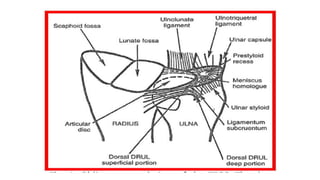
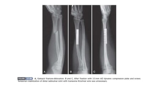
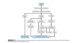
Galeazzi fracture dislocation
Dr. Ashiqur Rahman
Resident Orthopedics
Dhaka Medical college Hospital
Introduction
 The injury was first described in 1934 by Galleazzi.
 The usual cause is a fall on the hand, probably with a superimposed
rotation force.
 The Radius # in its lower third & it’s inferior radio-ulnar joint
subluxates & dislocates. (Apley - 794)
 The combination of fracture of the distal or middle third of the shaft
of the radius and dislocation of the distal radioulnar joint was called
“the fracture of necessity” by Campbell. Similar to Monteggia
fracture-dislocations, Galeazzi fracture dislocations often go
unrecognized.(Campbell’s – 2990)
 Dislocation of the distal radioulnar joint at the time of injury should
be suspected with a displaced fracture of the distal third of the shaft
of the radius.
TFCC: Triangular fibrocartilage complex is a major contributor to the
stability of the wrist.
Location: Ulnar aspect of the wrist between the lunate and
triquetrum of the proximal row.
Shape: Elongated triangular shape pointing at the radius
Consists of:
1. Triangular disc proper
2. Volar component
- Volar radioulnar ligament
- Ulnotriquetral ligament
- Ulnolunate ligament
3. Ulna components:
- Ulnar collateral ligament
- Triangular ligament
- Meniscal homologue
4. Dorsal component:
- Dorsal radioulnar ligament
- Extensor carpi ulnaris tendon.
 Blood Supply: Central and radial portions of the TFCC are avascular.
Functions of TFCC:
- Main stabilizer of the DRUJ
- Buttress to support proximal row
- 80% of the axial load transmitted through the radiocarpal
ligament and 20% through the TFCC from ulna to carpus.
Radiological evaluation
Not only for diagnosis; but also for:
- Displacement
- Angulation
- Rotation
- Comminution
Normal radiographic parameters of the wrist:
- Palmer tilt: 11°
- Radial inclination: 23°
- Radial height 12 mm
- Ulnar variance : 1mm
 Radiographic findings that suggest a distal radioulnar joint injury
include:
(1) fracture at the base of the ulnar styloid.
(2) widening of the distal radioulnar joint on the anteroposterior
view.
(3) dislocation of the ulna relative to the radius on a true
lateral view of the wrist; and
(4) more than 5 mm of shortening of the radius relative to the
ulna when compared with the contralateral wrist.
Clinical features:
(i) Swelling, deformity, and tenderness.
(ii) Painful forearm rotation and wrist motion.
(iii) Injury to the DRUJ:
- joint tenderness
- Ulnar head prominent dorsally or palmarly.
(v) Neurovascular injury is rare.
 It may be possible to demonstrate the instability of the radio-ulnar
joint by ‘balloting’ the distal end of the ulna (the ‘piano-key sign’) or
by rotating the wrist.
Treatment
 The important step is to restore the length of the fractured bone.
 In children reduction is often successful.
 In adult reduction is best achieved by open operation & internal
fixation with a 3.5mm AO dynamic compression plate.
 An X-ray is taken to ensure to ensure that the distal radio-ulnar joint
is reduced.
Treatment Cont’d
1. Distal radio ulnar is reduced and stable
a. No further action needed
b. Arm is rested for few days
c. Then gentle active movements are encouraged
d. R/U joint should be checked both clinically and radiologically,
during the last 06 weeks.
2. The distal radioulnar joint is reduced but unstable:
a. Immobilization in the position of stability ( supination)
b. Supplemented by a transverse k wire ( if required)
c. Supinated forearm and above elbow cast for 6 weeks
d. If large ulnar styloid fragment it should be reduced and fixed.
3. Distal radio ulnar joint is immobile:
a. Unusual
b. Open reduction is needed to remove the interposed soft
tissue.
c. TFCC and dorsal capsule is repaired.
d. Immobilization is supination and k wire if needed for 6 weeks.
Patients with malunited Galeazzi fracture may report:
- persistent pain in the DRUJ
- limited forearm rotation, and
- Loss of grip strength.
Complications:
1. Nerve compression,
2. tendon entrapment,
3. nonunion,
4. delayed union,
5. malunion, and
6. Infection.
Galeazzi fracture dislocation

Galeazzi fracture dislocation

  • 1.
    Galeazzi fracture dislocation Dr.Ashiqur Rahman Resident Orthopedics Dhaka Medical college Hospital
  • 2.
    Introduction  The injurywas first described in 1934 by Galleazzi.  The usual cause is a fall on the hand, probably with a superimposed rotation force.  The Radius # in its lower third & it’s inferior radio-ulnar joint subluxates & dislocates. (Apley - 794)  The combination of fracture of the distal or middle third of the shaft of the radius and dislocation of the distal radioulnar joint was called “the fracture of necessity” by Campbell. Similar to Monteggia fracture-dislocations, Galeazzi fracture dislocations often go unrecognized.(Campbell’s – 2990)
  • 3.
     Dislocation ofthe distal radioulnar joint at the time of injury should be suspected with a displaced fracture of the distal third of the shaft of the radius.
  • 4.
    TFCC: Triangular fibrocartilagecomplex is a major contributor to the stability of the wrist. Location: Ulnar aspect of the wrist between the lunate and triquetrum of the proximal row. Shape: Elongated triangular shape pointing at the radius Consists of: 1. Triangular disc proper 2. Volar component - Volar radioulnar ligament - Ulnotriquetral ligament - Ulnolunate ligament
  • 5.
    3. Ulna components: -Ulnar collateral ligament - Triangular ligament - Meniscal homologue 4. Dorsal component: - Dorsal radioulnar ligament - Extensor carpi ulnaris tendon.  Blood Supply: Central and radial portions of the TFCC are avascular.
  • 7.
    Functions of TFCC: -Main stabilizer of the DRUJ - Buttress to support proximal row - 80% of the axial load transmitted through the radiocarpal ligament and 20% through the TFCC from ulna to carpus.
  • 8.
    Radiological evaluation Not onlyfor diagnosis; but also for: - Displacement - Angulation - Rotation - Comminution Normal radiographic parameters of the wrist: - Palmer tilt: 11° - Radial inclination: 23° - Radial height 12 mm - Ulnar variance : 1mm
  • 9.
     Radiographic findingsthat suggest a distal radioulnar joint injury include: (1) fracture at the base of the ulnar styloid. (2) widening of the distal radioulnar joint on the anteroposterior view. (3) dislocation of the ulna relative to the radius on a true lateral view of the wrist; and (4) more than 5 mm of shortening of the radius relative to the ulna when compared with the contralateral wrist.
  • 16.
    Clinical features: (i) Swelling,deformity, and tenderness. (ii) Painful forearm rotation and wrist motion. (iii) Injury to the DRUJ: - joint tenderness - Ulnar head prominent dorsally or palmarly. (v) Neurovascular injury is rare.  It may be possible to demonstrate the instability of the radio-ulnar joint by ‘balloting’ the distal end of the ulna (the ‘piano-key sign’) or by rotating the wrist.
  • 20.
    Treatment  The importantstep is to restore the length of the fractured bone.  In children reduction is often successful.  In adult reduction is best achieved by open operation & internal fixation with a 3.5mm AO dynamic compression plate.  An X-ray is taken to ensure to ensure that the distal radio-ulnar joint is reduced.
  • 21.
    Treatment Cont’d 1. Distalradio ulnar is reduced and stable a. No further action needed b. Arm is rested for few days c. Then gentle active movements are encouraged d. R/U joint should be checked both clinically and radiologically, during the last 06 weeks.
  • 22.
    2. The distalradioulnar joint is reduced but unstable: a. Immobilization in the position of stability ( supination) b. Supplemented by a transverse k wire ( if required) c. Supinated forearm and above elbow cast for 6 weeks d. If large ulnar styloid fragment it should be reduced and fixed.
  • 23.
    3. Distal radioulnar joint is immobile: a. Unusual b. Open reduction is needed to remove the interposed soft tissue. c. TFCC and dorsal capsule is repaired. d. Immobilization is supination and k wire if needed for 6 weeks.
  • 24.
    Patients with malunitedGaleazzi fracture may report: - persistent pain in the DRUJ - limited forearm rotation, and - Loss of grip strength.
  • 25.
    Complications: 1. Nerve compression, 2.tendon entrapment, 3. nonunion, 4. delayed union, 5. malunion, and 6. Infection.