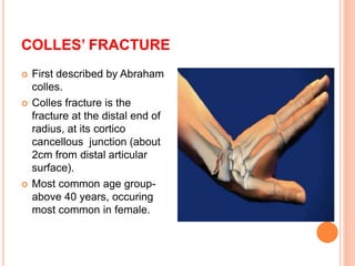
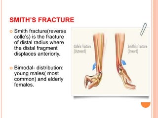
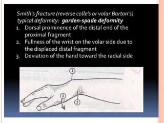
INJURIES DUE TO FALL
ON AN
OUTSTRETCHED HAND
SHAHID
 Colles’ fracture
 Smith’s fracture
 Galeazzi fracture
 Monteggia fracture
 Scaphoid fracture
 Supracondylar fracture of humerus
 Fracture of head of radius
 Clavicular fracture
COLLES’ FRACTURE
 First described by Abraham
colles.
 Colles fracture is the
fracture at the distal end of
radius, at its cortico
cancellous junction (about
2cm from distal articular
surface).
 Most common age group-
above 40 years, occuring
most common in female.
MECHANISM OF INJURY
PATHOANATOMY
 Displacement-The fracture line runs transversely
at cortico-cancellous junction.
In many cases one or more displacements may
occur as follows:
 Impaction of fragments
 Dorsal displacement
 Dorsal tilt
 Lateral displacement
 Lateral tilt
 Supination
CLINICAL FEATURES
 Pain
 Swelling
 Deformity-classical
‘dinner-fork
deformity’
 Radial styloid process
lies in the same level or
little higher than the
ulnar styloid process.
DIAGNOSIS
 Radiological
features:
It is important to
differentiate colles’
fracture from other
fractures occuring at
same time, such as
Smith’s fracture,
Barton’s fracture by
looking at the
displacements.
X-RAY
 Lateral view: Dorsal tilt- detected by looking
at the direction of distal articular surface of
radius.
 AP view: Lateral tilt- similarly it can be
detected by looking at articular surface if it
faces medially it is normal, if it becomes
horizontal or faces laterally, a lateral tilt is
present.
TREATMENT
 Conservative method:
For undisplaced fracture- immobilization in a
below-elbow plaster cast for 6 weeks.
For displaced fracture- Manipulative
reduction followed by immobilization
MANIPULATIVE REDUCTION
 Step 1- Disimpact the
fragments by firm longitudinal
traction against the counter-
traction by an assistant.
 Step 2- Press the distal
fragment into palmer flexion
and ulnar deviation by using
thumb of other hand.
 Step 3- Now hand is drawn in
pronation, palmer flexion and
ulnar deviation. A plaster cast is
applied extending from below
elbow to metacarpal heads.
SURGICAL METHODS
Closed reduction and per-cutaneous
fixation using K- wire.
Open reduction and plate fixation.
COMPLICATIONS
 Stiffness of joints
 Malunion
 Subluxation of inferior radio- ulnar joint
 Carpel-tunnel syndrome
 Sudeck’s osteodystrophy
 Rupture of extensor pollicis longus tendon.
SMITH’S FRACTURE
 Smith fracture(reverse
colle’s) is the fracture
of distal radius where
the distal fragment
displaces anteriorly.
 Bimodal- distribution:
young males( most
common) and elderly
females.
SCAPHOID FRACTURE
 A scaphoid fracture is
more common in young
adults. It is rare in
children and in elderly
people.
 Commonly, fracture
occurs through the waist
of scaphoid. Rarely it
occurs through the
tuberosity.
 It may be either a crack
fracture or a displaced
fracture.
BLOOD SUPPLY
CLINICAL FEATURES
Pain
Swelling
On/E-
 Tenderness at scaphoid fossa
(anatomical snuff box)
 Decrease thumb movement
X-RAY
 PA view
 Semipronated
view
 Semisupinated
view
 Ziter view-
TREATMENT
 Conservative treatment- Affected hand is
immobilized in a scaphoid cast for 3-4
months.
 Scaphoid cast is applied from below elbow
to metacarpel heads, includes thumb. Wrist
is maintained at little dorsiflexion and radial
deviation( glass holding position).
COMPLICATIONS
Avascular necrosis
Delayed union and non-union
Wrist osteoarthritis
MONTEGGIA FRACTURE
 This is a fracture of upper-third of
ulna with dislocation of head of
radius.
 Types:
 Extension type- where the
ulna fracture angulates anteriorly
and the radial head dislocates
anteriorly.
 Flexion type- where the ulna
fracture angulates posteriorly and
the radial head dislocates
posteriorly.
TREATMENT
This is very unstable fracture.
Reduction is attempted under general
aneasthesia. If closed reduction is not
possible, an open reduction and internal
fixation using a plate is performed.
GALEAZZI FRACTURE
 This is a fracture of
lower-third of radius
with dislocation or
subluxation of distal
radio-ulnar joint.
 Displacement-Radius
fracture is angulated
medially and anteriorly
and dorsal dislocation
of distal end of ulna.
TREATMENT
It is difficult to achieve and maintain
perfect reduction by conservative
methods( except in children).
Open-reduction and internal fixation of
radius with plate is done in adults.
THANK YOU

Fall on an outstrtched hand

  • 1.
    INJURIES DUE TOFALL ON AN OUTSTRETCHED HAND SHAHID
  • 2.
     Colles’ fracture Smith’s fracture  Galeazzi fracture  Monteggia fracture  Scaphoid fracture  Supracondylar fracture of humerus  Fracture of head of radius  Clavicular fracture
  • 3.
    COLLES’ FRACTURE  Firstdescribed by Abraham colles.  Colles fracture is the fracture at the distal end of radius, at its cortico cancellous junction (about 2cm from distal articular surface).  Most common age group- above 40 years, occuring most common in female.
  • 4.
  • 5.
    PATHOANATOMY  Displacement-The fractureline runs transversely at cortico-cancellous junction. In many cases one or more displacements may occur as follows:  Impaction of fragments  Dorsal displacement  Dorsal tilt  Lateral displacement  Lateral tilt  Supination
  • 6.
    CLINICAL FEATURES  Pain Swelling  Deformity-classical ‘dinner-fork deformity’  Radial styloid process lies in the same level or little higher than the ulnar styloid process.
  • 7.
    DIAGNOSIS  Radiological features: It isimportant to differentiate colles’ fracture from other fractures occuring at same time, such as Smith’s fracture, Barton’s fracture by looking at the displacements.
  • 9.
    X-RAY  Lateral view:Dorsal tilt- detected by looking at the direction of distal articular surface of radius.  AP view: Lateral tilt- similarly it can be detected by looking at articular surface if it faces medially it is normal, if it becomes horizontal or faces laterally, a lateral tilt is present.
  • 10.
    TREATMENT  Conservative method: Forundisplaced fracture- immobilization in a below-elbow plaster cast for 6 weeks. For displaced fracture- Manipulative reduction followed by immobilization
  • 11.
    MANIPULATIVE REDUCTION  Step1- Disimpact the fragments by firm longitudinal traction against the counter- traction by an assistant.  Step 2- Press the distal fragment into palmer flexion and ulnar deviation by using thumb of other hand.  Step 3- Now hand is drawn in pronation, palmer flexion and ulnar deviation. A plaster cast is applied extending from below elbow to metacarpal heads.
  • 12.
    SURGICAL METHODS Closed reductionand per-cutaneous fixation using K- wire. Open reduction and plate fixation.
  • 13.
    COMPLICATIONS  Stiffness ofjoints  Malunion  Subluxation of inferior radio- ulnar joint  Carpel-tunnel syndrome  Sudeck’s osteodystrophy  Rupture of extensor pollicis longus tendon.
  • 14.
    SMITH’S FRACTURE  Smithfracture(reverse colle’s) is the fracture of distal radius where the distal fragment displaces anteriorly.  Bimodal- distribution: young males( most common) and elderly females.
  • 21.
    SCAPHOID FRACTURE  Ascaphoid fracture is more common in young adults. It is rare in children and in elderly people.  Commonly, fracture occurs through the waist of scaphoid. Rarely it occurs through the tuberosity.  It may be either a crack fracture or a displaced fracture.
  • 23.
  • 25.
    CLINICAL FEATURES Pain Swelling On/E-  Tendernessat scaphoid fossa (anatomical snuff box)  Decrease thumb movement
  • 26.
    X-RAY  PA view Semipronated view  Semisupinated view  Ziter view-
  • 27.
    TREATMENT  Conservative treatment-Affected hand is immobilized in a scaphoid cast for 3-4 months.  Scaphoid cast is applied from below elbow to metacarpel heads, includes thumb. Wrist is maintained at little dorsiflexion and radial deviation( glass holding position).
  • 29.
    COMPLICATIONS Avascular necrosis Delayed unionand non-union Wrist osteoarthritis
  • 30.
    MONTEGGIA FRACTURE  Thisis a fracture of upper-third of ulna with dislocation of head of radius.  Types:  Extension type- where the ulna fracture angulates anteriorly and the radial head dislocates anteriorly.  Flexion type- where the ulna fracture angulates posteriorly and the radial head dislocates posteriorly.
  • 31.
    TREATMENT This is veryunstable fracture. Reduction is attempted under general aneasthesia. If closed reduction is not possible, an open reduction and internal fixation using a plate is performed.
  • 32.
    GALEAZZI FRACTURE  Thisis a fracture of lower-third of radius with dislocation or subluxation of distal radio-ulnar joint.  Displacement-Radius fracture is angulated medially and anteriorly and dorsal dislocation of distal end of ulna.
  • 33.
    TREATMENT It is difficultto achieve and maintain perfect reduction by conservative methods( except in children). Open-reduction and internal fixation of radius with plate is done in adults.
  • 34.