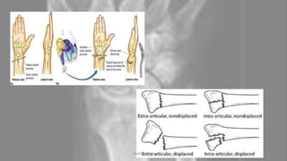
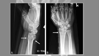
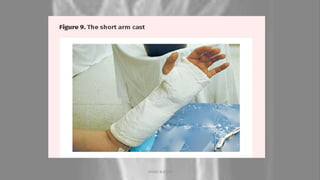
COLLES' FRACTURE
By Annie Blessie
04-10-2020 ANNIE BLESSIE 1
Introduction
• The Colles' fracture is named after Abraham Colles, an Irish surgeon,
who first described it in 1814 by simply looking at the classical
deformity before the advent of X-rays.
• The fracture originates from a fall on the outstretched hand and is
usually associated with dorsal and radial displacement of the distal
fragment, and disturbance of the radial-ulnar articulation.
04-10-2020 ANNIE BLESSIE 2
Definition :
A Colles' Fracture is a complete fracture of the radius
bone of the forearm close to the wrist resulting in an
upward (posterior) displacement of the radius and
obvious deformity. It is commonly called a “broken
wrist” in spite of the fact that the distal radius is the
location of the fracture, not the carpal bones of the
wrist.
04-10-2020 ANNIE BLESSIE 3
Etiology :
• The Colles' fracture is most commonly caused by a fall, landing on
an outstretched hand with the wrist in dorsiflexion-"FOOSH injury."
• Road traffic accident (RTA)
• Direct blow
04-10-2020 ANNIE BLESSIE 4
Risk factors :
• Osteoporosis.
• Elderly age group.
• Having low muscle mass or poor muscle strength,
or lack agility and have poor balance (these conditions
make you more likely to fall).
• Walking or doing other activities in snow or on ice,
or doing activities that require a lot of forward momentum,
such as in-line skating and skiing.
• Having an inadequate intake of calcium or vitamin D.
04-10-2020 ANNIE BLESSIE 5
Types
Types of fractures include:
• Open fracture: if the bone broke through your skin
• Comminuted fracture: if the bone broke into more than two pieces
• Intra-articular fracture: if the bone broke into your wrist joint
• Extra-articular fracture: if your joint isn’t affected
04-10-2020 ANNIE BLESSIE 6
04-10-2020 ANNIE BLESSIE 7
Clinical features :
• Dinner Fork" Deformity.
• History of fall on an outstretched hand.
• Dorsal wrist pain.
• Swelling of the wrist.
• Increased angulation of the distal radius.
• Inability to grasp object.
04-10-2020 ANNIE BLESSIE 8
FRYKMAN CLASSIFICATION
Gosta Frykman identified many
different forms of Colles fracture
and classified it into eight
different types based on the extra-
or intra-articular nature of
fractures involving the distal ends
of the radius and ulna.
04-10-2020 ANNIE BLESSIE 9
• Type I: transverse metaphyseal fracture
• includes both Colles and Smith fractures as angulation is not a feature
• Type II: type I + ulnar styloid fracture
• Type III: fracture involves the radiocarpal joint
• includes both Barton and reverse Barton fractures
• includes Chauffeur fractures
• Type IV: type III + ulnar styloid fracture
• Type V: transverse fracture involves distal radioulnar joint
• Type VI: type V + ulnar styloid fracture
• Type VII: comminuted fracture with the involvement of both the
radiocarpal and radioulnar joints
• Type VIII: type VII + ulnar styloid fracture
04-10-2020 ANNIE BLESSIE 10
04-10-2020 ANNIE BLESSIE 11
Complications :
• Malunion.
• Persistent translation of the carpus.
• Shortening of radius.
• Stiffness of the wrist and the forearm
04-10-2020 ANNIE BLESSIE 12
Diagnosis
Diagnosis is most often made
upon interpretation of
posteroanterior and lateral views
alone.
(X-ray of a Colles' fracture of the
left wrist accompanied by an ulnar
styloid fracture.)
04-10-2020 ANNIE BLESSIE 13
The classic Colles fracture has the following characteristics;
• Transverse fracture of the radius.
• 2.5 cm (0.98 inches) proximal to the radiocarpal joint.
• dorsal displacement and dorsal angulation, together with the radial tilt.
Other characteristics on plain radiographs may include:
• Radial shortening.
• Loss of ulnar inclination.
• Radial angulation of the wrist.
• Comminution at the fracture site.
• Associated fracture of the ulnar styloid process in more than 60% of cases.
04-10-2020 ANNIE BLESSIE 14
Differential diagnosis/associated injuries
• Scapholunate ligament tear.
• Median nerve injury.
• TFCC (triangular fibrocartilage complex) injury, up to 50% when ulnar styloid fx
also present.
• Carpal ligament injury: Scapholunate Instability(most common), lunotriquetral
ligament.
• Tendon injury, attritional EPL rupture, usually treated with EIP tendon transfer.
• Compartment syndrome.
• Ulnar styloid fracture.
• DRUJ (Distal Radial Ulnar Joint) Instability.
• Galeazzi Fracture: highly associated with distal 1/3 radial shaft fractures.
04-10-2020 ANNIE BLESSIE 15
Outcome measures
• DASH.
• Patient Rated Wrist Evaluation (PRWE).
• Green Score.
• O'Brian Score.
04-10-2020 ANNIE BLESSIE 16
Medical management :
• The treatment will depend on the type of Colle's fracture present,
the age and activity level of the patient.
• Management of a Colle's fracture depends on the severity of the fracture.
An undisplaced fracture may be treated conservatively with a cast alone.
The cast is applied with the distal fragment in palmar flexion and ulnar deviation.
• Surgical options- external fixation, internal fixation, percutaneous pinning,
and bone substitutes.
• A fracture with mild angulation and displacement may require closed reduction.
• Significant angulation and deformity may require an open reduction and
internal fixation or external fixation. The volar forearm splint is best for temporary
immobilisation of forearm, wrist and hand fractures, including Colles fracture.
04-10-2020 ANNIE BLESSIE 17
Physiotherapy management
• Patients will present to a physiotherapist with pain, oedema,
decreased ROM, decreased strength, and decreased functional abilities.
• Once a Colles’ fracture has healed rehabilitation is recommended
in an attempt to restore function and strength to the fractured wrist.
• The primary focus in early rehabilitation is to mobilise the wrist,
which is indicated approximately 7-8 weeks post-fracture.
• If the fracture has been managed using an internal fixation device,
early mobilisation can begin as early as 1-week post-surgery.
• Fractures that have been treated with external fixation as the wrist is
often held in a pronated position. This can predispose the patient to a contracture
at the distal radioulnar joint.
04-10-2020 ANNIE BLESSIE 18
Initial rehabilitation :
• To restore normal range of motion (ROM) at the wrist with both
passive ROM and progression to active ROM.
• Wrist flexion and extension are often the first motions emphasised
working within the patient's pain-free available range.
• It is also important to emphasise motion at the joints above and
below (shoulder, elbow, and fingers) during all phases of rehab.
• To limit the pain and the amount of oedema present in the wrist and
hand region.
04-10-2020 ANNIE BLESSIE 19
Sub acute phase :
The next phase of rehab continues to focus on increasing wrist ROM and the
commencement of strengthening exercises.
For fractures that were surgically treated, ROM should be regained between 6 to 8 weeks
post-op. Examples of ROM exercises that can be performed include:
• Wrist flexion/extension.
• Radial/ulnar deviation.
• Pronation/supination.
• Making a fist and opening.
In the sub-acute phase, ROM exercises can progress into strengthening by performing all
exercises with a weight in the hand or performing grip squeeze with a foam ball or a towel
roll. During this phase, progressive stretching can begin to increase available ROM. Each
stretch should be held for 30-60 seconds for 3 repetitions. If the patient is unable to
tolerate a slow, prolonged stretch, shorter stretches of 10 seconds can be performed for
10 repetitions.
04-10-2020 ANNIE BLESSIE 20
Modalities /Other method:
• Heat/Parrafin wax.
• Massage.
• Cryotherapy.
• Electrical stimulation-TENS
• Exercise-Isometric exercises.
Active ROM exercises.
Intrinsic hand muscle exercises.
04-10-2020 ANNIE BLESSIE 21
Prevention :
• Proper intake of nutritious food.
• Exercise.
• Wrist guard.
04-10-2020 ANNIE BLESSIE 22
04-10-2020 ANNIE BLESSIE 23
04-10-2020 ANNIE BLESSIE 24
04-10-2020 ANNIE BLESSIE 25
Thank you
04-10-2020 ANNIE BLESSIE 26

Colles' fracture & physiotherapy management

  • 1.
    COLLES' FRACTURE By AnnieBlessie 04-10-2020 ANNIE BLESSIE 1
  • 2.
    Introduction • The Colles'fracture is named after Abraham Colles, an Irish surgeon, who first described it in 1814 by simply looking at the classical deformity before the advent of X-rays. • The fracture originates from a fall on the outstretched hand and is usually associated with dorsal and radial displacement of the distal fragment, and disturbance of the radial-ulnar articulation. 04-10-2020 ANNIE BLESSIE 2
  • 3.
    Definition : A Colles'Fracture is a complete fracture of the radius bone of the forearm close to the wrist resulting in an upward (posterior) displacement of the radius and obvious deformity. It is commonly called a “broken wrist” in spite of the fact that the distal radius is the location of the fracture, not the carpal bones of the wrist. 04-10-2020 ANNIE BLESSIE 3
  • 4.
    Etiology : • TheColles' fracture is most commonly caused by a fall, landing on an outstretched hand with the wrist in dorsiflexion-"FOOSH injury." • Road traffic accident (RTA) • Direct blow 04-10-2020 ANNIE BLESSIE 4
  • 5.
    Risk factors : •Osteoporosis. • Elderly age group. • Having low muscle mass or poor muscle strength, or lack agility and have poor balance (these conditions make you more likely to fall). • Walking or doing other activities in snow or on ice, or doing activities that require a lot of forward momentum, such as in-line skating and skiing. • Having an inadequate intake of calcium or vitamin D. 04-10-2020 ANNIE BLESSIE 5
  • 6.
    Types Types of fracturesinclude: • Open fracture: if the bone broke through your skin • Comminuted fracture: if the bone broke into more than two pieces • Intra-articular fracture: if the bone broke into your wrist joint • Extra-articular fracture: if your joint isn’t affected 04-10-2020 ANNIE BLESSIE 6
  • 7.
  • 8.
    Clinical features : •Dinner Fork" Deformity. • History of fall on an outstretched hand. • Dorsal wrist pain. • Swelling of the wrist. • Increased angulation of the distal radius. • Inability to grasp object. 04-10-2020 ANNIE BLESSIE 8
  • 9.
    FRYKMAN CLASSIFICATION Gosta Frykmanidentified many different forms of Colles fracture and classified it into eight different types based on the extra- or intra-articular nature of fractures involving the distal ends of the radius and ulna. 04-10-2020 ANNIE BLESSIE 9
  • 10.
    • Type I:transverse metaphyseal fracture • includes both Colles and Smith fractures as angulation is not a feature • Type II: type I + ulnar styloid fracture • Type III: fracture involves the radiocarpal joint • includes both Barton and reverse Barton fractures • includes Chauffeur fractures • Type IV: type III + ulnar styloid fracture • Type V: transverse fracture involves distal radioulnar joint • Type VI: type V + ulnar styloid fracture • Type VII: comminuted fracture with the involvement of both the radiocarpal and radioulnar joints • Type VIII: type VII + ulnar styloid fracture 04-10-2020 ANNIE BLESSIE 10
  • 11.
  • 12.
    Complications : • Malunion. •Persistent translation of the carpus. • Shortening of radius. • Stiffness of the wrist and the forearm 04-10-2020 ANNIE BLESSIE 12
  • 13.
    Diagnosis Diagnosis is mostoften made upon interpretation of posteroanterior and lateral views alone. (X-ray of a Colles' fracture of the left wrist accompanied by an ulnar styloid fracture.) 04-10-2020 ANNIE BLESSIE 13
  • 14.
    The classic Collesfracture has the following characteristics; • Transverse fracture of the radius. • 2.5 cm (0.98 inches) proximal to the radiocarpal joint. • dorsal displacement and dorsal angulation, together with the radial tilt. Other characteristics on plain radiographs may include: • Radial shortening. • Loss of ulnar inclination. • Radial angulation of the wrist. • Comminution at the fracture site. • Associated fracture of the ulnar styloid process in more than 60% of cases. 04-10-2020 ANNIE BLESSIE 14
  • 15.
    Differential diagnosis/associated injuries •Scapholunate ligament tear. • Median nerve injury. • TFCC (triangular fibrocartilage complex) injury, up to 50% when ulnar styloid fx also present. • Carpal ligament injury: Scapholunate Instability(most common), lunotriquetral ligament. • Tendon injury, attritional EPL rupture, usually treated with EIP tendon transfer. • Compartment syndrome. • Ulnar styloid fracture. • DRUJ (Distal Radial Ulnar Joint) Instability. • Galeazzi Fracture: highly associated with distal 1/3 radial shaft fractures. 04-10-2020 ANNIE BLESSIE 15
  • 16.
    Outcome measures • DASH. •Patient Rated Wrist Evaluation (PRWE). • Green Score. • O'Brian Score. 04-10-2020 ANNIE BLESSIE 16
  • 17.
    Medical management : •The treatment will depend on the type of Colle's fracture present, the age and activity level of the patient. • Management of a Colle's fracture depends on the severity of the fracture. An undisplaced fracture may be treated conservatively with a cast alone. The cast is applied with the distal fragment in palmar flexion and ulnar deviation. • Surgical options- external fixation, internal fixation, percutaneous pinning, and bone substitutes. • A fracture with mild angulation and displacement may require closed reduction. • Significant angulation and deformity may require an open reduction and internal fixation or external fixation. The volar forearm splint is best for temporary immobilisation of forearm, wrist and hand fractures, including Colles fracture. 04-10-2020 ANNIE BLESSIE 17
  • 18.
    Physiotherapy management • Patientswill present to a physiotherapist with pain, oedema, decreased ROM, decreased strength, and decreased functional abilities. • Once a Colles’ fracture has healed rehabilitation is recommended in an attempt to restore function and strength to the fractured wrist. • The primary focus in early rehabilitation is to mobilise the wrist, which is indicated approximately 7-8 weeks post-fracture. • If the fracture has been managed using an internal fixation device, early mobilisation can begin as early as 1-week post-surgery. • Fractures that have been treated with external fixation as the wrist is often held in a pronated position. This can predispose the patient to a contracture at the distal radioulnar joint. 04-10-2020 ANNIE BLESSIE 18
  • 19.
    Initial rehabilitation : •To restore normal range of motion (ROM) at the wrist with both passive ROM and progression to active ROM. • Wrist flexion and extension are often the first motions emphasised working within the patient's pain-free available range. • It is also important to emphasise motion at the joints above and below (shoulder, elbow, and fingers) during all phases of rehab. • To limit the pain and the amount of oedema present in the wrist and hand region. 04-10-2020 ANNIE BLESSIE 19
  • 20.
    Sub acute phase: The next phase of rehab continues to focus on increasing wrist ROM and the commencement of strengthening exercises. For fractures that were surgically treated, ROM should be regained between 6 to 8 weeks post-op. Examples of ROM exercises that can be performed include: • Wrist flexion/extension. • Radial/ulnar deviation. • Pronation/supination. • Making a fist and opening. In the sub-acute phase, ROM exercises can progress into strengthening by performing all exercises with a weight in the hand or performing grip squeeze with a foam ball or a towel roll. During this phase, progressive stretching can begin to increase available ROM. Each stretch should be held for 30-60 seconds for 3 repetitions. If the patient is unable to tolerate a slow, prolonged stretch, shorter stretches of 10 seconds can be performed for 10 repetitions. 04-10-2020 ANNIE BLESSIE 20
  • 21.
    Modalities /Other method: •Heat/Parrafin wax. • Massage. • Cryotherapy. • Electrical stimulation-TENS • Exercise-Isometric exercises. Active ROM exercises. Intrinsic hand muscle exercises. 04-10-2020 ANNIE BLESSIE 21
  • 22.
    Prevention : • Properintake of nutritious food. • Exercise. • Wrist guard. 04-10-2020 ANNIE BLESSIE 22
  • 23.
  • 24.
  • 25.
  • 26.