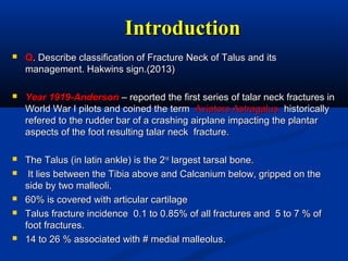
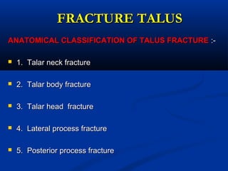
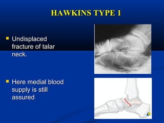
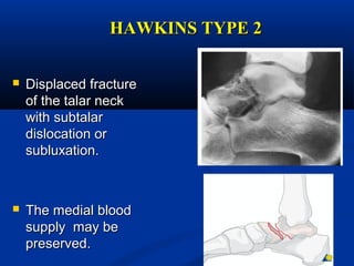
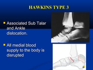
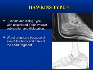
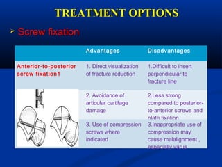
Dr. Manoj Kumar ParidaDr. Manoj Kumar Parida
DNB OrthopedicsDNB Orthopedics
Apollo HospitalsApollo Hospitals
BhubaneswarBhubaneswar
Fracture TalusFracture Talus
IntroductionIntroduction
 QQ. Describe classification of Fracture Neck of Talus and its. Describe classification of Fracture Neck of Talus and its
management. Hakwins sign.(2013)management. Hakwins sign.(2013)
 Year 1919-AndersonYear 1919-Anderson – reported the first series of talar neck fractures in– reported the first series of talar neck fractures in
World War I pilots and coined the termWorld War I pilots and coined the term Aviators Astragalus .Aviators Astragalus .historicallyhistorically
refered to the rudder bar of a crashing airplane impacting the plantarrefered to the rudder bar of a crashing airplane impacting the plantar
aspects of the foot resulting talar neck fracture.aspects of the foot resulting talar neck fracture.
 The Talus (in latin ankle) is the 2The Talus (in latin ankle) is the 2ndnd
largest tarsal bone.largest tarsal bone.
 It lies between the Tibia above and Calcanium below, gripped on theIt lies between the Tibia above and Calcanium below, gripped on the
side by two malleoli.side by two malleoli.
 60% is covered with articular cartilage60% is covered with articular cartilage
 Talus fracture incidence 0.1 to 0.85% of all fractures and 5 to 7 % ofTalus fracture incidence 0.1 to 0.85% of all fractures and 5 to 7 % of
foot fractures.foot fractures.
 14 to 26 % associated with # medial malleolus.14 to 26 % associated with # medial malleolus.
Brief AnatomyBrief Anatomy
 PARTS OF TALUSPARTS OF TALUS
1.1. HEADHEAD
2.2. NECKNECK
3.3. BODYBODY
4.4. LATERALLATERAL
PROCESSPROCESS
5.5. POSTERIORPOSTERIOR
PROCESSPROCESS
AnatomyAnatomy
 NECK OF TALUSNECK OF TALUS
 Constricted potionConstricted potion of bone between theof bone between the
body and the oval head .body and the oval head .
 Directed forward , medial and downwardDirected forward , medial and downward
 Angle of medial deviation is 15 to 20Angle of medial deviation is 15 to 20
degree in adultsdegree in adults
 Plantar deviation is 24 degree approxPlantar deviation is 24 degree approx
 Neck body angle is 150 degree in adultsNeck body angle is 150 degree in adults
 RelativelyRelatively thin diameterthin diameter makes it weakermakes it weaker
area and hence more vulnerable toarea and hence more vulnerable to
fracturesfractures
AnatomyAnatomy
 HEAD OF TALUSHEAD OF TALUS
 Anterior articular surface is large , oval and convex articulating withAnterior articular surface is large , oval and convex articulating with
navicular bonenavicular bone
 Inferior surface have two facets medial and lateral for articulation withInferior surface have two facets medial and lateral for articulation with
calcaneumcalcaneum
AnatomyAnatomy
 TARSAL CANALTARSAL CANAL
 Formed of sulcus of inferiorFormed of sulcus of inferior
surface of talus and superiorsurface of talus and superior
sulcus of calcaneumsulcus of calcaneum
 Contents-Contents- artery of tarsalartery of tarsal
canal and talocalcanealcanal and talocalcaneal
interosseous ligamentinterosseous ligament
 Posterior process has a medialPosterior process has a medial
and lateral tubercle separatedand lateral tubercle separated
by a groove for theby a groove for the flexorflexor
hallucis longus tendonhallucis longus tendon
AnatomyAnatomy
 Talus Articulates with 4Talus Articulates with 4
bonesbones
 1.Tibia1.Tibia
 2. Fibula2. Fibula
 3. Calcaneus3. Calcaneus
 4. Navicular4. Navicular
AnatomyAnatomy
 ATTACHMENTSATTACHMENTS
 NO MUSCLENO MUSCLE
ATTACHMENTSATTACHMENTS
 Medial sideMedial side
 Anterior Tibio talar ligamentAnterior Tibio talar ligament
 Posterior tibio talar ligamentPosterior tibio talar ligament
• Lateral sideLateral side
 Anterior talo fibular ligamentAnterior talo fibular ligament
 PosteriorlyPosteriorly
 Posterior talo fibularPosterior talo fibular
ligamentligament
Blood supply of talus -Blood supply of talus -
EXTRAOSEOUSEXTRAOSEOUS
ANTERIOR TIBIAL
ARTERY(36.2%)
POSTETIOR TIBIAL
ARTERY(47 %)
PERFORATING
PERONEAL
ARTERIES(16.9 %)
EXTRAOSSEOUS ARTERIES INCLUDE
ANTERIR TIBIAL OR DORSALIS PEDIS
ARTERY WHICH IS SMALLER TERMINAL
BRANCH OF POPLITEAL ARTERY.
POSTERIOR TIBIAL ARTERY WHICH IS
LARGER TERMINAL BRANCH OF POPLITEAL
ARTERY .
PEROFORATING PERONEAL ARTERY
BRANCH OF POSTERIOR TIBIAL ARTEY
THESE ARTEIES ANASTOMOSE TO FORM
SLING AROUND THE TALUS WHIS IS
SOURCE OF INTERAOSSEOUS BLOOD
SUPPLY OF TALLUS .
ARTERY TO
TARSAL SINUS
Blood supply to Tarsal CanalBlood supply to Tarsal Canal
This arises from the
Posterior tibial artery
1cm proximal to the
origin of medial and
lateral plantar artery
MEDIAL AND
LATERLA PLANTAR
ARTERIES
ARTERY TO TARSAL
CANAL
Blood supply of talus-Blood supply of talus-
INTERAOSSEOUSINTERAOSSEOUS
CORONAL SECTION
FRACTURE TALUSFRACTURE TALUS
ANATOMICAL CLASSIFICATION OF TALUS FRACTUREANATOMICAL CLASSIFICATION OF TALUS FRACTURE :-:-
 1. Talar neck fracture1. Talar neck fracture
 2. Talar body fracture2. Talar body fracture
 3. Talar head fracture3. Talar head fracture
 4. Lateral process fracture4. Lateral process fracture
 5. Posterior process fracture5. Posterior process fracture
FRACTURE NECK OF TALUSFRACTURE NECK OF TALUS
 ConstitueConstitue 30 %30 % of talar fractures.of talar fractures.
 MECHANISM OF INJURYMECHANISM OF INJURY
 Forced hyperdorsiflexion of the ankle andForced hyperdorsiflexion of the ankle and
impingement of the taller neck on the distalimpingement of the taller neck on the distal
anterior tibia .anterior tibia .
 In children, mc cause - Fall from heightIn children, mc cause - Fall from height
 Talus fractures frequently occur inTalus fractures frequently occur in a young anda young and
active populationactive population
CLINICAL EVALUATIONCLINICAL EVALUATION
• Clinical Features:Clinical Features:
 Intense pain , unable to move ankle,Intense pain , unable to move ankle,
 Gross edema and echymosis usually presentGross edema and echymosis usually present
 When there is subluxation or dislocation the normalWhen there is subluxation or dislocation the normal
contours of ankle and hind foot are distortedcontours of ankle and hind foot are distorted
 Diffuse swelling of hind foot with tenderness at the talar andDiffuse swelling of hind foot with tenderness at the talar and
subtalar joint.subtalar joint.
 Neuro vasular examination should be performed.Neuro vasular examination should be performed.
 Associated #Associated # of the foot and ankle are commonly seen withof the foot and ankle are commonly seen with
# of talar neck and body.# of talar neck and body.
HAWKIN CLASSIFICATION OFHAWKIN CLASSIFICATION OF
TALAR NECK FRACTURETALAR NECK FRACTURE
 Hawkins 1970 - talar neck fractures into fourHawkins 1970 - talar neck fractures into four
typestypes
 Canale and Kelly added type IVCanale and Kelly added type IV
 Based on displacement of body of talus.Based on displacement of body of talus.
 Useful to predict long term outcome andUseful to predict long term outcome and
development of avn of talar bodydevelopment of avn of talar body
HAWKINS TYPE 1HAWKINS TYPE 1
 UndisplacedUndisplaced
fracture of talarfracture of talar
neckneck..
 Here medial bloodHere medial blood
supply is stillsupply is still
assuredassured
HAWKINS TYPE 2HAWKINS TYPE 2
 Displaced fractureDisplaced fracture
of the talar neckof the talar neck
with subtalarwith subtalar
dislocation ordislocation or
subluxation.subluxation.
 The medial bloodThe medial blood
supply may besupply may be
preserved.preserved.
HAWKINS TYPE 3HAWKINS TYPE 3
 Associated Sub TalarAssociated Sub Talar
and Ankleand Ankle
dislocation.dislocation.
 All medial bloodAll medial blood
supply to the body issupply to the body is
disrupteddisrupted
HAWKINS TYPE 4HAWKINS TYPE 4
 (Canale and Kelly) Type 3(Canale and Kelly) Type 3
with associated Talonavicularwith associated Talonavicular
sublaxation and dislocation.sublaxation and dislocation.
 Worst prognosis because ofWorst prognosis because of
avn of the body and often ofavn of the body and often of
the head fragmentthe head fragment
RADIOGRAPHICRADIOGRAPHIC
EVALUATIONEVALUATION
 XRAYSXRAYS
 AP VIEW – Alignment of Talar bodyAP VIEW – Alignment of Talar body
 ANKLE MORTISE VIEW -ANKLE MORTISE VIEW -
 LATERAL VIEW – Talar neck and alignment of posteriorLATERAL VIEW – Talar neck and alignment of posterior
facet of subtalar jointfacet of subtalar joint
 CANALE VIEW – This provides optimum view of TalarCANALE VIEW – This provides optimum view of Talar
neckneck
RADIOGRAPHIC XRAYSRADIOGRAPHIC XRAYS
 CANALE AND KELLYCANALE AND KELLY
VIEWVIEW
 view of the talar neckview of the talar neck
achieved by ankle inachieved by ankle in
maximum equinus, footmaximum equinus, foot
placed on a cassette,placed on a cassette,
pronated 15 deg and thepronated 15 deg and the
radiographic source isradiographic source is
directed 15 deg from vertical.directed 15 deg from vertical.
 This view described forThis view described for
evaluation of post traumaticevaluation of post traumatic
deformity and is difficult todeformity and is difficult to
obtain in the acute setting.obtain in the acute setting.
DIAGNOSISDIAGNOSIS
 CT SCANCT SCAN
 give excellent visualization ofgive excellent visualization of
the congruity of the subtalarthe congruity of the subtalar
joint and provide superiorjoint and provide superior
details of fracture.details of fracture.
 small but significant fracturessmall but significant fractures
of the inferior aspect of theof the inferior aspect of the
talus, are better appreciatedtalus, are better appreciated
on CT scans compared toon CT scans compared to
plain xray films alone.plain xray films alone.
DIAGNOSISDIAGNOSIS
 MRI SCANMRI SCAN
 demonstratesdemonstrates
osteonecrosis mostosteonecrosis most
effectively.effectively.
 Use of titanium screwsUse of titanium screws
have been preffered ifhave been preffered if
AVN of bone isAVN of bone is
suspected.suspected.
 ..
TREATMENTTREATMENT
 Goals of treatment:Goals of treatment:
1.1. Early anatomic reduction of the neck fractureEarly anatomic reduction of the neck fracture
2.2. Reduction of dislocated jointsReduction of dislocated joints
3.3. Stable fixationStable fixation
4.4. Avoidance of complicationsAvoidance of complications
TREATMENTTREATMENT
 Non displaced # (Hawkins Type I )Non displaced # (Hawkins Type I )
 Non operative managementNon operative management
 Treated with below knee non weight bearing cast with ankle in slightTreated with below knee non weight bearing cast with ankle in slight
equinus for 1 monthequinus for 1 month
 Cast should be removed and short leg walking cast is applied for 2 moreCast should be removed and short leg walking cast is applied for 2 more
months until Clinical and x-ray signs of healing appears.months until Clinical and x-ray signs of healing appears.
 Once secure union is achieved active range of motion and progressiveOnce secure union is achieved active range of motion and progressive
weight bearing as tolerated is startedweight bearing as tolerated is started
SURGICAL TECHNIQUESSURGICAL TECHNIQUES
 Displaced # (Hawkins Type II to IV)Displaced # (Hawkins Type II to IV)
 SURGICAL APPROACH –SURGICAL APPROACH –
1.1. AntromedialAntromedial::
This approach may be extended from limited capsulotomy to wideThis approach may be extended from limited capsulotomy to wide
exposure with malleolur Osteotomy (as the # progress towards theexposure with malleolur Osteotomy (as the # progress towards the
body).body).
This approach allows visualization of talar neck and body. Care must beThis approach allows visualization of talar neck and body. Care must be
taken to preserve the Sephenous vein ,nerve and deltoid artery.taken to preserve the Sephenous vein ,nerve and deltoid artery.
2.2. Posteriolateral:Posteriolateral:
This approach provide posterior process and talar body. The interval isThis approach provide posterior process and talar body. The interval is
between the Peroneus brevis and FHL. Sural N must be protected. It isbetween the Peroneus brevis and FHL. Sural N must be protected. It is
usually necessary to displace the FHL from its group in the posteriorusually necessary to displace the FHL from its group in the posterior
process to facilitate exposureprocess to facilitate exposure
SURGICAL TECHNIQUESSURGICAL TECHNIQUES
 Displaced # (Hawkins Type II to IV)Displaced # (Hawkins Type II to IV)
 SURGICAL APPROACH –SURGICAL APPROACH –
3. Antrolateral:3. Antrolateral:
This approach allows visualization of Sinus Tarsi,This approach allows visualization of Sinus Tarsi,
Lateral Talar neck and subtalar joint. InadvertentLateral Talar neck and subtalar joint. Inadvertent
damage to the artery of tarsal sinus.damage to the artery of tarsal sinus.
4. Combined Anteriomedial and Anteriolateral:4. Combined Anteriomedial and Anteriolateral:
This is often used to allow maximum visualizationThis is often used to allow maximum visualization
of talar neck.of talar neck.
TREATMENTTREATMENT
 Hawkins Type III and Type IV fractureHawkins Type III and Type IV fracture
 Is Orthopaedic Emergency for two reasons:Is Orthopaedic Emergency for two reasons:
 1. Pressure from the dislocated body on the skin and neurovascular1. Pressure from the dislocated body on the skin and neurovascular
structure can lead to skin slough, neurovascular insult.structure can lead to skin slough, neurovascular insult.
 2. Blood supply to the talus is compromised and leads to avascular2. Blood supply to the talus is compromised and leads to avascular
necrosis.necrosis.
 Almost all requireAlmost all require surgical stabilizationsurgical stabilization
 SCREW FIXATIONSCREW FIXATION
 ANTERIOR TO POSTERORANTERIOR TO POSTEROR
 POSTERIOR TO ANTERIORPOSTERIOR TO ANTERIOR
• DIRECT PLATE FIXATIONDIRECT PLATE FIXATION
TREATMENT OPTIONSTREATMENT OPTIONS
 Screw fixationScrew fixation
Advantages Disadvantages
Anterior-to-posterior
screw fixation1
1. Direct visualization
of fracture reduction
1.Difficult to insert
perpendicular to
fracture line
2. Avoidance of
articular cartilage
damage
2.Less strong
compared to posterior-
to-anterior screws and
plate fixation
3. Use of compression
screws where
indicated
3.Inappropriate use of
compression may
cause malalignment ,
especially varus
TREATMENT OPTIONSTREATMENT OPTIONS
 Screw fixationScrew fixation
Advantages Disadvantages
Posterior-to-Anterior
screw fixation1
Stronger fixation
compared with anterior
screw fixation
Indirect visualization of
reduction; may require
change in positioning
Easily inserted
perpendicular to
fracture line
Some cartilage
damage to posterior
talus.
May cause less soft
tissue disruption
Risk of iatrogenic
nerve damage
TREATMENT OPTIONSTREATMENT OPTIONS
 Plate fixationPlate fixation
Advantages Disadvantages
Direct Plate Fixation
1. Strong
fixation
1. Extensive
soft tissue
dissection
2. Useful to
buttress
comminuted
columns
2. Risk of
hardware
prominence
HAWKINS SIGNHAWKINS SIGN
 Osteonecrosis is identified based on AP
radiograph between 6 and 8 week
 Subchondral lucency is indicative of
relative osteopenia secondary to bony
resorption and an intact blood supply
Progresses from medial to lateral due to
vascular re-establishing from medial side of
dome through deltoid ligament
 Indicative of diffuse osteopenia with
vascular congestion suggests continuity of
blood supply.
However the presence of sign doesn’t rule
out the Osteonecrosis, its absence also not
diagnostic for Osteonecrosis.
COMPLICATIONSCOMPLICATIONS
 Infection
Post Traumatic Arthritis
Delayed Union or Non Union
Malunion
Skin Slough
Inter position of long flexor tendon
Food Compartment syndrome
Osteonecrosis –
Hawkins I – 0 to 15%
Hawkins II – 20 to 50%
Hawkins III – 50 to 100%
Hawkins IV – Upto 100%
TALAR HEAD FRACTURETALAR HEAD FRACTURE
 Plantar Flexion and Longitudinal
compression
Non displaced # - Short Leg Cast, Partial
weight bearing for 6 weeks to preserve
longitudinal arch
Displaced # - ORIF
LATERAL PROCESSLATERAL PROCESS
FRACTUREFRACTURE
 Foot is dorsiflexed and inverted
# are often missed on initial presentation. Misinterpreted
as ankle sprain
 CT Scan should be performed
 Less than 2mm displacement - Short leg cast or boot for
6 weeks and non weight bearing atleast 4 weeks.
More than 2mm displacement – ORIF through lat.
Approach.
Comminuted # - Non viable fragment are excised.
POSTERIOR PROCESSPOSTERIOR PROCESS
FRACTUREFRACTURE
 Foot is inverted
Diagnosis of # can be difficult in part relating to
the presence of an Os-Trigunm
 Non displaced or minimal displaced - short leg
cast for 6 weeks and NBW for 4 weeks.
Displaced # - ORIF through Osterio lateral
approach.
Thank you.

Fracture Talus

  • 1.
    Dr. Manoj KumarParidaDr. Manoj Kumar Parida DNB OrthopedicsDNB Orthopedics Apollo HospitalsApollo Hospitals BhubaneswarBhubaneswar Fracture TalusFracture Talus
  • 2.
    IntroductionIntroduction  QQ. Describeclassification of Fracture Neck of Talus and its. Describe classification of Fracture Neck of Talus and its management. Hakwins sign.(2013)management. Hakwins sign.(2013)  Year 1919-AndersonYear 1919-Anderson – reported the first series of talar neck fractures in– reported the first series of talar neck fractures in World War I pilots and coined the termWorld War I pilots and coined the term Aviators Astragalus .Aviators Astragalus .historicallyhistorically refered to the rudder bar of a crashing airplane impacting the plantarrefered to the rudder bar of a crashing airplane impacting the plantar aspects of the foot resulting talar neck fracture.aspects of the foot resulting talar neck fracture.  The Talus (in latin ankle) is the 2The Talus (in latin ankle) is the 2ndnd largest tarsal bone.largest tarsal bone.  It lies between the Tibia above and Calcanium below, gripped on theIt lies between the Tibia above and Calcanium below, gripped on the side by two malleoli.side by two malleoli.  60% is covered with articular cartilage60% is covered with articular cartilage  Talus fracture incidence 0.1 to 0.85% of all fractures and 5 to 7 % ofTalus fracture incidence 0.1 to 0.85% of all fractures and 5 to 7 % of foot fractures.foot fractures.  14 to 26 % associated with # medial malleolus.14 to 26 % associated with # medial malleolus.
  • 3.
    Brief AnatomyBrief Anatomy PARTS OF TALUSPARTS OF TALUS 1.1. HEADHEAD 2.2. NECKNECK 3.3. BODYBODY 4.4. LATERALLATERAL PROCESSPROCESS 5.5. POSTERIORPOSTERIOR PROCESSPROCESS
  • 4.
    AnatomyAnatomy  NECK OFTALUSNECK OF TALUS  Constricted potionConstricted potion of bone between theof bone between the body and the oval head .body and the oval head .  Directed forward , medial and downwardDirected forward , medial and downward  Angle of medial deviation is 15 to 20Angle of medial deviation is 15 to 20 degree in adultsdegree in adults  Plantar deviation is 24 degree approxPlantar deviation is 24 degree approx  Neck body angle is 150 degree in adultsNeck body angle is 150 degree in adults  RelativelyRelatively thin diameterthin diameter makes it weakermakes it weaker area and hence more vulnerable toarea and hence more vulnerable to fracturesfractures
  • 5.
    AnatomyAnatomy  HEAD OFTALUSHEAD OF TALUS  Anterior articular surface is large , oval and convex articulating withAnterior articular surface is large , oval and convex articulating with navicular bonenavicular bone  Inferior surface have two facets medial and lateral for articulation withInferior surface have two facets medial and lateral for articulation with calcaneumcalcaneum
  • 6.
    AnatomyAnatomy  TARSAL CANALTARSALCANAL  Formed of sulcus of inferiorFormed of sulcus of inferior surface of talus and superiorsurface of talus and superior sulcus of calcaneumsulcus of calcaneum  Contents-Contents- artery of tarsalartery of tarsal canal and talocalcanealcanal and talocalcaneal interosseous ligamentinterosseous ligament  Posterior process has a medialPosterior process has a medial and lateral tubercle separatedand lateral tubercle separated by a groove for theby a groove for the flexorflexor hallucis longus tendonhallucis longus tendon
  • 7.
    AnatomyAnatomy  Talus Articulateswith 4Talus Articulates with 4 bonesbones  1.Tibia1.Tibia  2. Fibula2. Fibula  3. Calcaneus3. Calcaneus  4. Navicular4. Navicular
  • 8.
    AnatomyAnatomy  ATTACHMENTSATTACHMENTS  NOMUSCLENO MUSCLE ATTACHMENTSATTACHMENTS  Medial sideMedial side  Anterior Tibio talar ligamentAnterior Tibio talar ligament  Posterior tibio talar ligamentPosterior tibio talar ligament • Lateral sideLateral side  Anterior talo fibular ligamentAnterior talo fibular ligament  PosteriorlyPosteriorly  Posterior talo fibularPosterior talo fibular ligamentligament
  • 9.
    Blood supply oftalus -Blood supply of talus - EXTRAOSEOUSEXTRAOSEOUS ANTERIOR TIBIAL ARTERY(36.2%) POSTETIOR TIBIAL ARTERY(47 %) PERFORATING PERONEAL ARTERIES(16.9 %) EXTRAOSSEOUS ARTERIES INCLUDE ANTERIR TIBIAL OR DORSALIS PEDIS ARTERY WHICH IS SMALLER TERMINAL BRANCH OF POPLITEAL ARTERY. POSTERIOR TIBIAL ARTERY WHICH IS LARGER TERMINAL BRANCH OF POPLITEAL ARTERY . PEROFORATING PERONEAL ARTERY BRANCH OF POSTERIOR TIBIAL ARTEY THESE ARTEIES ANASTOMOSE TO FORM SLING AROUND THE TALUS WHIS IS SOURCE OF INTERAOSSEOUS BLOOD SUPPLY OF TALLUS . ARTERY TO TARSAL SINUS
  • 10.
    Blood supply toTarsal CanalBlood supply to Tarsal Canal This arises from the Posterior tibial artery 1cm proximal to the origin of medial and lateral plantar artery MEDIAL AND LATERLA PLANTAR ARTERIES ARTERY TO TARSAL CANAL
  • 11.
    Blood supply oftalus-Blood supply of talus- INTERAOSSEOUSINTERAOSSEOUS CORONAL SECTION
  • 12.
    FRACTURE TALUSFRACTURE TALUS ANATOMICALCLASSIFICATION OF TALUS FRACTUREANATOMICAL CLASSIFICATION OF TALUS FRACTURE :-:-  1. Talar neck fracture1. Talar neck fracture  2. Talar body fracture2. Talar body fracture  3. Talar head fracture3. Talar head fracture  4. Lateral process fracture4. Lateral process fracture  5. Posterior process fracture5. Posterior process fracture
  • 13.
    FRACTURE NECK OFTALUSFRACTURE NECK OF TALUS  ConstitueConstitue 30 %30 % of talar fractures.of talar fractures.  MECHANISM OF INJURYMECHANISM OF INJURY  Forced hyperdorsiflexion of the ankle andForced hyperdorsiflexion of the ankle and impingement of the taller neck on the distalimpingement of the taller neck on the distal anterior tibia .anterior tibia .  In children, mc cause - Fall from heightIn children, mc cause - Fall from height  Talus fractures frequently occur inTalus fractures frequently occur in a young anda young and active populationactive population
  • 14.
    CLINICAL EVALUATIONCLINICAL EVALUATION •Clinical Features:Clinical Features:  Intense pain , unable to move ankle,Intense pain , unable to move ankle,  Gross edema and echymosis usually presentGross edema and echymosis usually present  When there is subluxation or dislocation the normalWhen there is subluxation or dislocation the normal contours of ankle and hind foot are distortedcontours of ankle and hind foot are distorted  Diffuse swelling of hind foot with tenderness at the talar andDiffuse swelling of hind foot with tenderness at the talar and subtalar joint.subtalar joint.  Neuro vasular examination should be performed.Neuro vasular examination should be performed.  Associated #Associated # of the foot and ankle are commonly seen withof the foot and ankle are commonly seen with # of talar neck and body.# of talar neck and body.
  • 15.
    HAWKIN CLASSIFICATION OFHAWKINCLASSIFICATION OF TALAR NECK FRACTURETALAR NECK FRACTURE  Hawkins 1970 - talar neck fractures into fourHawkins 1970 - talar neck fractures into four typestypes  Canale and Kelly added type IVCanale and Kelly added type IV  Based on displacement of body of talus.Based on displacement of body of talus.  Useful to predict long term outcome andUseful to predict long term outcome and development of avn of talar bodydevelopment of avn of talar body
  • 16.
    HAWKINS TYPE 1HAWKINSTYPE 1  UndisplacedUndisplaced fracture of talarfracture of talar neckneck..  Here medial bloodHere medial blood supply is stillsupply is still assuredassured
  • 17.
    HAWKINS TYPE 2HAWKINSTYPE 2  Displaced fractureDisplaced fracture of the talar neckof the talar neck with subtalarwith subtalar dislocation ordislocation or subluxation.subluxation.  The medial bloodThe medial blood supply may besupply may be preserved.preserved.
  • 18.
    HAWKINS TYPE 3HAWKINSTYPE 3  Associated Sub TalarAssociated Sub Talar and Ankleand Ankle dislocation.dislocation.  All medial bloodAll medial blood supply to the body issupply to the body is disrupteddisrupted
  • 19.
    HAWKINS TYPE 4HAWKINSTYPE 4  (Canale and Kelly) Type 3(Canale and Kelly) Type 3 with associated Talonavicularwith associated Talonavicular sublaxation and dislocation.sublaxation and dislocation.  Worst prognosis because ofWorst prognosis because of avn of the body and often ofavn of the body and often of the head fragmentthe head fragment
  • 20.
    RADIOGRAPHICRADIOGRAPHIC EVALUATIONEVALUATION  XRAYSXRAYS  APVIEW – Alignment of Talar bodyAP VIEW – Alignment of Talar body  ANKLE MORTISE VIEW -ANKLE MORTISE VIEW -  LATERAL VIEW – Talar neck and alignment of posteriorLATERAL VIEW – Talar neck and alignment of posterior facet of subtalar jointfacet of subtalar joint  CANALE VIEW – This provides optimum view of TalarCANALE VIEW – This provides optimum view of Talar neckneck
  • 21.
    RADIOGRAPHIC XRAYSRADIOGRAPHIC XRAYS CANALE AND KELLYCANALE AND KELLY VIEWVIEW  view of the talar neckview of the talar neck achieved by ankle inachieved by ankle in maximum equinus, footmaximum equinus, foot placed on a cassette,placed on a cassette, pronated 15 deg and thepronated 15 deg and the radiographic source isradiographic source is directed 15 deg from vertical.directed 15 deg from vertical.  This view described forThis view described for evaluation of post traumaticevaluation of post traumatic deformity and is difficult todeformity and is difficult to obtain in the acute setting.obtain in the acute setting.
  • 22.
    DIAGNOSISDIAGNOSIS  CT SCANCTSCAN  give excellent visualization ofgive excellent visualization of the congruity of the subtalarthe congruity of the subtalar joint and provide superiorjoint and provide superior details of fracture.details of fracture.  small but significant fracturessmall but significant fractures of the inferior aspect of theof the inferior aspect of the talus, are better appreciatedtalus, are better appreciated on CT scans compared toon CT scans compared to plain xray films alone.plain xray films alone.
  • 23.
    DIAGNOSISDIAGNOSIS  MRI SCANMRISCAN  demonstratesdemonstrates osteonecrosis mostosteonecrosis most effectively.effectively.  Use of titanium screwsUse of titanium screws have been preffered ifhave been preffered if AVN of bone isAVN of bone is suspected.suspected.  ..
  • 24.
    TREATMENTTREATMENT  Goals oftreatment:Goals of treatment: 1.1. Early anatomic reduction of the neck fractureEarly anatomic reduction of the neck fracture 2.2. Reduction of dislocated jointsReduction of dislocated joints 3.3. Stable fixationStable fixation 4.4. Avoidance of complicationsAvoidance of complications
  • 25.
    TREATMENTTREATMENT  Non displaced# (Hawkins Type I )Non displaced # (Hawkins Type I )  Non operative managementNon operative management  Treated with below knee non weight bearing cast with ankle in slightTreated with below knee non weight bearing cast with ankle in slight equinus for 1 monthequinus for 1 month  Cast should be removed and short leg walking cast is applied for 2 moreCast should be removed and short leg walking cast is applied for 2 more months until Clinical and x-ray signs of healing appears.months until Clinical and x-ray signs of healing appears.  Once secure union is achieved active range of motion and progressiveOnce secure union is achieved active range of motion and progressive weight bearing as tolerated is startedweight bearing as tolerated is started
  • 26.
    SURGICAL TECHNIQUESSURGICAL TECHNIQUES Displaced # (Hawkins Type II to IV)Displaced # (Hawkins Type II to IV)  SURGICAL APPROACH –SURGICAL APPROACH – 1.1. AntromedialAntromedial:: This approach may be extended from limited capsulotomy to wideThis approach may be extended from limited capsulotomy to wide exposure with malleolur Osteotomy (as the # progress towards theexposure with malleolur Osteotomy (as the # progress towards the body).body). This approach allows visualization of talar neck and body. Care must beThis approach allows visualization of talar neck and body. Care must be taken to preserve the Sephenous vein ,nerve and deltoid artery.taken to preserve the Sephenous vein ,nerve and deltoid artery. 2.2. Posteriolateral:Posteriolateral: This approach provide posterior process and talar body. The interval isThis approach provide posterior process and talar body. The interval is between the Peroneus brevis and FHL. Sural N must be protected. It isbetween the Peroneus brevis and FHL. Sural N must be protected. It is usually necessary to displace the FHL from its group in the posteriorusually necessary to displace the FHL from its group in the posterior process to facilitate exposureprocess to facilitate exposure
  • 27.
    SURGICAL TECHNIQUESSURGICAL TECHNIQUES Displaced # (Hawkins Type II to IV)Displaced # (Hawkins Type II to IV)  SURGICAL APPROACH –SURGICAL APPROACH – 3. Antrolateral:3. Antrolateral: This approach allows visualization of Sinus Tarsi,This approach allows visualization of Sinus Tarsi, Lateral Talar neck and subtalar joint. InadvertentLateral Talar neck and subtalar joint. Inadvertent damage to the artery of tarsal sinus.damage to the artery of tarsal sinus. 4. Combined Anteriomedial and Anteriolateral:4. Combined Anteriomedial and Anteriolateral: This is often used to allow maximum visualizationThis is often used to allow maximum visualization of talar neck.of talar neck.
  • 28.
    TREATMENTTREATMENT  Hawkins TypeIII and Type IV fractureHawkins Type III and Type IV fracture  Is Orthopaedic Emergency for two reasons:Is Orthopaedic Emergency for two reasons:  1. Pressure from the dislocated body on the skin and neurovascular1. Pressure from the dislocated body on the skin and neurovascular structure can lead to skin slough, neurovascular insult.structure can lead to skin slough, neurovascular insult.  2. Blood supply to the talus is compromised and leads to avascular2. Blood supply to the talus is compromised and leads to avascular necrosis.necrosis.  Almost all requireAlmost all require surgical stabilizationsurgical stabilization  SCREW FIXATIONSCREW FIXATION  ANTERIOR TO POSTERORANTERIOR TO POSTEROR  POSTERIOR TO ANTERIORPOSTERIOR TO ANTERIOR • DIRECT PLATE FIXATIONDIRECT PLATE FIXATION
  • 29.
    TREATMENT OPTIONSTREATMENT OPTIONS Screw fixationScrew fixation Advantages Disadvantages Anterior-to-posterior screw fixation1 1. Direct visualization of fracture reduction 1.Difficult to insert perpendicular to fracture line 2. Avoidance of articular cartilage damage 2.Less strong compared to posterior- to-anterior screws and plate fixation 3. Use of compression screws where indicated 3.Inappropriate use of compression may cause malalignment , especially varus
  • 30.
    TREATMENT OPTIONSTREATMENT OPTIONS Screw fixationScrew fixation Advantages Disadvantages Posterior-to-Anterior screw fixation1 Stronger fixation compared with anterior screw fixation Indirect visualization of reduction; may require change in positioning Easily inserted perpendicular to fracture line Some cartilage damage to posterior talus. May cause less soft tissue disruption Risk of iatrogenic nerve damage
  • 31.
    TREATMENT OPTIONSTREATMENT OPTIONS Plate fixationPlate fixation Advantages Disadvantages Direct Plate Fixation 1. Strong fixation 1. Extensive soft tissue dissection 2. Useful to buttress comminuted columns 2. Risk of hardware prominence
  • 32.
    HAWKINS SIGNHAWKINS SIGN Osteonecrosis is identified based on AP radiograph between 6 and 8 week  Subchondral lucency is indicative of relative osteopenia secondary to bony resorption and an intact blood supply Progresses from medial to lateral due to vascular re-establishing from medial side of dome through deltoid ligament  Indicative of diffuse osteopenia with vascular congestion suggests continuity of blood supply. However the presence of sign doesn’t rule out the Osteonecrosis, its absence also not diagnostic for Osteonecrosis.
  • 33.
    COMPLICATIONSCOMPLICATIONS  Infection Post TraumaticArthritis Delayed Union or Non Union Malunion Skin Slough Inter position of long flexor tendon Food Compartment syndrome Osteonecrosis – Hawkins I – 0 to 15% Hawkins II – 20 to 50% Hawkins III – 50 to 100% Hawkins IV – Upto 100%
  • 34.
    TALAR HEAD FRACTURETALARHEAD FRACTURE  Plantar Flexion and Longitudinal compression Non displaced # - Short Leg Cast, Partial weight bearing for 6 weeks to preserve longitudinal arch Displaced # - ORIF
  • 35.
    LATERAL PROCESSLATERAL PROCESS FRACTUREFRACTURE Foot is dorsiflexed and inverted # are often missed on initial presentation. Misinterpreted as ankle sprain  CT Scan should be performed  Less than 2mm displacement - Short leg cast or boot for 6 weeks and non weight bearing atleast 4 weeks. More than 2mm displacement – ORIF through lat. Approach. Comminuted # - Non viable fragment are excised.
  • 36.
    POSTERIOR PROCESSPOSTERIOR PROCESS FRACTUREFRACTURE Foot is inverted Diagnosis of # can be difficult in part relating to the presence of an Os-Trigunm  Non displaced or minimal displaced - short leg cast for 6 weeks and NBW for 4 weeks. Displaced # - ORIF through Osterio lateral approach.
  • 37.