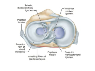
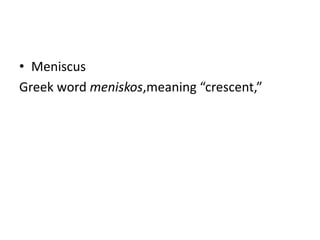
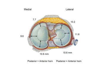
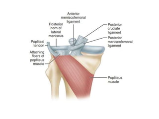
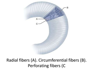
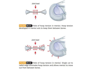
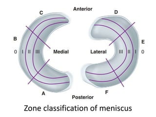
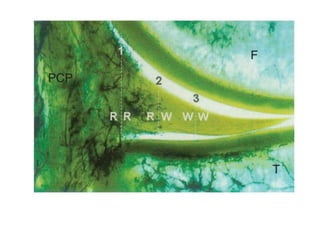
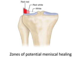
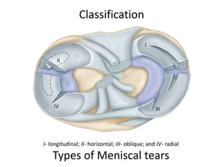
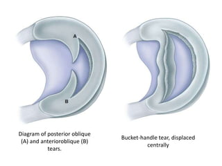
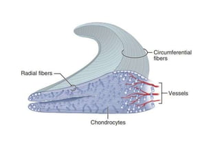
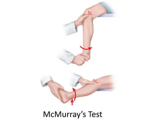
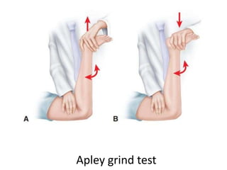
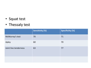
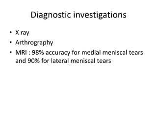
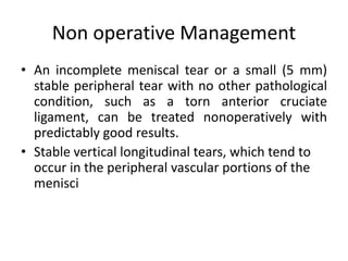
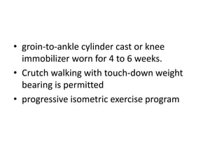
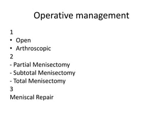
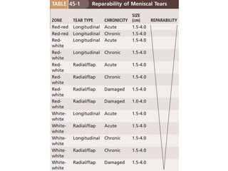
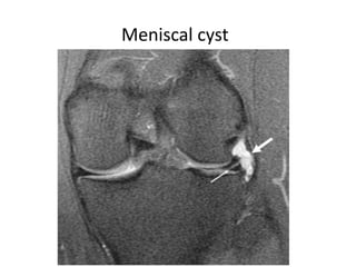
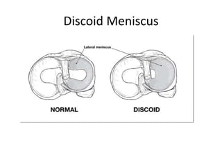
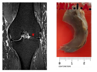
Meniscus
Anatomy, Function and Injury
Dr Asish Rajak
Fellow Sports Medicine and
Arthroplasty
• Meniscus
Greek word meniskos,meaning “crescent,”
Embryology and Development
• characteristic shape of the lateral and medial
menisci is attained between the 8th and 10th
week of gestation
• Highly cellular and vascular, with the blood
supply entering from the periphery and
extending through the entire width of the
menisci
Radial fibers (A). Circumferential fibers (B).
Perforating fibers (C
Zone classification of meniscus
Zones of potential meniscal healing
Function
• Joint filler
• Prevent capsular and synovial impingement
• joint lubrication
• contribute to stability
• Nutrition
• Load bearing / Weight bearing
• Stabilization of the joint
Tears of Menisci
• most commonly by rotation as the flexed knee
moves toward an extended position
• most common location for injury is the
posterior horn of the meniscus, and
longitudinal tears are the most common type
of injury
• More with
- peripheral cystic formation
- previous injury or disease
- discoid lateral meniscus
- Aging
- Incongruities or ligamentous disruptions
- Congenitally relaxed joints
- Inadequate musculature, especially the
quadriceps
Mechanism of tear
• rotational force incurred while the joint is
partially flexed
• the meniscus is entrapped between the
femoral and tibial condyles in flexion, tearing
as the knee is extended
I- longitudinal; II- horizontal; III- oblique; and IV- radial
Types of Meniscal tears
Classification
Diagram of posterior oblique
(A) and anterioroblique (B)
tears.
Bucket-handle tear, displaced
centrally
Radial tears
Presentation
• catching, snapping, or clicking
• occasional pain and mild swelling
• Locking/ No locking
• Atrophy of the Quadriceps
• Significant : giving way and locking
Diagnosis
• Difficult
• Careful history, physical examination, standard
X rays , MRI and Arthroscopy – reduces errors
to <5%
McMurray’s Test
Apley grind test
Caution:
• Tears of one meniscus can produce pain in the
opposite compartment of the knee. This is
most commonly seen with posterior tears of
the lateral meniscus.
• Squat test
• Thessaly test
Sensitivity (%) Specificiity (%)
McMurray’s test 70 71
Apley 60 70
Joint line tenderness 63 77
Diagnostic investigations
• X ray
• Arthrography
• MRI : 98% accuracy for medial meniscal tears
and 90% for lateral meniscal tears
Non operative Management
• An incomplete meniscal tear or a small (5 mm)
stable peripheral tear with no other pathological
condition, such as a torn anterior cruciate
ligament, can be treated nonoperatively with
predictably good results.
• Stable vertical longitudinal tears, which tend to
occur in the peripheral vascular portions of the
menisci
• groin-to-ankle cylinder cast or knee
immobilizer worn for 4 to 6 weeks.
• Crutch walking with touch-down weight
bearing is permitted
• progressive isometric exercise program
Operative management
1
• Open
• Arthroscopic
2
- Partial Menisectomy
- Subtotal Menisectomy
- Total Menisectomy
3
Meniscal Repair
Meniscal cyst
Discoid Meniscus
Watanabe Classification
A. Incomplete B. Complete C. Wrisberg
Thank you
Questions
• ? Difference between Radial and Horizontal tear
• ? Why Meniscal cyst and Discoid meniscus is more in
the lateral mensicus
• ? Why MRI accuracy differs between lateral and medial
meniscus
• ? How does Medial meniscus contribute to stability
• ? What is Pisani's Sign
• ? Function and clinical significance of Humphry’s and
Wrisberg Ligament
• ? ISAKOS classification
• ? History of Meniscus procedures

Meniscus - Anatomy, function and injury

  • 1.
    Meniscus Anatomy, Function andInjury Dr Asish Rajak Fellow Sports Medicine and Arthroplasty
  • 3.
    • Meniscus Greek wordmeniskos,meaning “crescent,”
  • 4.
    Embryology and Development •characteristic shape of the lateral and medial menisci is attained between the 8th and 10th week of gestation • Highly cellular and vascular, with the blood supply entering from the periphery and extending through the entire width of the menisci
  • 7.
    Radial fibers (A).Circumferential fibers (B). Perforating fibers (C
  • 9.
  • 12.
    Zones of potentialmeniscal healing
  • 13.
    Function • Joint filler •Prevent capsular and synovial impingement • joint lubrication • contribute to stability • Nutrition • Load bearing / Weight bearing • Stabilization of the joint
  • 14.
    Tears of Menisci •most commonly by rotation as the flexed knee moves toward an extended position • most common location for injury is the posterior horn of the meniscus, and longitudinal tears are the most common type of injury
  • 15.
    • More with -peripheral cystic formation - previous injury or disease - discoid lateral meniscus - Aging - Incongruities or ligamentous disruptions - Congenitally relaxed joints - Inadequate musculature, especially the quadriceps
  • 16.
    Mechanism of tear •rotational force incurred while the joint is partially flexed • the meniscus is entrapped between the femoral and tibial condyles in flexion, tearing as the knee is extended
  • 17.
    I- longitudinal; II-horizontal; III- oblique; and IV- radial Types of Meniscal tears Classification
  • 18.
    Diagram of posterioroblique (A) and anterioroblique (B) tears. Bucket-handle tear, displaced centrally
  • 21.
  • 22.
    Presentation • catching, snapping,or clicking • occasional pain and mild swelling • Locking/ No locking • Atrophy of the Quadriceps • Significant : giving way and locking
  • 23.
    Diagnosis • Difficult • Carefulhistory, physical examination, standard X rays , MRI and Arthroscopy – reduces errors to <5%
  • 24.
  • 25.
  • 26.
    Caution: • Tears ofone meniscus can produce pain in the opposite compartment of the knee. This is most commonly seen with posterior tears of the lateral meniscus.
  • 27.
    • Squat test •Thessaly test Sensitivity (%) Specificiity (%) McMurray’s test 70 71 Apley 60 70 Joint line tenderness 63 77
  • 28.
    Diagnostic investigations • Xray • Arthrography • MRI : 98% accuracy for medial meniscal tears and 90% for lateral meniscal tears
  • 29.
    Non operative Management •An incomplete meniscal tear or a small (5 mm) stable peripheral tear with no other pathological condition, such as a torn anterior cruciate ligament, can be treated nonoperatively with predictably good results. • Stable vertical longitudinal tears, which tend to occur in the peripheral vascular portions of the menisci
  • 30.
    • groin-to-ankle cylindercast or knee immobilizer worn for 4 to 6 weeks. • Crutch walking with touch-down weight bearing is permitted • progressive isometric exercise program
  • 31.
    Operative management 1 • Open •Arthroscopic 2 - Partial Menisectomy - Subtotal Menisectomy - Total Menisectomy 3 Meniscal Repair
  • 33.
  • 34.
  • 36.
  • 37.
  • 38.
    Questions • ? Differencebetween Radial and Horizontal tear • ? Why Meniscal cyst and Discoid meniscus is more in the lateral mensicus • ? Why MRI accuracy differs between lateral and medial meniscus • ? How does Medial meniscus contribute to stability • ? What is Pisani's Sign • ? Function and clinical significance of Humphry’s and Wrisberg Ligament • ? ISAKOS classification • ? History of Meniscus procedures

Editor's Notes

  • #2 The menisci are described as moving with the femoral condyles with flexion and extension but moving with the tibia with rotary movements. The articular surface of the medial condyle is prolonged anteriorly, and as the knee comes into the fully extended position, the femur internally rotates until the remaining articular surface on the medial condyle is in contact. The posterior portion of the lateral condyle rotates forward laterally, thus producing a “screwing home” movement, locking the knee in the fully extended position. When flexion is initiated, unscrewing of the joint occurs by external rotation of the femur on the tibia.
  • #3 Superior view of tibial condyles after removal of femur. Lateral meniscus is smaller in diameter, thicker around its periphery, wider in body, and more mobile; posteriorly, it is attached to medial femoral condyle by either anterior or posterior meniscofemoral ligament, depending on which is present, and to popliteus muscle. - The medial meniscus is a C-shaped structure larger in radius than the lateral meniscus, with the posterior horn being wider than the anterior. The anterior horn is attached firmly to the tibia anterior to the intercondylar eminence and to the anterior cruciate ligament. Most of the weight is borne on the posterior portion of the meniscus. The posterior horn is anchored immediately in front of the attachments of the posterior cruciate ligament posterior to the intercondylar eminence. Its entire peripheral border is firmly attached to the medial capsule and through the coronary ligament to the upper border of the tibia. - The lateral meniscus is more circular in form, covering up to two thirds of the articular surface of the underlying tibial plateau. The anterior horn is attached to the tibia medially in front of the intercondylar eminence, whereas the posterior horn inserts into the posterior aspect of the intercondylar eminence and in front of the posterior attachment of the medial meniscus. The posterior horn often receives anchorage also to the femur by the ligament of Wrisberg and the ligament of Humphry and from fascia covering the popliteus muscle and the arcuate complex at the posterolateral corner of the knee. The inner border, like that of the medial meniscus, is thin, concave, and free. The tendon of the popliteus muscle separates the posterolateral periphery of the lateral meniscus from the joint capsule and the fibular collateral ligament. The tendon of the popliteus is enveloped in a synovial membrane and forms an oblique groove on the lateral border of the meniscus. - The lateral meniscus is smaller in diameter, thicker in periphery, wider in body, and more mobile than the medial meniscus. It is attached to both cruciate ligaments and posteriorly to the medial femoral condyle by either the ligament of Humphry or the ligament of Wrisberg, depending on which is present; it is also attached posteriorly to the popliteus muscle (see Figs. 45-26 and 45-27). It is separated from the lateral collateral ligament by the popliteal tendon. In contrast, the medial meniscus is much larger in diameter, is thinner in its periphery and narrower in body, and does not attach to either cruciate ligament. It is loosely attached to the medial capsular ligaments.
  • #5 By adulthood, only the peripheral 10% to 30% have a blood supply.
  • #7 Posterior view of knee after removal of femur. Posteriorly, lateral meniscus is attached to either anterior or posterior meniscofemoral ligament, depending on which is present, and to popliteus muscle.
  • #8 Pattern of collagen fibers within meniscus. Radial fibers (A). Circumferential fibers (B). Perforating fibers (C). - In an anterior cruciate ligament–deficient knee, medial meniscectomy has been shown to increase tibial translation by 58% at 90 degrees. - Biomechanical studies have shown that under loads of up to 150 kg, the lateral meniscus appears to carry 70% of the load on that side of the joint; whereas on the medial side, the load is shared approximately equally by the meniscus and the exposed articular cartilage. Medial meniscectomy decreases contact area by 50% to 70% and increases contact stress by 100%. Lateral meniscectomy decreases contact area by 40% to 50% but dramatically increases contact stress by 200% to 300% because of the relative convex surface of the lateral tibial plateau.
  • #10 Zone classification of meniscus (modified from Cooper et al.). Most anterior zone of medial meniscus is labeled C, whereas most anterior zone of lateral meniscus is labeled D. 0 is meniscosynovial junction; I is outer third, II is middle third, and III is inner third of each meniscus.
  • #11 Frontal section of medial compartment of knee. Branching radial vessels from perimeniscal capillary plexus (PCP) can be seen penetrating peripheral border of medial meniscus. F, Femur; T, tibia. Three zones of meniscal vascularity are shown: 1 RR, red-red is fully within vascular area; 2 RW, red-white is at border of vascular area; and 3 WW, white-white is within avascular area. - Microinjection techniques have shown that the depth of peripheral vascular penetration is 10% to 30% of the width of the medial meniscus and 10% to 25% of the width of the lateral meniscus. - Meniscal tears have been classified on the basis of their location in three zones of vascularity—red (fully within the vascular area), red-white (at the border of the vascular area), and white (within the avascular area)—and this classification indicates the potential for healing after repair.
  • #12 Superior aspect of medial (A) and lateral (B) menisci after vascular perfusion with India ink and tissue clearing by modified Spalteholz technique. Note vascularity at periphery of meniscus as well as at anterior and posterior horn attachments. Absence of peripheral vasculature at posterolateral corner of lateral meniscus (arrow) represents area of passage of popliteal tendon. - The vascular supply to the medial and lateral menisci originates predominantly from the lateral and medial geniculate vessels (both inferior and superior). Branches from these vessels give rise to a perimeniscal capillary plexus within the synovial and capsular tissue.
  • #14 The menisci act as a joint filler (approx. 1mm), compensating for gross incongruity between femoral and tibial articulating surfaces. The menisci are believed to have a joint lubrication function, helping to distribute synovial fluid throughout the joint and aiding the nutrition of the articular cartilage. They undoubtedly contribute to stability in all planes but are especially important rotary stabilizers and are probably essential for the smooth transition from a pure hinge to a gliding or rotary motion as the knee moves from flexion to extension. The larger contact area provided by the meniscus reduces the average contact stress acting between the bones. The menisci are thus important in reducing the stress on the articular cartilage; they prevent mechanical damage to both the chondrocytes and the extracellular matrix.
  • #18 O’Connor Four basic patterns of meniscal tears: I, longitudinal; II, horizontal; III, oblique; and IV, radial. - variations include flap tears, complex tears, and degenerative meniscal tears .
  • #19 Diagram of posterior oblique (A) and anterioroblique (B) tears. Bucket-handle tear, displaced centrally
  • #20 Cross section of meniscus showing horizontal cleavage split.
  • #21 Two tears of medial meniscus: classic buckethandle tear and tear of posterior peripheral part.
  • #22 Radial tears: incomplete radial tear involves part of width of meniscus (A); complete radial tear extends to periphery (B); and incomplete tear extending posteriorly or anteriorly is called “parrot beak” tear (C).
  • #23 - Locking may not be recognized unless the injured knee is compared with the opposite knee, which should exhibit the 5 to 10 degrees of recurvatum that normally is present. The injured knee can be locked and still extend to neutral position. Locking usually occurs only with longitudinal tears and is much more common with bucket-handle tears, usually of the medial meniscus. Locking of the knee must not be considered pathognomonic of a bucket-handle tear of a meniscus; an intraarticular tumor, an osteocartilaginous loose body, and other conditions can cause locking . - False locking occurs most often soon after an injury in which hemorrhage around the posterior part of the capsule or a collateral ligament with associated hamstring spasm prevents complete extension of the knee. - Aspiration and a short period of rest until the reaction has partially subsided usually will differentiate locking from false locking of the joint. - The meniscus itself is without nerve fibers except at its periphery; therefore, the tenderness or pain is related to synovitis in the adjacent capsular and synovial tissues. - Clicks, snaps, or catches, either audible or detected by palpation during flexion, extension, and rotary motions of the joint, can be valuable diagnostically, and efforts should be made to reproduce and accurately locate them. If these noises are localized to the joint line, the meniscus most likely contains a tear.
  • #32 Anteroposterior, lateral, and intercondylar notch views with a tangential view of the inferior surface of the patella should be routine. Ordinary radiographs will not confirm the diagnosis of a torn meniscus but are essential to exclude osteocartilaginous loose bodies, osteochondritis dissecans, and other pathological processes that can mimic a torn meniscus
  • #33 - A vertical longitudinal tear involving the body of the meniscus was classified as stable when the portion of the meniscus that was central to the tear could not be displaced more than 3 mm from the intact peripheral rim. - Chronic tears with a superimposed acute injury cannot be expected to heal with nonoperative treatment.
  • #35  In partial meniscectomies, a intact, stable and balanced peripheral rim of healthy meniscal tissue is preserved. Subtotal : In this type of meniscectomy, the type and extent of the tear require excision of a portion of the peripheral rim of the meniscus. It is termed subtotal because in most cases most of the anterior horn and a portion of the middle third of the meniscus are not resected. Total removal of the meniscus is required when it is detached from its peripheral meniscosynovial attachment, and intrameniscal damage and tears are extensive.
  • #37 - Fat-suppressed coronal T2-weighted MR sequence shows high-signal intensity meniscal cyst (large white arrow) with an associated oblique tear (small white arrow) in the body of the lateral meniscus. - When of average size, they are characteristically more prominent when the knee is extended and less prominent when the knee is flexed; small cysts may disappear within the joint on flexion (Pisani sign).
  • #39 The transverse diameter of a normal meniscus is approximately 10 to 11 mm; therefore a normal meniscus body will be visible on only 2 slices of a MR with 4-5-mm sagittal slices. A discoid meniscus should be considered if more than two contiguous body segments are present. However, this method may lead to a false negative when evaluating people with the Wrisberg variant of discoid meniscus since it maintains a narrow crescent shape. Coronal and radial images of the meniscus are useful to demonstrate the extension of the aberrant meniscus into the joint as seen here. On coronal images, it is diagnosed when the horizontal measurement between the free margin and the periphery of the body is more than 1.4 cm. Rarely, X-ray may show lateral joint space widening, squaring of the lateral condyle, cupping of the lateral tibial plateau and hypoplasia of the lateral tibial spine that suggest discoid meniscus.
  • #40 Discoid meniscus on coronal proton-density weighted MRI Photograph of a congenital discoid menisci specimen from the University of Cape Town Pathology Learning Centre teaching collection.
  • #41 Wrisberg-type discoid menisci usually are nearly normal in size and shape and have no posterior attachment except the ligament of Wrisberg (Fig. 45-44). Because this type is not disc shaped, Neuschwander et al. described it as a “lateral meniscal variant with absence of the posterior coronary ligament” to distinguish it from a truly discoid meniscus. Wrisberg-type discoid menisci often occur at a younger age than complete or incomplete types and are unassociated with trauma. Abnormal motion of this type of discoid meniscus results in a popping sound during knee flexion and extension (“snapping knee syndrome”).