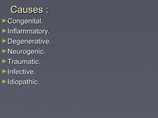
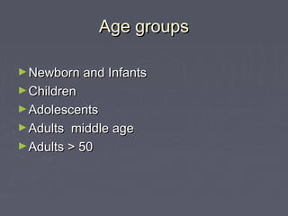
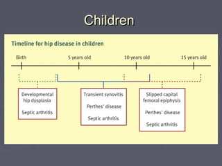
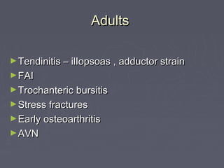
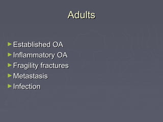
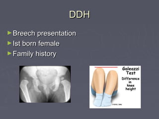
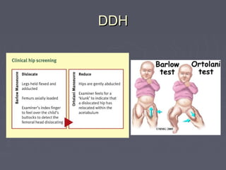
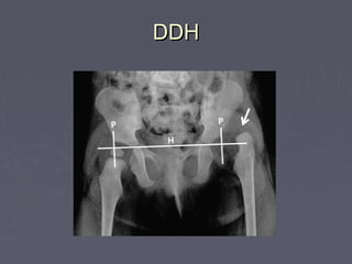
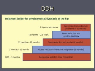
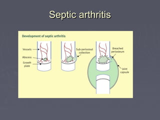
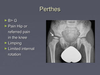
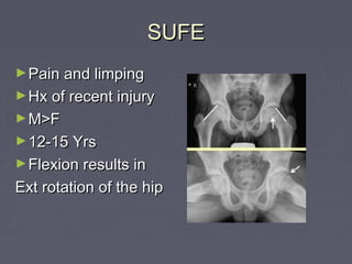
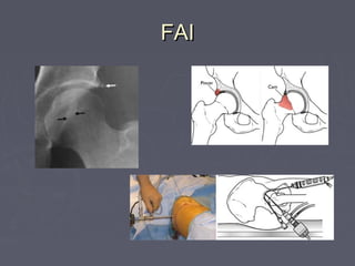
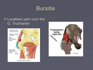
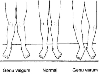
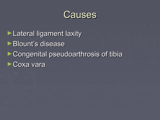
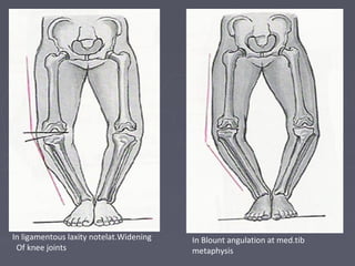
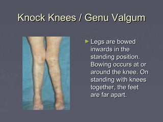
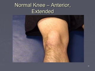
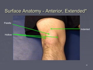
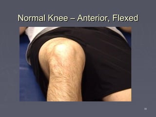
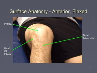
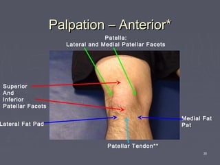
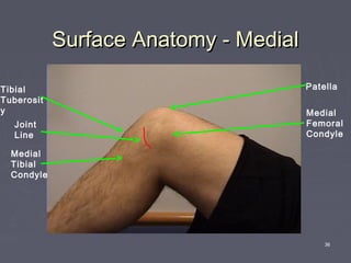
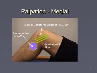
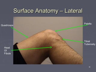
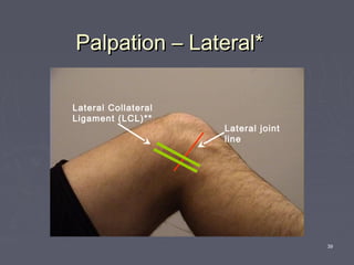
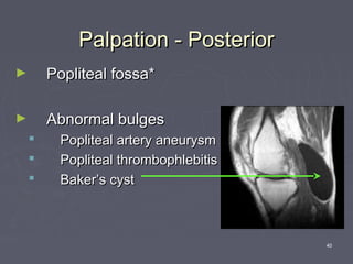
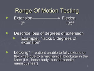
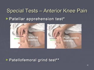
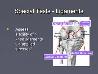
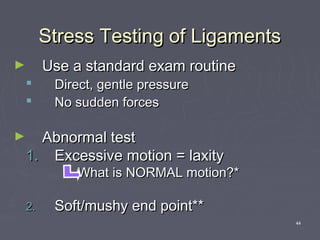
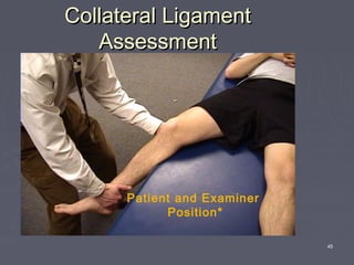
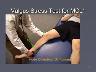
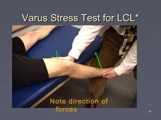
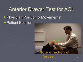
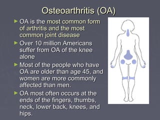
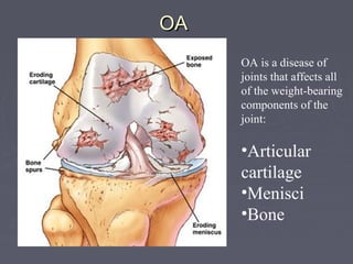
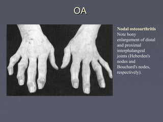
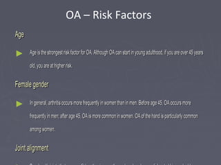
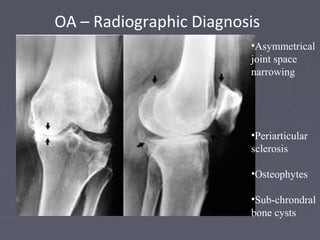
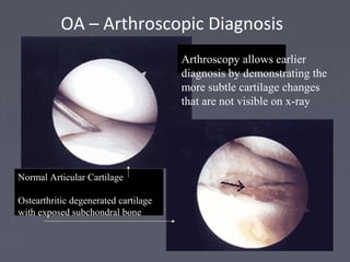
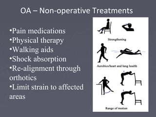
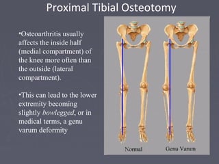
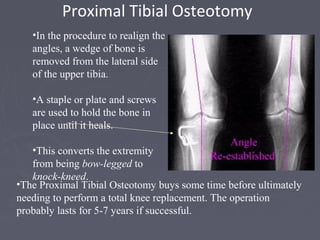
Disorders of lower limbDisorders of lower limb
Causes :Causes :
►Congenital.Congenital.
►Inflammatory.Inflammatory.
►Degenerative.Degenerative.
►Neurogenic.Neurogenic.
►Traumatic.Traumatic.
►Infective.Infective.
►Idiopathic.Idiopathic.
Age groupsAge groups
►Newborn and InfantsNewborn and Infants
►ChildrenChildren
►AdolescentsAdolescents
►Adults middle ageAdults middle age
►Adults > 50Adults > 50
1) Hip Pathologies1) Hip Pathologies
ChildrenChildren
AdultsAdults
►Tendinitis – ilIopsoas , adductor strainTendinitis – ilIopsoas , adductor strain
►FAIFAI
►Trochanteric bursitisTrochanteric bursitis
►Stress fracturesStress fractures
►Early osteoarthritisEarly osteoarthritis
►AVNAVN
AdultsAdults
►Established OAEstablished OA
►Inflammatory OAInflammatory OA
►Fragility fracturesFragility fractures
►MetastasisMetastasis
►InfectionInfection
DDHDDH
►Breech presentationBreech presentation
►Ist born femaleIst born female
►Family historyFamily history
DDHDDH
DDHDDH
DDHDDH
Septic arthritisSeptic arthritis
PerthesPerthes
► B> GB> G
► Pain Hip orPain Hip or
referred painreferred pain
in the kneein the knee
► LimpingLimping
► Limited internalLimited internal
rotationrotation
SUFESUFE
►Pain and limpingPain and limping
►Hx of recent injuryHx of recent injury
►M>FM>F
►12-15 Yrs12-15 Yrs
►Flexion results inFlexion results in
Ext rotation of the hipExt rotation of the hip
FAIFAI
BursitisBursitis
► Localised pain over theLocalised pain over the
G. TrochanterG. Trochanter
Stress fractureStress fracture
OAOA
►Pain, poorly localisedPain, poorly localised
►StiffnessStiffness
►Lose rotation firstLose rotation first
AVNAVN
►Alcohol and steroidsAlcohol and steroids
►Xrays may be normalXrays may be normal
METASTASISMETASTASIS
► Hx of Primary CaHx of Primary Ca
(thyroid, breast,lung(thyroid, breast,lung
kidneys, prostate)kidneys, prostate)
► Unrelenting painUnrelenting pain
► Multiple myelomaMultiple myeloma
► ChondrosarcomaChondrosarcoma
2) The knee2) The knee
GENU VALGUS & GENUGENU VALGUS & GENU
VARUSVARUS
CausesCauses
►Lateral ligament laxityLateral ligament laxity
►Blount’s diseaseBlount’s disease
►Congenital pseudoarthrosis of tibiaCongenital pseudoarthrosis of tibia
►Coxa varaCoxa vara
In ligamentous laxity notelat.Widening
Of knee joints
In Blount angulation at med.tib
metaphysis
InIn coxa vara ,angulation at the neckcoxa vara ,angulation at the neck
shaft levelshaft level
In cong. Pseudarthrosis of tibia,the
angulation is in the distal ⅓
►Gait:Gait: intoeing, lateral thrust-the fibular headintoeing, lateral thrust-the fibular head
and upper tibia shift laterally in Blount due toand upper tibia shift laterally in Blount due to
laxity and incompetence of the lat. Collat. Lig.laxity and incompetence of the lat. Collat. Lig.
►StabilityStability
►SymmetrySymmetry
►Level of fibular head,Level of fibular head, normally at thenormally at the
level of the upper tibial growth plate, while it islevel of the upper tibial growth plate, while it is
proximal in Blount, cong.longitudinal dificiencyproximal in Blount, cong.longitudinal dificiency
of the tibia and achondroplasiaof the tibia and achondroplasia
X-rayX-ray
► 3 years and older3 years and older
► Getting worseGetting worse
► Abnormal site ofAbnormal site of
angulationangulation
► Large physis andLarge physis and
epiphysisepiphysis
► History –History – taruma, infection,taruma, infection,
possible metal intoxication(lead orpossible metal intoxication(lead or
floride)floride)
Metaphysial/diaphysial
angle ≥ 18°
FindingFinding
► Metaphysis,Metaphysis, thick andthick and
frayed in ricketsfrayed in rickets
► InIn physiologic genuphysiologic genu
varumvarum no intrinsic boneno intrinsic bone
disease, gentle curve, medialdisease, gentle curve, medial
cortices thickening, horizontalcortices thickening, horizontal
joint lines of the knee & anklejoint lines of the knee & ankle
are tilted mediallyare tilted medially
Knock Knees / Genu ValgumKnock Knees / Genu Valgum
► Legs are bowedLegs are bowed
inwards in theinwards in the
standing position.standing position.
Bowing occurs at orBowing occurs at or
around the knee. Onaround the knee. On
standing with kneesstanding with knees
together, the feettogether, the feet
are far apart.are far apart.
31
Normal Knee – Anterior,Normal Knee – Anterior,
ExtendedExtended
32
Surface Anatomy - Anterior, Extended*Surface Anatomy - Anterior, Extended*
Patella
Hollow
Indented
33
Normal Knee – Anterior, FlexedNormal Knee – Anterior, Flexed
34
Surface Anatomy - Anterior, FlexedSurface Anatomy - Anterior, Flexed
Head
Of
Fibula
Patella
Tibial
Tuberosity
35
Palpation – Anterior*Palpation – Anterior*
Patella:
Lateral and Medial Patellar Facets
Superior
And
Inferior
Patellar Facets
Patellar Tendon**
Lateral Fat Pad
Medial Fat
Pat
36
Surface Anatomy - MedialSurface Anatomy - Medial
Medial
Femoral
Condyle
Patella
Joint
Line
Medial
Tibial
Condyle
Tibial
Tuberosit
y
37
Palpation - MedialPalpation - Medial
Medial Collateral Ligament (MCL)*
Pes anserine
bursa**
Medial joint
line
38
Surface Anatomy – LateralSurface Anatomy – Lateral
Patella
Head
Of
Fibula
Tibial
Tuberosity
Quadriceps
39
Palpation – Lateral*Palpation – Lateral*
Lateral joint
line
Lateral Collateral
Ligament (LCL)**
40
Palpation - PosteriorPalpation - Posterior
► Popliteal fossa*Popliteal fossa*
► Abnormal bulgesAbnormal bulges
 Popliteal artery aneurysmPopliteal artery aneurysm
 Popliteal thrombophlebitisPopliteal thrombophlebitis
 Baker’s cystBaker’s cyst
41
Range Of Motion TestingRange Of Motion Testing
► ExtensionExtension FlexionFlexion
00ºº 135º135º
► Describe loss of degrees of extensionDescribe loss of degrees of extension
► Example:Example: “lacks 5 degrees of“lacks 5 degrees of
extension”extension”
► Locking* =Locking* = patient unable to fully extend orpatient unable to fully extend or
flex knee due to a mechanical blockage in theflex knee due to a mechanical blockage in the
knee (i.e., loose body, bucket-handleknee (i.e., loose body, bucket-handle
meniscus tear)meniscus tear)
42
Special Tests – Anterior Knee PainSpecial Tests – Anterior Knee Pain
► Patellar apprehension test*Patellar apprehension test*
► Patellofemoral grind test**Patellofemoral grind test**
Starting
position
Push patella
laterally
43
Special Tests - LigamentsSpecial Tests - Ligaments
► AssessAssess
stability of 4stability of 4
knee ligamentsknee ligaments
via appliedvia applied
stresses*stresses*
Anterior Cruciate
Posterior
Cruciate
Lateral Collateral
Medial Collateral
44
Stress Testing of LigamentsStress Testing of Ligaments
► Use a standard exam routineUse a standard exam routine
 Direct, gentle pressureDirect, gentle pressure
 No sudden forcesNo sudden forces
► Abnormal testAbnormal test
1.1. Excessive motion = laxityExcessive motion = laxity
What is NORMAL motion?*What is NORMAL motion?*
2.2. Soft/mushy end point**Soft/mushy end point**
45
Collateral LigamentCollateral Ligament
AssessmentAssessment
Patient and Examiner
Position*
46
Valgus Stress Test for MCL*Valgus Stress Test for MCL*
Note Direction Of Forces
47
Varus Stress Test for LCL*Varus Stress Test for LCL*
Note direction of
forces
48
Anterior Drawer Test for ACLAnterior Drawer Test for ACL
►Physician Position & Movements*Physician Position & Movements*
►Patient PositionPatient Position
Note direction of
forces
OsteoarthritisOsteoarthritis
Osteoarthritis (OA)Osteoarthritis (OA)
► OA is theOA is the most common formmost common form
of arthritis and the mostof arthritis and the most
common joint diseasecommon joint disease
► Over 10 million AmericansOver 10 million Americans
suffer from OA of the kneesuffer from OA of the knee
alonealone
► Most of the people who haveMost of the people who have
OA are older than age 45, andOA are older than age 45, and
women are more commonlywomen are more commonly
affected than men.affected than men.
► OA most often occurs at theOA most often occurs at the
ends of the fingers, thumbs,ends of the fingers, thumbs,
neck, lower back, knees, andneck, lower back, knees, and
hips.hips.
OAOA
OA is a disease of
joints that affects all
of the weight-bearing
components of the
joint:
•Articular
cartilage
•Menisci
•Bone
OAOA
Nodal osteoarthritis
Note bony
enlargement of distal
and proximal
interphalangeal
joints (Heberden's
nodes and
Bouchard's nodes,
respectively).
AgeAge
► Age is the strongest risk factor for OA. Although OA can start in young adulthood, if you are over 45 yearsAge is the strongest risk factor for OA. Although OA can start in young adulthood, if you are over 45 years
old, you are at higher risk.old, you are at higher risk.
Female genderFemale gender
► In general, arthritis occurs more frequently in women than in men. Before age 45, OA occurs moreIn general, arthritis occurs more frequently in women than in men. Before age 45, OA occurs more
frequently in men; after age 45, OA is more common in women. OA of the hand is particularly commonfrequently in men; after age 45, OA is more common in women. OA of the hand is particularly common
among women.among women.
Joint alignmentJoint alignment
OA – Risk Factors
Hereditary gene defectHereditary gene defect
► A defect in one of theA defect in one of the genes responsible for the cartilage component collagen can cause deterioration ofgenes responsible for the cartilage component collagen can cause deterioration of
cartilage.cartilage.
Joint injury or overuse caused by physical labor or sportsJoint injury or overuse caused by physical labor or sports
► Traumatic injury (ex. Ligament or meniscal tears) to the knee or hip increases your risk for developing OATraumatic injury (ex. Ligament or meniscal tears) to the knee or hip increases your risk for developing OA
in these joints. Joints that are used repeatedly in certain jobs may be more likely to develop OA because ofin these joints. Joints that are used repeatedly in certain jobs may be more likely to develop OA because of
injury or overuse.injury or overuse.
ObesityObesity
► Being overweight during midlife or the later years is among the strongest risk factors for OA of the knee.Being overweight during midlife or the later years is among the strongest risk factors for OA of the knee.
OA – Risk Factors
Osteoarthritis (OA) - DefinitionOsteoarthritis (OA) - Definition
Osteoarthritis may result from wear and tear
on the joint
•The normal
cartilage lining
is gradually
worn away and
the underlying
bone is
exposed.
Osteoarthritis (OA) - DefinitionOsteoarthritis (OA) - Definition
•The repair mechanisms of tissue absorption and
synthesis get out of balance and result in
osteophyte formation (bone spurs) and bone cysts
A case of the, “Which
came first? The
chicken or the egg?”
Asymmetrical joint space narrowing from loss of
articular cartilage
The medial (inside) part of the knee is most commonly affected by osteoarthritis.
OA – Radiographic Diagnosis
•Asymmetrical
joint space
narrowing
•Periarticular
sclerosis
•Osteophytes
•Sub-chrondral
bone cysts
OA – Arthroscopic Diagnosis
Normal Articular Cartilage
Ostearthritic degenerated cartilage
with exposed subchondral bone
Arthroscopy allows earlier
diagnosis by demonstrating the
more subtle cartilage changes
that are not visible on x-ray
•OA is a condition which progresses slowly over a
period of many years and cannot be cured
•Treatment is directed at decreasing the symptoms of
the condition, and slowing the progress of the
condition
•Functional treatment goals:
•Limit pain
•Increase range of motion
•Increase muscle strength
OA – Disease Management
OA – Non-operative Treatments
•Pain medications
•Physical therapy
•Walking aids
•Shock absorption
•Re-alignment through
orthotics
•Limit strain to affected
areas
•Osteoarthritis usually
affects the inside half
(medial compartment) of
the knee more often than
the outside (lateral
compartment).
•This can lead to the lower
extremity becoming
slightly bowlegged, or in
medical terms, a genu
varum deformity
Proximal Tibial Osteotomy
•In the procedure to realign the
angles, a wedge of bone is
removed from the lateral side
of the upper tibia.
•A staple or plate and screws
are used to hold the bone in
place until it heals.
•This converts the extremity
from being bow-legged to
knock-kneed.
•The Proximal Tibial Osteotomy buys some time before ultimately
needing to perform a total knee replacement. The operation
probably lasts for 5-7 years if successful.
3) The foot3) The foot
Case1 varus deformityCase1 varus deformity
Case 2 calcaneus deformityCase 2 calcaneus deformity
Case 3 equinus deformityCase 3 equinus deformity
Thank youThank you

Disorders of Lower Limb

  • 1.
    Disorders of lowerlimbDisorders of lower limb
  • 2.
  • 3.
    Age groupsAge groups ►Newbornand InfantsNewborn and Infants ►ChildrenChildren ►AdolescentsAdolescents ►Adults middle ageAdults middle age ►Adults > 50Adults > 50
  • 4.
    1) Hip Pathologies1)Hip Pathologies
  • 5.
  • 6.
    AdultsAdults ►Tendinitis – ilIopsoas, adductor strainTendinitis – ilIopsoas , adductor strain ►FAIFAI ►Trochanteric bursitisTrochanteric bursitis ►Stress fracturesStress fractures ►Early osteoarthritisEarly osteoarthritis ►AVNAVN
  • 7.
    AdultsAdults ►Established OAEstablished OA ►InflammatoryOAInflammatory OA ►Fragility fracturesFragility fractures ►MetastasisMetastasis ►InfectionInfection
  • 8.
    DDHDDH ►Breech presentationBreech presentation ►Istborn femaleIst born female ►Family historyFamily history
  • 9.
  • 10.
  • 11.
  • 12.
  • 13.
    PerthesPerthes ► B> GB>G ► Pain Hip orPain Hip or referred painreferred pain in the kneein the knee ► LimpingLimping ► Limited internalLimited internal rotationrotation
  • 14.
    SUFESUFE ►Pain and limpingPainand limping ►Hx of recent injuryHx of recent injury ►M>FM>F ►12-15 Yrs12-15 Yrs ►Flexion results inFlexion results in Ext rotation of the hipExt rotation of the hip
  • 15.
  • 16.
    BursitisBursitis ► Localised painover theLocalised pain over the G. TrochanterG. Trochanter
  • 17.
  • 18.
    OAOA ►Pain, poorly localisedPain,poorly localised ►StiffnessStiffness ►Lose rotation firstLose rotation first
  • 19.
    AVNAVN ►Alcohol and steroidsAlcoholand steroids ►Xrays may be normalXrays may be normal
  • 20.
    METASTASISMETASTASIS ► Hx ofPrimary CaHx of Primary Ca (thyroid, breast,lung(thyroid, breast,lung kidneys, prostate)kidneys, prostate) ► Unrelenting painUnrelenting pain ► Multiple myelomaMultiple myeloma ► ChondrosarcomaChondrosarcoma
  • 21.
    2) The knee2)The knee
  • 22.
    GENU VALGUS &GENUGENU VALGUS & GENU VARUSVARUS
  • 24.
    CausesCauses ►Lateral ligament laxityLateralligament laxity ►Blount’s diseaseBlount’s disease ►Congenital pseudoarthrosis of tibiaCongenital pseudoarthrosis of tibia ►Coxa varaCoxa vara
  • 25.
    In ligamentous laxitynotelat.Widening Of knee joints In Blount angulation at med.tib metaphysis
  • 26.
    InIn coxa vara,angulation at the neckcoxa vara ,angulation at the neck shaft levelshaft level In cong. Pseudarthrosis of tibia,the angulation is in the distal ⅓
  • 27.
    ►Gait:Gait: intoeing, lateralthrust-the fibular headintoeing, lateral thrust-the fibular head and upper tibia shift laterally in Blount due toand upper tibia shift laterally in Blount due to laxity and incompetence of the lat. Collat. Lig.laxity and incompetence of the lat. Collat. Lig. ►StabilityStability ►SymmetrySymmetry ►Level of fibular head,Level of fibular head, normally at thenormally at the level of the upper tibial growth plate, while it islevel of the upper tibial growth plate, while it is proximal in Blount, cong.longitudinal dificiencyproximal in Blount, cong.longitudinal dificiency of the tibia and achondroplasiaof the tibia and achondroplasia
  • 28.
    X-rayX-ray ► 3 yearsand older3 years and older ► Getting worseGetting worse ► Abnormal site ofAbnormal site of angulationangulation ► Large physis andLarge physis and epiphysisepiphysis ► History –History – taruma, infection,taruma, infection, possible metal intoxication(lead orpossible metal intoxication(lead or floride)floride) Metaphysial/diaphysial angle ≥ 18°
  • 29.
    FindingFinding ► Metaphysis,Metaphysis, thickandthick and frayed in ricketsfrayed in rickets ► InIn physiologic genuphysiologic genu varumvarum no intrinsic boneno intrinsic bone disease, gentle curve, medialdisease, gentle curve, medial cortices thickening, horizontalcortices thickening, horizontal joint lines of the knee & anklejoint lines of the knee & ankle are tilted mediallyare tilted medially
  • 30.
    Knock Knees /Genu ValgumKnock Knees / Genu Valgum ► Legs are bowedLegs are bowed inwards in theinwards in the standing position.standing position. Bowing occurs at orBowing occurs at or around the knee. Onaround the knee. On standing with kneesstanding with knees together, the feettogether, the feet are far apart.are far apart.
  • 31.
    31 Normal Knee –Anterior,Normal Knee – Anterior, ExtendedExtended
  • 32.
    32 Surface Anatomy -Anterior, Extended*Surface Anatomy - Anterior, Extended* Patella Hollow Indented
  • 33.
    33 Normal Knee –Anterior, FlexedNormal Knee – Anterior, Flexed
  • 34.
    34 Surface Anatomy -Anterior, FlexedSurface Anatomy - Anterior, Flexed Head Of Fibula Patella Tibial Tuberosity
  • 35.
    35 Palpation – Anterior*Palpation– Anterior* Patella: Lateral and Medial Patellar Facets Superior And Inferior Patellar Facets Patellar Tendon** Lateral Fat Pad Medial Fat Pat
  • 36.
    36 Surface Anatomy -MedialSurface Anatomy - Medial Medial Femoral Condyle Patella Joint Line Medial Tibial Condyle Tibial Tuberosit y
  • 37.
    37 Palpation - MedialPalpation- Medial Medial Collateral Ligament (MCL)* Pes anserine bursa** Medial joint line
  • 38.
    38 Surface Anatomy –LateralSurface Anatomy – Lateral Patella Head Of Fibula Tibial Tuberosity Quadriceps
  • 39.
    39 Palpation – Lateral*Palpation– Lateral* Lateral joint line Lateral Collateral Ligament (LCL)**
  • 40.
    40 Palpation - PosteriorPalpation- Posterior ► Popliteal fossa*Popliteal fossa* ► Abnormal bulgesAbnormal bulges  Popliteal artery aneurysmPopliteal artery aneurysm  Popliteal thrombophlebitisPopliteal thrombophlebitis  Baker’s cystBaker’s cyst
  • 41.
    41 Range Of MotionTestingRange Of Motion Testing ► ExtensionExtension FlexionFlexion 00ºº 135º135º ► Describe loss of degrees of extensionDescribe loss of degrees of extension ► Example:Example: “lacks 5 degrees of“lacks 5 degrees of extension”extension” ► Locking* =Locking* = patient unable to fully extend orpatient unable to fully extend or flex knee due to a mechanical blockage in theflex knee due to a mechanical blockage in the knee (i.e., loose body, bucket-handleknee (i.e., loose body, bucket-handle meniscus tear)meniscus tear)
  • 42.
    42 Special Tests –Anterior Knee PainSpecial Tests – Anterior Knee Pain ► Patellar apprehension test*Patellar apprehension test* ► Patellofemoral grind test**Patellofemoral grind test** Starting position Push patella laterally
  • 43.
    43 Special Tests -LigamentsSpecial Tests - Ligaments ► AssessAssess stability of 4stability of 4 knee ligamentsknee ligaments via appliedvia applied stresses*stresses* Anterior Cruciate Posterior Cruciate Lateral Collateral Medial Collateral
  • 44.
    44 Stress Testing ofLigamentsStress Testing of Ligaments ► Use a standard exam routineUse a standard exam routine  Direct, gentle pressureDirect, gentle pressure  No sudden forcesNo sudden forces ► Abnormal testAbnormal test 1.1. Excessive motion = laxityExcessive motion = laxity What is NORMAL motion?*What is NORMAL motion?* 2.2. Soft/mushy end point**Soft/mushy end point**
  • 45.
  • 46.
    46 Valgus Stress Testfor MCL*Valgus Stress Test for MCL* Note Direction Of Forces
  • 47.
    47 Varus Stress Testfor LCL*Varus Stress Test for LCL* Note direction of forces
  • 48.
    48 Anterior Drawer Testfor ACLAnterior Drawer Test for ACL ►Physician Position & Movements*Physician Position & Movements* ►Patient PositionPatient Position Note direction of forces
  • 49.
  • 50.
    Osteoarthritis (OA)Osteoarthritis (OA) ►OA is theOA is the most common formmost common form of arthritis and the mostof arthritis and the most common joint diseasecommon joint disease ► Over 10 million AmericansOver 10 million Americans suffer from OA of the kneesuffer from OA of the knee alonealone ► Most of the people who haveMost of the people who have OA are older than age 45, andOA are older than age 45, and women are more commonlywomen are more commonly affected than men.affected than men. ► OA most often occurs at theOA most often occurs at the ends of the fingers, thumbs,ends of the fingers, thumbs, neck, lower back, knees, andneck, lower back, knees, and hips.hips.
  • 51.
    OAOA OA is adisease of joints that affects all of the weight-bearing components of the joint: •Articular cartilage •Menisci •Bone
  • 52.
    OAOA Nodal osteoarthritis Note bony enlargementof distal and proximal interphalangeal joints (Heberden's nodes and Bouchard's nodes, respectively).
  • 53.
    AgeAge ► Age isthe strongest risk factor for OA. Although OA can start in young adulthood, if you are over 45 yearsAge is the strongest risk factor for OA. Although OA can start in young adulthood, if you are over 45 years old, you are at higher risk.old, you are at higher risk. Female genderFemale gender ► In general, arthritis occurs more frequently in women than in men. Before age 45, OA occurs moreIn general, arthritis occurs more frequently in women than in men. Before age 45, OA occurs more frequently in men; after age 45, OA is more common in women. OA of the hand is particularly commonfrequently in men; after age 45, OA is more common in women. OA of the hand is particularly common among women.among women. Joint alignmentJoint alignment OA – Risk Factors
  • 54.
    Hereditary gene defectHereditarygene defect ► A defect in one of theA defect in one of the genes responsible for the cartilage component collagen can cause deterioration ofgenes responsible for the cartilage component collagen can cause deterioration of cartilage.cartilage. Joint injury or overuse caused by physical labor or sportsJoint injury or overuse caused by physical labor or sports ► Traumatic injury (ex. Ligament or meniscal tears) to the knee or hip increases your risk for developing OATraumatic injury (ex. Ligament or meniscal tears) to the knee or hip increases your risk for developing OA in these joints. Joints that are used repeatedly in certain jobs may be more likely to develop OA because ofin these joints. Joints that are used repeatedly in certain jobs may be more likely to develop OA because of injury or overuse.injury or overuse. ObesityObesity ► Being overweight during midlife or the later years is among the strongest risk factors for OA of the knee.Being overweight during midlife or the later years is among the strongest risk factors for OA of the knee. OA – Risk Factors
  • 55.
    Osteoarthritis (OA) -DefinitionOsteoarthritis (OA) - Definition Osteoarthritis may result from wear and tear on the joint •The normal cartilage lining is gradually worn away and the underlying bone is exposed.
  • 56.
    Osteoarthritis (OA) -DefinitionOsteoarthritis (OA) - Definition •The repair mechanisms of tissue absorption and synthesis get out of balance and result in osteophyte formation (bone spurs) and bone cysts A case of the, “Which came first? The chicken or the egg?”
  • 57.
    Asymmetrical joint spacenarrowing from loss of articular cartilage The medial (inside) part of the knee is most commonly affected by osteoarthritis. OA – Radiographic Diagnosis
  • 58.
    OA – RadiographicDiagnosis •Asymmetrical joint space narrowing •Periarticular sclerosis •Osteophytes •Sub-chrondral bone cysts
  • 59.
    OA – ArthroscopicDiagnosis Normal Articular Cartilage Ostearthritic degenerated cartilage with exposed subchondral bone Arthroscopy allows earlier diagnosis by demonstrating the more subtle cartilage changes that are not visible on x-ray
  • 60.
    •OA is acondition which progresses slowly over a period of many years and cannot be cured •Treatment is directed at decreasing the symptoms of the condition, and slowing the progress of the condition •Functional treatment goals: •Limit pain •Increase range of motion •Increase muscle strength OA – Disease Management
  • 61.
    OA – Non-operativeTreatments •Pain medications •Physical therapy •Walking aids •Shock absorption •Re-alignment through orthotics •Limit strain to affected areas
  • 62.
    •Osteoarthritis usually affects theinside half (medial compartment) of the knee more often than the outside (lateral compartment). •This can lead to the lower extremity becoming slightly bowlegged, or in medical terms, a genu varum deformity Proximal Tibial Osteotomy
  • 63.
    Proximal Tibial Osteotomy •Inthe procedure to realign the angles, a wedge of bone is removed from the lateral side of the upper tibia. •A staple or plate and screws are used to hold the bone in place until it heals. •This converts the extremity from being bow-legged to knock-kneed. •The Proximal Tibial Osteotomy buys some time before ultimately needing to perform a total knee replacement. The operation probably lasts for 5-7 years if successful.
  • 64.
    3) The foot3)The foot
  • 65.
  • 69.
    Case 2 calcaneusdeformityCase 2 calcaneus deformity
  • 72.
    Case 3 equinusdeformityCase 3 equinus deformity
  • 76.

Editor's Notes

  • #33 Appears hollow on either side of patella There is a slight indentation above the patella A small amount of fluid will make these hollow-appearing areas disappear. Larger effusions are most conspicuous as a fullness proximal to the patella.
  • #36 *Assess for tenderness, edema, warmth **Palpate the insertion of the patellar tendon on tibial tubercle in adolescents (location of pain in Osgood-Schlatter syndrome in adolescents)
  • #38 *Assess for tenderness along entire course of ligament from origin on medial femoral condyle to insertion on proximal tibia. **Pes anserine bursa is about 3 finger widths inferior to the medial joint line and contains the insertion site for the sartorius, gracilis, and semitendinosis muscles
  • #40 * The LCL and joint line are more easily palpated with the knee in 90 degrees of flexion. ** LCL originates on lateral femoral epicondyle and inserts on fibular head
  • #41 *Popliteal artery is only palpable structure normally in this area
  • #42 *Locking vs Effusion Effusion can hinder extension and is often confused with locking
  • #43 *Patellar apprehension test: Apply firm, laterally-directed force toward medial aspect of patella Positive test is trepidation of the patient (pain or fear that patella will dislocate) Positive test implies a preceding episode of patellar instability (subluxation or dislocation) **Patellofemoral grind test Patient supine with knees extended Examiner’s thumb on superior patella Pt. contracts quadriceps muscle Examiner applies downward and inferior pressure Positive - pain with movement or unable to complete test Positive test suggests patellofemoral dysfunction (patellofemoral stress syndrome)
  • #44 * The stabilizing roles of each ligament include: The medial collateral ligament (MCL) prevents the knee from buckling inwards (valgus injury) The lateral collateral ligament (LCL) prevents the knee from buckling outwards (varus injury) The anterior cruciate ligament (ACL) prevents the tibia from sliding forward under the femur The posterior cruciate ligament (PCL) prevents the tibial from sliding backward under the femur
  • #45 *Normal Stability Medial and Lateral collateral ligaments Normal test is no motion with varus and/or valgus stress with knee in neutral and 30 degrees of flexion Anterior and Posterior Cruciate Ligements control anterior/posterior motion Lachman’s test assesses Anterior Cruciate Ligament: Normal test is <5mm of forward movement of tibia on femur with knee at 30 degrees of flexion Anterior and posterior drawer testing assesses ACL and PCL With knee in 90 degrees of flexion and foot stabilized, normal test will have <5mm of anterior motion (assessing ACL) or <5mm of posterior motion (assessing PCL) ** Normal end point of ligament that examiner feels with applied stress is FIRM. A soft or mushy end point implies ligament damage (stretching or complete tear).
  • #46 *Position patient supine on table with thigh resting on edge of exam table and foot supported by examiner Knee in 30 degrees of flexion – WHY? Increased laxity of medial side of knee in extension may indicate additional damage to posterior structures (posterior joint capsule & PCL)
  • #47 *VALGUS (MCL) stress Proximal hand on lateral aspect of knee holds and stabilizes thigh Distal hand directs ankle laterally Attempt to open knee joint on medial side Estimate the medial joint space and evaluate the stiffness of motion. Positive test = Significant gap in medial aspect of knee with valgus stress = MCL injury. Laxity is graded on a 1 to 4 scale: 1+, 5mm of medial joint space opening with a firm but abnormal endpoint; 2+, 10mm medial opening with a soft endpoint; 3+ (15mm) and 4+ (20mm) may be indicative of an associated cruciate ligament injury and must be carefully examined.
  • #48 *VARUS (LCL) Stress Supine position, with knee at 20 to 30 degrees of flexion and thigh supported. Stabilize medial aspect of knee and push ankle medially, trying to open knee joint on lateral side Disruption of LCL is indicated by difference in degree of lateral knee tautness with varus stress. Compare affected knee to uninjured side
  • #49 *Patient Position Supine Flex hip of affected knee to 45 degrees Bend knee to 90 degrees Patient's foot planted firmly on examination table Physician position: Sitting on dorsum of foot, place both hands behind knee Once hamstrings relaxed, try to displace proximal leg anteriorly Anterior drawer test is LESS SENSITIVE for ACL damage than Lachman’s Maneuver