hip osteoarthritis is most disabling condition and surgery is a consequence of the same. but if this condition can assess on time so it can be manageable with conservative treatment and decrease the prevalence of AVN. further life of an individual become better.
references:
Campbell’s operative orthopaedics 11th edition
Text book of orthopaedics & fractures 5th edition Dr B. Aalami Harandi
Gray’s anatomy 2nd edition
Clinical anatomy Richard S. Snell
Collapse of medial longitudinal arch, with the entire sole of the foot coming into complete or near-complete contact with the ground.
Books Refered :
Text Book Of ANATOMY - Vishram Singh
Joint Structure And Function – Cynthia Norkin
Therapeutic Exrercise – Carolyn Kisner
Orthopaedic Physical Assessment – Magee
Orthopaedic Medicine – L. Ombregt
Campbell’s Operative Orthopaedics
Goniometry of lower limb joints/ROM of lower limb jointsShalu Thariwal
Goniometer, goniometry, hip joint, knee joint, ankle, ROM, range of motion, hip flexion, knee extension, ankle dorsiflexion and planter flexion, inversion, eversion, alignment, position, fulcrum, stationary arm, moving arm, normal range of motion.
Describing some of the most important disorders of the shoulder area: frozen shoulder, biceps tenosynovitis, biceps tendon tear, rotator cuff tear, impingement syndrome, Rotator Cuff Calcified Tendonitis
Evidence-based Interventional Pain Medicine
according to Clinical Diagnoses
13. Sacroiliac Joint Pain
Pascal Vanelderen, MD, FIPP*,†; Karolina Szadek, MD‡; Steven P. Cohen, MD§;
Jan De Witte, MD¶; Arno Lataster, MSc**; Jacob Patijn, MD, PHD††;
Nagy Mekhail, MD PhD, FIPP‡‡; Maarten van Kleef, MD, PhD, FIPP††;
Jan Van Zundert, MD, PhD, FIPP*,††
3. Pain
•ACJ pain usually well localised
•Neck pain, pain over trapezius or medial border of
scapula usually cervical in origin
•Associated pain in wrist or hand +/- parasthesiae
usually neurogenic
•Poorly localised pain from deltoid region usually
subacromial and rotator cuff pathology
4. Pain
•Night pain often rotator cuff disease,
glenohumeral arthritis and frozen shoulder
•Sudden onset excruciating pain is typical of
acute calcific tendonitis
•Pain occurring in part of the range of
shoulder abduction is termed painful arc
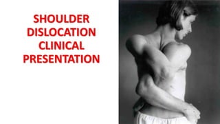
5. Instability
• Any history of trauma?
• Was shoulder dislocated?
• How many dislocations since then?
• Was dislocation spontaneous? If atraumatic
dislocation is there history of joint laxity?
• Painless clicks in the shoulder are common and
usually have no significance
6. Weakness
•Following traumatic event - important to
exclude brachial plexus injury
•May be due to pain (would examine
with local anaesthetic joint examination
in shoulder clinic)
7. Stiffness
•Restriction of both passive and active
movements
•Usually associated with frozen shoulder,
osteoarthritis, rheumatoid arthritis,
chronic dislocation and cuff tear
8. Inspection (Look)
•Undressed to the waist
•Observe for difficulty getting undressed
•Scars
•Asymmetry or deformity of sternoclavicular
joints
•Outline and contour of clavicles and ACJ
compared
9. Inspection (Look)
• Squaring off of the shoulder profile from anterior dislocation,
deltoid wasting or erosive arthritis with medialisation of
humeral head
• Bulk of pectoral and trapezius muscles should be compared
• “Pop-Eye” appearance of biceps might signify rupture of long
head of biceps
• Winging of scapula caused by injury to long thoracic nerve
10. Palpation (Feel)
•Start with sternoclavicular joint medially
•Move along clavicle to ACJ
•Tenderness over ACJ associated with
degenerative change (common and not nec.
abnormal) and traumatic subluxation
11. Palpation (Feel)
• Palpation lateral and inferior to coracoid assoc with
inflammatory arthropathy or primary frozen shoulder
• Palpation should continue anterolaterally to
intertubercular sulcus on humerus. Pain here may
suggest biciptal tendonitis
• Palpation of posterior joint line, where pain is more
typical of osteoarthritis
12. Movement
•Assess cervical spine to see if neck movements
recreate shoulder symptoms
•In full extension of C spine nose parallels the
floor and in full flexion chin should rest on chest
•Lateral rotation approx 80o
•Lateral flexion 40o
13. Movement
• Active and Passive range of movements of shoulder
• Forward elevation (0-170)
• Abduction (0-170)
• External rotation with abduction (0-90)
• Internal rotation (behind the back)
• Comparison with contralateral side
14. Neurovascular assessment
•Sensation dermatomes C4 toT2
•Power around elbow, wrist and hand
•Shoulder power tested separately
•Test peripheral nerves, esp. Axillary nerve
•Biceps and triceps reflexes
•Radial pulses
15. Special Tests
• Deltoid
• Arm in 90o abduction, neutral rotation
• With resistance deltoid can be felt to contract
• Supraspinatus
• “empty cans test”
• Subscapularis
• “Push-off test”
• Infraspinatus
• “Swinging doors test”
16. Empty Cans Test
Supraspinatus
• The purpose of this test is to assist the
integrity of the rotator cuff muscles.
• It specifically targets the supraspinatus
muscles.
• Aids in the diagnosis of rotator cuff tears
and shoulder impegment syndrome (SIS).
1. The patient is standing facing the
examiner
2. In this test, the examiner resists
abduction with the arm of the
patient eleveted to 90˚ combined
with internally rotation and 30˚ in
the forward plane.
3. If the patient gives away, the test is
considered positive.
17. Internal rotation lag
sign test
The purpose of this test is to assess tears of
the subscapularis muscle.
1. The patient is asked to bring the arm
behind the back with the palm facing
outwards.
2. The arm is held in near maximum
internal rotation and with the hand
away from the back by approximately
20˚ of extension.
3. The patient is asked to hold the position
while the examiner supports the elbow
but releases the wrist hold.
4. If the patient is unable to hold the
position, the lag sign is positive.
19. Other tests.
Painful arc/Impingement test
Internally rotated arm passively abducted in scapula plane (20-30 deg
off coronal). Pain usually elicited in arc between 70-120o
Scarf test
ACJ injury
Anterior apprehension test
Arm externally rotated and shoulder abducted to 90 and elbow flexed.
With gentle external pressure on back of humeral head, arm is
externally rotated further
20. Scarf Test ACJ
1. The examiner forward flexes the involved
arm of the patient to 90˚ (with elbow to 90˚)
2. A flexed arm is then passively adducted
across the body putting an adduction stress
on the acromioclavicular joint.
3. This position results in compression of the
medial acromial facet against the distal
clavicle to provoke symptoms at the
acromioclavicular joint.
4. A positive test is indicated by reproduction
of the patients symptoms.
21. Anterior
Apprehension Test
This test assists with the diagnosis of
glenohumeral joint instability which can
be caused by pathology to the labrum,
rotator cuff muscles or joint capsule.
This test evaluates specifically anterior
joint instability.
• As the shoulder is moved passively
into maximum external rotation in
abduction and forward pressure is
applied to the posterior aspect of
the humeral head, the patient
complains of pain or instability.