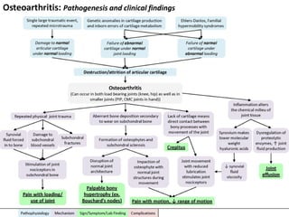
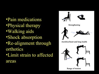
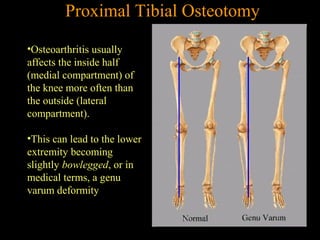
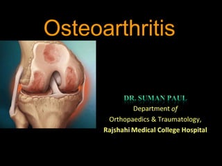
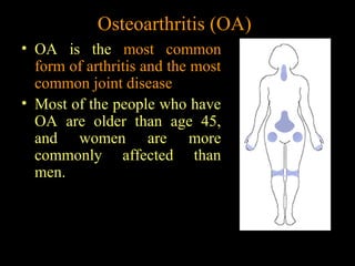
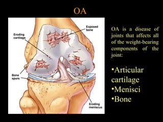
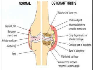
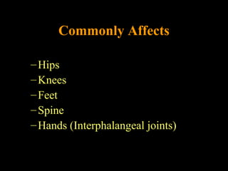
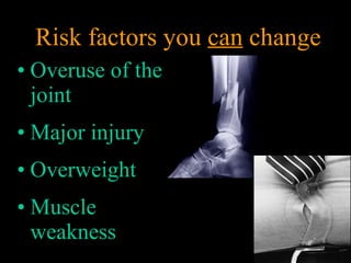
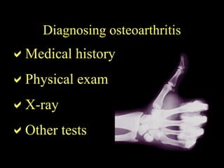
Osteoarthritis
Osteoarthritis (OA)
• OA is the most common
form of arthritis and the most
common joint disease
• Most of the people who have
OA are older than age 45,
and women are more
commonly affected than
men.
Definition
• Osteoarthritis is an idiopathic disease
characterized by degeneration of articular
cartilage leads to fibrillation, fissures, gross
ulceration and finally disappearance of the
full thickness of articular cartilage.
• As defined by the American College of
Rheumatology (ACR), OA is a
heterogeneous group of condition that leads
to joint signs and symptoms which are
associated with defective integrity of
articular cartilage, in addition to related
changes in the underlying bone at the joint
margins.
•The repair mechanisms of tissue absorption and
synthesis get out of balance and result in
osteophyte formation (bone spurs) and bone cysts
OA
OA is a disease of
joints that affects all
of the weight-bearing
components of the
joint:
•Articular
cartilage
•Menisci
•Bone
OA – Articular Cartilage
Articular cartilage is the main tissue affected
OA results in:
•Increased tissue swelling
•Change in color
•Cartilage fibrillation
•Cartilage erosion down to subchondral bone
OA – Articular Cartilage
A) Normal articular
cartilage from 21-year
old adult (3000X)
B) Osteoarthritic
cartilage (3000X)
Commonly AffectsCommonly Affects
–Hips
–Knees
–Feet
–Spine
–Hands (Interphalangeal joints)
Uncommonly Affected Joints
• Shoulder
• Wrist
• Elbow
• Metacarpophalangeal joint
• TMJ
• SI
• Ankle
OA
Nodal osteoarthritis
Note bony
enlargement of distal
and proximal
interphalangeal
joints (Heberden's
nodes and
Bouchard's nodes,
respectively).
Risk factors –
Conditions that contribute to
osteoarthritis
Age
• Ageisthestrongestrisk factorforOA.AlthoughOAcanstartinyoungadulthood,ifyouareover45yearsold,you
areathigherrisk.
Femalegender
• Ingeneral,arthritisoccursmorefrequentlyinwomenthaninmen.Beforeage45,OAoccursmorefrequently inmen;
afterage45,OAismorecommonin women.OAofthehandisparticularlycommonamongwomen.
Jointalignment
• Peoplewithjointsthatmoveorfittogetherincorrectly,suchasbowlegs,adislocatedhip,ordouble-jointedness,are
OA – Risk Factors
Hereditarygenedefect
• Adefectinoneofthe genesresponsibleforthecartilagecomponentcollagen cancausedeterioration ofcartilage.
Jointinjury oroverusecausedby physicallabororsports
• Traumaticinjury(ex.Ligamentormeniscaltears)tothekneeorhipincreasesyourriskfordevelopingOAinthese
joints.Jointsthatareusedrepeatedlyincertainjobsmay bemore likelytodevelopOAbecauseofinjuryoroveruse.
Obesity
• BeingoverweightduringmidlifeorthelateryearsisamongthestrongestriskfactorsforOAoftheknee.
Pathogenesis of Osteoarthritis
An Interplay of Factors
• Family history
of disease
• Increasing Age
• Being female
Risk factors you cannot change
• Overuse of the
joint
• Major injury
• Overweight
• Muscle
weakness
Risk factors you can change
Medical history
Physical exam
X-ray
Other tests
Diagnosing osteoarthritis
OA – Symptoms
• OA usually occurs slowly -
It may be many years before
the damage to the joint
becomes noticeable
• Only a third of people
whose X-rays show
OA report pain or
other symptoms:
– Steady or intermittent pain in a joint
– Stiffness that tends to follow periods of inactivity, such as sleep
or sitting
– Swelling or tenderness in one or more joints [not necessarily
occurring on both sides of the body at the same time]
– Crunching feeling or sound of bone rubbing on bone (called
crepitus) when the joint is used
Two Major Types of OA
• Primary or Idiopathic
– Most common type
– Diagnosed when there is no known cause for
the symptoms
• Secondary
– Diagnosed when there is an identifiable cause
• Trauma or Underlying joint disorder
• Each of these major types has subtypes
OA vs. Aging
Unlike aging, OA is progressive and a significantly
more active process
Osteoarthritis with lateral osteophyte, loss of articular cartilage and
some subchondral bony sclerosis- X-ray shows loss of joint space
OA – Overall Changes
Laboratory findings in OA
• THERE ARE NO DIAGNOSTIC LAB
TESTS FOR OSTEOARTHRITIS
• OA is not a systemic disease, therefore:
– ESR, Chem 7, CBC, and UA all WNL
• Synovial fluid
• Mild leukocytosis (<2000 WBC/microliter)
• Can be used to exclude gout, CPPD, or septic
arthritis if diagnosis is in doubt
Synovial fluid analysis
• Severe, acute joint pain is an
uncommon manifestation of OA
• Clear fluidWBC <2000/mm3
• Normal viscosity
Differential Diagnosis
• Rheumatoid Arthritis
• Gout
• CPPD (Calcium pyrophosphate crystal
deposition disease)
• Septic Joint
• Polymyalgia Rheumatica
Asymmetrical joint space narrowing from loss of
articular cartilage
The medial (inside) part of the knee is most commonly affected by osteoarthritis.
OA – Radiographic Diagnosis
•Asymmetrical
joint space
narrowing
•Periarticular
sclerosis
•Osteophytes
•Sub-chrondral
bone cysts
OA – Arthroscopic Diagnosis
Normal Articular Cartilage
Ostearthritic degenerated cartilage
with exposed subchondral bone
Arthroscopy allows earlier
diagnosis by demonstrating the
more subtle cartilage changes
that are not visible on x-ray
•OA is a condition which progresses slowly over a
period of many years and cannot be cured
•Treatment is directed at decreasing the symptoms of
the condition, and slowing the progress of the
condition
•Functional treatment goals:
•Limit pain
•Increase range of motion
•Increase muscle strength
OA – Disease Management
Management/Treatment of OA
Conservative…..
– Educate patient about disease and management
– Improve function
– Control pain
– Alter disease process and its consequences
• Pharmacologic
– Acetaminophen
– NSAIDS
• Cox-2 specific
inhibitors
• With PPI or
misoprostol
– Nonacetylated
salicylate
– Tramadol
– Opioids
• Topical
– Capsaicin
– Methylsalicylate
– NSAIDS
• Intra-articular
– Corticosteroids
– Hyaluronic acid
• Possible structure/disease modifying stuff
– Glucosamine
– Diacerein
– Cytokine inhibitors
– Cartilage repair
– Bisphosphonates
– Degradative enzyme inhibitors
• Tetracyclines, metalloproteinase inhibitors
• Pharmacologic
– Glucosamine 20 studies with >2500 patients
• If only high quality studies evaluated:
– No benefit over placebo on pain
• If all studies included:
– Pain may improve by as much as 13 points
• 2 RCT’s using Rotta preparation:
– Demonstrated slowing of radiological progression of OA
over a 3 year period
• Pharmacologic
– Diacerein
• Pain improved 5 points compared to placebo
• Over 3 years,
– Slowed progress of OA in the hip compared to placebo
– Did not slow progress of OA in the knee
• Diarrhea is most common side effect
– 42 out of 100 had diarrhea in the first 2 weeks
– 18 discontinued because of side effects (13 in placebo)
• Non-pharmacologic
– Patient education
– Self-management
programs
– Weight loss
– PT/OT
– ROM exercises
– Muscle strengthening
• Non-pharmacologic
– Assistive devices
– Patellar taping
– Appropriate footwear
– Lateral-wedged insoles
– Bracing
– Joint protection and
energy conservation
•Pain medications
•Physical therapy
•Walking aids
•Shock absorption
•Re-alignment through
orthotics
•Limit strain to affected
areas
Operative options…
•Osteoarthritis usually
affects the inside half
(medial compartment) of
the knee more often than
the outside (lateral
compartment).
•This can lead to the lower
extremity becoming
slightly bowlegged, or in
medical terms, a genu
varum deformity
Proximal Tibial Osteotomy
•The result is that the weight bearing line of the lower
extremity moves more medially (towards the medial
compartment of the knee).
•The end result is that there is more pressure on the medial
joint surfaces, which leads to more pain and faster
degeneration.
•In some cases, re-aligning the angles in the lower extremity
can result in shifting the weight-bearing line to the lateral
compartment of the knee. This, presumably, places the
majority of the weight-bearing force into a healthier
compartment. The result is to reduce the pain and delay the
progression of the degeneration of the medial compartment.
Proximal Tibial Osteotomy
•In the procedure to realign the
angles, a wedge of bone is
removed from the lateral side
of the upper tibia.
•A staple or plate and screws
are used to hold the bone in
place until it heals.
•This converts the extremity
from being bow-legged to
knock-kneed.
•The Proximal Tibial Osteotomy buys some time before ultimately
needing to perform a total knee replacement. The operation
probably lasts for 5-7 years if successful.
The ultimate solution for osteoarthritis of the knee is to
replace the joint surfaces with an artificial knee joint:
•Usually only considered in people over the age of 60
•Artificial knee joints last about 12 years in an elderly population
•Not recommended in younger patients because:
•The younger the patient, the more likely the artificial joint will fail
•Replacing the knee the second and third time is much harder and much
less likely to succeed.
•Younger patients are more active and place more stress on the artificial
joint, that can lead to loosening and failure earlier
•Younger patients are also more likely to outlive their artificial joint, and
will almost surely require a revision at some point down the road.
•Younger patients sometimes require the surgery (simply because
no other acceptable solution is available to treat their condition)
Total Knee Replacement
•The ends of the femur, tibia, and patella are shaped to accept
the artificial surfaces.
•The end result is that all moving surfaces of the knee are
metal against plastic
Total Knee Replacement
Thank you all……

Osteoarthritis

  • 1.
  • 2.
    Osteoarthritis (OA) • OAis the most common form of arthritis and the most common joint disease • Most of the people who have OA are older than age 45, and women are more commonly affected than men.
  • 3.
    Definition • Osteoarthritis isan idiopathic disease characterized by degeneration of articular cartilage leads to fibrillation, fissures, gross ulceration and finally disappearance of the full thickness of articular cartilage.
  • 4.
    • As definedby the American College of Rheumatology (ACR), OA is a heterogeneous group of condition that leads to joint signs and symptoms which are associated with defective integrity of articular cartilage, in addition to related changes in the underlying bone at the joint margins.
  • 5.
    •The repair mechanismsof tissue absorption and synthesis get out of balance and result in osteophyte formation (bone spurs) and bone cysts
  • 6.
    OA OA is adisease of joints that affects all of the weight-bearing components of the joint: •Articular cartilage •Menisci •Bone
  • 7.
    OA – ArticularCartilage Articular cartilage is the main tissue affected OA results in: •Increased tissue swelling •Change in color •Cartilage fibrillation •Cartilage erosion down to subchondral bone
  • 8.
    OA – ArticularCartilage A) Normal articular cartilage from 21-year old adult (3000X) B) Osteoarthritic cartilage (3000X)
  • 11.
  • 12.
    Uncommonly Affected Joints •Shoulder • Wrist • Elbow • Metacarpophalangeal joint • TMJ • SI • Ankle
  • 14.
    OA Nodal osteoarthritis Note bony enlargementof distal and proximal interphalangeal joints (Heberden's nodes and Bouchard's nodes, respectively).
  • 15.
    Risk factors – Conditionsthat contribute to osteoarthritis
  • 16.
    Age • Ageisthestrongestrisk factorforOA.AlthoughOAcanstartinyoungadulthood,ifyouareover45yearsold,you areathigherrisk. Femalegender •Ingeneral,arthritisoccursmorefrequentlyinwomenthaninmen.Beforeage45,OAoccursmorefrequently inmen; afterage45,OAismorecommonin women.OAofthehandisparticularlycommonamongwomen. Jointalignment • Peoplewithjointsthatmoveorfittogetherincorrectly,suchasbowlegs,adislocatedhip,ordouble-jointedness,are OA – Risk Factors
  • 17.
    Hereditarygenedefect • Adefectinoneofthe genesresponsibleforthecartilagecomponentcollagencancausedeterioration ofcartilage. Jointinjury oroverusecausedby physicallabororsports • Traumaticinjury(ex.Ligamentormeniscaltears)tothekneeorhipincreasesyourriskfordevelopingOAinthese joints.Jointsthatareusedrepeatedlyincertainjobsmay bemore likelytodevelopOAbecauseofinjuryoroveruse. Obesity • BeingoverweightduringmidlifeorthelateryearsisamongthestrongestriskfactorsforOAoftheknee.
  • 18.
  • 19.
    • Family history ofdisease • Increasing Age • Being female Risk factors you cannot change
  • 20.
    • Overuse ofthe joint • Major injury • Overweight • Muscle weakness Risk factors you can change
  • 21.
  • 22.
    OA – Symptoms •OA usually occurs slowly - It may be many years before the damage to the joint becomes noticeable • Only a third of people whose X-rays show OA report pain or other symptoms: – Steady or intermittent pain in a joint – Stiffness that tends to follow periods of inactivity, such as sleep or sitting – Swelling or tenderness in one or more joints [not necessarily occurring on both sides of the body at the same time] – Crunching feeling or sound of bone rubbing on bone (called crepitus) when the joint is used
  • 23.
    Two Major Typesof OA • Primary or Idiopathic – Most common type – Diagnosed when there is no known cause for the symptoms • Secondary – Diagnosed when there is an identifiable cause • Trauma or Underlying joint disorder • Each of these major types has subtypes
  • 25.
    OA vs. Aging Unlikeaging, OA is progressive and a significantly more active process
  • 26.
    Osteoarthritis with lateralosteophyte, loss of articular cartilage and some subchondral bony sclerosis- X-ray shows loss of joint space OA – Overall Changes
  • 28.
    Laboratory findings inOA • THERE ARE NO DIAGNOSTIC LAB TESTS FOR OSTEOARTHRITIS • OA is not a systemic disease, therefore: – ESR, Chem 7, CBC, and UA all WNL • Synovial fluid • Mild leukocytosis (<2000 WBC/microliter) • Can be used to exclude gout, CPPD, or septic arthritis if diagnosis is in doubt
  • 29.
    Synovial fluid analysis •Severe, acute joint pain is an uncommon manifestation of OA • Clear fluidWBC <2000/mm3 • Normal viscosity
  • 30.
    Differential Diagnosis • RheumatoidArthritis • Gout • CPPD (Calcium pyrophosphate crystal deposition disease) • Septic Joint • Polymyalgia Rheumatica
  • 31.
    Asymmetrical joint spacenarrowing from loss of articular cartilage The medial (inside) part of the knee is most commonly affected by osteoarthritis. OA – Radiographic Diagnosis
  • 32.
    OA – RadiographicDiagnosis •Asymmetrical joint space narrowing •Periarticular sclerosis •Osteophytes •Sub-chrondral bone cysts
  • 34.
    OA – ArthroscopicDiagnosis Normal Articular Cartilage Ostearthritic degenerated cartilage with exposed subchondral bone Arthroscopy allows earlier diagnosis by demonstrating the more subtle cartilage changes that are not visible on x-ray
  • 35.
    •OA is acondition which progresses slowly over a period of many years and cannot be cured •Treatment is directed at decreasing the symptoms of the condition, and slowing the progress of the condition •Functional treatment goals: •Limit pain •Increase range of motion •Increase muscle strength OA – Disease Management
  • 36.
    Management/Treatment of OA Conservative….. –Educate patient about disease and management – Improve function – Control pain – Alter disease process and its consequences
  • 37.
    • Pharmacologic – Acetaminophen –NSAIDS • Cox-2 specific inhibitors • With PPI or misoprostol – Nonacetylated salicylate – Tramadol – Opioids • Topical – Capsaicin – Methylsalicylate – NSAIDS • Intra-articular – Corticosteroids – Hyaluronic acid
  • 38.
    • Possible structure/diseasemodifying stuff – Glucosamine – Diacerein – Cytokine inhibitors – Cartilage repair – Bisphosphonates – Degradative enzyme inhibitors • Tetracyclines, metalloproteinase inhibitors
  • 39.
    • Pharmacologic – Glucosamine20 studies with >2500 patients • If only high quality studies evaluated: – No benefit over placebo on pain • If all studies included: – Pain may improve by as much as 13 points • 2 RCT’s using Rotta preparation: – Demonstrated slowing of radiological progression of OA over a 3 year period
  • 40.
    • Pharmacologic – Diacerein •Pain improved 5 points compared to placebo • Over 3 years, – Slowed progress of OA in the hip compared to placebo – Did not slow progress of OA in the knee • Diarrhea is most common side effect – 42 out of 100 had diarrhea in the first 2 weeks – 18 discontinued because of side effects (13 in placebo)
  • 41.
    • Non-pharmacologic – Patienteducation – Self-management programs – Weight loss – PT/OT – ROM exercises – Muscle strengthening • Non-pharmacologic – Assistive devices – Patellar taping – Appropriate footwear – Lateral-wedged insoles – Bracing – Joint protection and energy conservation
  • 42.
    •Pain medications •Physical therapy •Walkingaids •Shock absorption •Re-alignment through orthotics •Limit strain to affected areas
  • 43.
  • 44.
    •Osteoarthritis usually affects theinside half (medial compartment) of the knee more often than the outside (lateral compartment). •This can lead to the lower extremity becoming slightly bowlegged, or in medical terms, a genu varum deformity Proximal Tibial Osteotomy
  • 45.
    Proximal Tibial Osteotomy •Theresult is that the weight bearing line of the lower extremity moves more medially (towards the medial compartment of the knee). •The end result is that there is more pressure on the medial joint surfaces, which leads to more pain and faster degeneration. •In some cases, re-aligning the angles in the lower extremity can result in shifting the weight-bearing line to the lateral compartment of the knee. This, presumably, places the majority of the weight-bearing force into a healthier compartment. The result is to reduce the pain and delay the progression of the degeneration of the medial compartment.
  • 46.
    Proximal Tibial Osteotomy •Inthe procedure to realign the angles, a wedge of bone is removed from the lateral side of the upper tibia. •A staple or plate and screws are used to hold the bone in place until it heals. •This converts the extremity from being bow-legged to knock-kneed. •The Proximal Tibial Osteotomy buys some time before ultimately needing to perform a total knee replacement. The operation probably lasts for 5-7 years if successful.
  • 47.
    The ultimate solutionfor osteoarthritis of the knee is to replace the joint surfaces with an artificial knee joint: •Usually only considered in people over the age of 60 •Artificial knee joints last about 12 years in an elderly population •Not recommended in younger patients because: •The younger the patient, the more likely the artificial joint will fail •Replacing the knee the second and third time is much harder and much less likely to succeed. •Younger patients are more active and place more stress on the artificial joint, that can lead to loosening and failure earlier •Younger patients are also more likely to outlive their artificial joint, and will almost surely require a revision at some point down the road. •Younger patients sometimes require the surgery (simply because no other acceptable solution is available to treat their condition) Total Knee Replacement
  • 48.
    •The ends ofthe femur, tibia, and patella are shaped to accept the artificial surfaces. •The end result is that all moving surfaces of the knee are metal against plastic Total Knee Replacement
  • 49.
  • 50.

Editor's Notes

  • #16 What are risk factors? Risk factors are certain characteristics that, if present, may increase the chance of developing a disease, in this case osteoarthritis. Not everyone with a risk factor for a disease will develop that disease. Risk factors simply increase your chance of developing the disease in the long-run. Conversely, some people without risk factors for a disease end up getting the disease anyway. There are 2 types of risk factors – those that you cannot change and those that you can change.
  • #21 Other risk factors for osteoarthritis can be changed. In many cases you can lower your risk for developing osteoarthritis by avoiding or making improvements in these factors. One risk factor that can be changed is overuse of the joint. Constant, repetitive use of the joints can increase the chances of developing osteoarthritis. For example, certain work environments can lead to an increased chance of having osteoarthritis. Construction workers have been found to have increased chances of developing osteoarthritis of the knee or the hip. Also, repeated heavy lifting can lead to the development of osteoarthritis. With awareness, you can change the way you do things to prevent overuse of your joints.
  • #22 How is osteoarthritis diagnosed? Your healthcare provider will use your medical history and a physical exam to determine whether you have osteoarthritis. Other procedures, such as an x-ray or a joint aspiration, may be used. A joint aspiration, in which synovial fluid is drawn from around the affected joint and examined, can be used to rule out other possible diseases. These procedures confirm the diagnosis, determine how much damage has occurred, and rule out other possible causes of the pain.