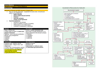
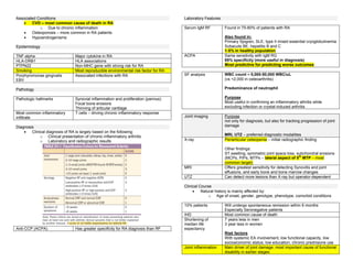
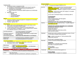
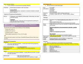
This document provides an overview of approaches to articular and musculoskeletal disorders, including osteoarthritis. It discusses evaluating patients for red flag diagnoses like infection or fracture. Key factors to determine include if the pain is articular or non-articular, acute or chronic, inflammatory or non-inflammatory. Risk factors for osteoarthritis include age, obesity, injury, and joint alignment. Symptoms include joint pain and stiffness. Treatment involves exercise, weight loss, bracing, acetaminophen, NSAIDs, and surgery for advanced cases.
This document provides information on diseases of the locomotor system, including the musculoskeletal system, manifestations of musculoskeletal disorders, anatomy, types of joints, and an algorithm for evaluating musculoskeletal complaints. It then discusses specific conditions like osteoarthritis, rheumatoid arthritis, ankylosing spondylitis, and their signs, symptoms, risk factors, investigations, and management. Key points covered include the hallmarks and prevalence of osteoarthritis and rheumatoid arthritis, as well as extra-articular manifestations, diagnostic criteria, differential diagnosis, and treatment approaches for rheumatoid arthritis.
Osteoarthritis is the most common form of arthritis, affecting over 60% of people over 65 years old. It involves the breakdown and eventual loss of cartilage in one or more joints. Risk factors include age, female gender, joint injuries, genetics, and obesity. Symptoms include joint pain, stiffness, swelling, and loss of motion. Treatment focuses on pain management through medications, physical therapy, weight loss, and sometimes joint replacement surgery.
This document provides information about seronegative arthritis, also known as spondyloarthritis. It discusses the characteristics and hallmarks of spondyloarthritis such as inflammatory back pain and enthesitis. The document covers the pathology, clinical manifestations, extra-articular features, diagnostic criteria, treatment options including NSAIDs and anti-TNF drugs, and complications such as fractures for this group of arthritic conditions.
All about Spondyloarthropaties also known as Seronegative Arthritis in a nutshell....includes Pathology,signs and symptoms, investigations, and latest approved treatment of all subtypes....compiled from Turek and Harrisons textbook.
Osteoarthritis is the most common form of joint disease. It is a non-inflammatory disorder affecting synovial joints that results from cartilage damage within the joints. Over time, the damaged cartilage leads to thinning of cartilage and bone growths around the joint margins. This causes pain, stiffness, and deformity of the joints. The most commonly affected joints are those in the hands, hips, knees, and lower back. Symptoms include joint pain that worsens with use and is relieved by rest, crepitus, and morning stiffness.
This document provides information on osteoarthritis (OA) and rheumatoid arthritis (RA). It defines OA as the most common form of joint disease, affecting over 90% of adults by age 40. Risk factors include aging, obesity, and joint injury from overuse. RA is a chronic systemic inflammatory disease that affects the synovial joints, with potential extra-articular manifestations. RA is believed to have an autoimmune cause and is associated with the presence of rheumatoid factor. Both diseases can cause pain, stiffness, and loss of physical function in affected joints. Treatment involves managing symptoms, maintaining function, and may include medications, exercise, joint protection, and sometimes surgery.
Osteoarthritis is a chronic degenerative disorder of synovial joints in which there is progressive softening and erosion/disintegration of the articular cartilage. In the presentation, I will deal in detail about the condition in every dimension with the most recent evidence.
This document provides an overview of how to approach a patient presenting with joint pain. It discusses how the pain may occur only at rest or only with activity. The physical exam aims to localize the source and understand the pathophysiology by assessing for signs of inflammation, functional impairment, swelling, range of motion, and tendon issues. Taking a thorough history is important to generate differential diagnoses and evaluate features like onset, duration, joint involvement, and extra-articular manifestations that may indicate underlying conditions like rheumatoid arthritis or lupus.
Osteoarthritis is a degenerative joint disease characterized by breakdown of cartilage and bone changes. It most commonly affects weight-bearing joints like the hips and knees. Risk factors include obesity, joint injury, genetics, and age. Symptoms include joint pain, stiffness, swelling, and decreased range of motion. Diagnosis is made based on clinical features and confirmed with x-rays showing cartilage loss, bone spurs, and bone changes. Treatment focuses on education, exercises, braces, medications, and sometimes surgery to relieve symptoms and improve function.
Osteoarthritis is a progressive joint disease caused by the gradual loss of cartilage. It is the most common form of arthritis and joint disease. The commonly affected joints are those in the hands, feet, spine, and knees. Risk factors include age, female gender, genetics, obesity, joint injuries, and overuse. Symptoms include joint pain, stiffness, and loss of function. Diagnosis involves physical exam, x-rays showing bone spurs and joint space narrowing, and ruling out other arthritic conditions. Treatment focuses on reducing joint stress through exercise, weight loss, and assistive devices. Medications may include acetaminophen, NSAIDs, and opioids for pain. Injections and surgery are
This document provides information on types of arthritis, signs and symptoms, treatment, and specific types like rheumatoid arthritis and osteoarthritis. It discusses normal joint structure, causes of osteoarthritis related to disparity between stress on cartilage and cartilage strength. Radiographic features and treatment options for osteoarthritis are outlined. Rheumatoid arthritis signs on imaging and physical exam are also summarized. Treatment of arthritis focuses on reducing inflammation and pain through medications like NSAIDs, DMARDs, steroids, and biologics that target proteins like TNF.
Rheumatoid arthritis is a chronic autoimmune disease that causes inflammation of the synovium and leads to joint damage over time. It is influenced by genetic, environmental, and hormonal factors. The inflammation causes swelling, stiffness, and pain in the joints. If left untreated, it can result in joint deformity and erosion of cartilage and bone. Treatment focuses on controlling inflammation, preventing deformity, and rehabilitation. Medications include NSAIDs, DMARDs, and biologics, along with physical therapy and surgery in some cases.
The document discusses various types of rheumatic and musculoskeletal disorders including rheumatoid arthritis, osteoarthritis, gout, and septic arthritis. It provides information on the symptoms, physical exam findings, laboratory/radiological findings, and treatments for each condition. Rheumatoid arthritis is described as a chronic inflammatory joint disease while osteoarthritis is degenerative joint disease more common in older individuals. Gout is caused by urate crystal deposition in joints. Septic arthritis is a medical emergency due to bacterial joint infection.
Osteoarthritis is a slowly progressive degenerative disease leading to gradual loss of articular cartilage. It affects not only cartilage but other joint tissues as well, including bone, ligaments, capsules, and synovial membrane. Risk factors include age, obesity, joint injury, genetics, and occupational factors. Symptoms include joint pain, stiffness, crepitus, and deformity. Diagnosis is based on clinical features and confirmed with x-rays showing bone spurs and joint space narrowing. Treatment involves lifestyle changes, medications for pain/inflammation, injections, and surgery for advanced cases.
This document discusses seronegative arthropathies (spondyloarthropathies), which are a group of inflammatory arthritides characterized by involvement of the sacroiliac joint and peripheral joints in the absence of rheumatoid factor. Key features include predominant involvement of the axial skeleton, association with HLA-B27 positivity, and extra-articular manifestations affecting the eyes, skin, gut and genitourinary system. Diagnosis involves clinical features, imaging such as X-ray showing sacroiliitis and spondylitis, and lab tests including elevated ESR/CRP and HLA-B27 testing. Treatment involves medications such as NSAIDs, DMARDs and TNF inhibitors as well
This document provides an overview of various rheumatologic disorders including their classification, symptoms, physical exam findings, and management considerations for dental patients. Key points include:
1. Rheumatologic disorders are classified as joint, degenerative, inflammatory, connective tissue, spondarthritis, autoimmune, or crystal arthropathy diseases. Common conditions discussed are osteoarthritis, rheumatoid arthritis, and gout.
2. Physical exam findings help differentiate conditions based on factors like joint involvement, inflammation signs, range of motion, and presence of extra-articular manifestations.
3. Laboratory/radiology tests are used to confirm diagnoses but can be misleading. Synovial fluid analysis and antibody/acute phase reactant testing
This document provides an overview of orthopaedics and the key components of taking a patient history. It describes the musculoskeletal system and main divisions of orthopaedics, including common diseases and injuries. The importance of systematically and carefully compiling a patient's history is emphasized. Key elements to cover include presenting complaints, symptoms of pain, stiffness, swelling, instability, and deformity. Details on grading and characterizing pain and assessing loss of function are also outlined.
Visco supplymentation in osteoarthrosis of knee and autologous copypunithpc605
The document discusses viscosupplementation for osteoarthritis of the knee. It provides background on osteoarthritis, describing how it leads to cartilage degeneration and bone changes. Viscosupplementation involves injecting hyaluronic acid into joints to restore the properties of synovial fluid and produce analgesic, anti-inflammatory, and chondroprotective effects by maintaining fluid viscosity and joint integrity. Hyaluronic acid is a natural polysaccharide in synovial fluid that lubricates joints and retains moisture.
This document provides an overview of knee anatomy, common causes of knee pain, physical exam techniques for evaluating knee pain, and differential diagnoses for various knee conditions. It discusses topics like intrinsic vs extrinsic knee pain, intra-articular vs peri-articular pain, inflammatory vs structural knee injuries, and common causes of knee pain like osteoarthritis, meniscal tears, ACL injuries, septic arthritis, and Baker's cysts. Physical exam maneuvers for assessing different knee structures and conditions are outlined. The prevalence of knee pain in the general population is cited.
This document provides information about arthritis and occupational therapy treatment. It defines arthritis as a group of disorders affecting the joints. The main types of arthritis discussed are osteoarthritis, rheumatoid arthritis, gouty arthritis, psoriatic arthritis, septic arthritis, and spinal arthritis. Occupational therapy aims to maintain function and mobility through techniques like splinting, exercises, ergonomics, fatigue management, and assistive devices. Proper evaluation and a multidisciplinary approach are important to arthritis treatment.
Cervical spondylosis is a common cause of neck pain, radiculopathy, and myelopathy. It involves chronic degenerative changes in the cervical discs and vertebrae due to aging. Common symptoms include neck pain, headaches, and radiating arm pain. Diagnosis involves clinical exam showing signs of radiculopathy or myelopathy as well as imaging like x-rays, CT, and MRI to identify areas of nerve root or spinal cord compression. Treatment options include conservative measures or surgery to decompress the spinal cord if conservative treatment fails.
Low back pain is very common, affecting 50-80% of adults at some point. It is the leading cause of disability in the US, costing $50 billion annually. While most cases resolve within 6 weeks, pain and disability may persist longer in up to 12-72% of patients. Risk factors include poor physical fitness, obesity, smoking, and hard physical labor. Mechanical low back pain makes up 90% of cases and involves overuse or injury of back structures, while 10% have non-mechanical systemic causes. Diagnosis involves history, exam, and sometimes imaging to identify pain generators and rule out serious causes requiring prompt treatment. Initial treatment focuses on remaining active, over-the-counter medications,
The document discusses various chronic pain syndromes including low back pain, sciatica, complex regional pain syndrome, trigeminal neuralgia, and cancer pain. It provides details on the definition, causes, symptoms, diagnostic tools and treatment options for low back pain and sciatica, which are the most commonly discussed chronic pain conditions. The treatment sections cover medications, physical therapy, injections including epidural steroid injections, radiofrequency ablation, and other minimally invasive procedures.
- Fractures are breaks or disruptions in the continuity of bone structure. They are commonly caused by traumatic injuries and are described and classified based on type, location, appearance of fragments, and stability.
- Clinical manifestations of fractures include immediate pain, inability to use the affected area, and potential deformity. Fracture healing involves hematoma formation, granulation tissue, callus formation, ossification, and remodeling over weeks to months.
- Treatment goals are anatomic realignment, immobilization to maintain alignment, and restoring function. This may involve closed or open reduction, traction, casting, external or internal fixation, and surgery. Nursing care focuses on assessment, pain management, prevention of complications, and
Arthritis and arthroplasty- dr. Mahmoud Abdel KareemAhmed-shedeed
This document provides information about osteoarthritis (OA), including its definition, prevalence, risk factors, pathology, diagnosis, natural history, differential diagnosis, and treatment. It notes that OA is the most common form of arthritis, affecting over 20 million people in the US. Risk factors include age, obesity, family history, and previous joint injury or disorder. Diagnosis is typically based on symptoms like pain and stiffness, physical exam findings, and x-ray evidence of cartilage loss, bone spurs, and bone changes. Treatment includes conservative options like medications, exercise, and weight loss, as well as intra-articular injections or surgery for advanced cases.
Osteoarthritis is a progressive degenerative joint disease characterized by the breakdown of cartilage. As the cartilage breaks down, the bone ends are no longer protected and begin to rub together causing pain, swelling and stiffness. Risk factors include age, obesity, joint injury and genetics. The most common symptoms are joint pain and stiffness that worsens with use. Treatment focuses on reducing pain and inflammation, maintaining joint mobility and managing risk factors.
Fractures medical surgical nursing .pptssuser47b89a
Fractures are breaks or disruptions in bone continuity. They are commonly caused by trauma and are classified based on type, location, stability, and communication with the external environment. The goals of treatment are to realign bone fragments through reduction, immobilize the bone to maintain alignment through various fixation methods like casting or traction, and restore normal function. Complications can include infection, compartment syndrome, fat embolism, and venous thrombosis. Hip fractures commonly occur in the elderly and require surgical repair and careful nursing management to prevent complications and promote mobility.
This document provides an overview of low back pain (LBP), including prevalence, classifications, types, and key points regarding evaluation and management. Some key points:
- 60-80% of people experience LBP at some point, though 90% resolves within 6 weeks. Recurrence is common and LBP is a major cause of disability.
- LBP can be classified as mechanical, traumatic, infectious, neoplastic, and more. 97% are considered mechanical.
- Types include discogenic, radicular, facet joint, sacroiliac joint, muscular/myofascial, and others. Herniated discs can cause radicular symptoms.
- Evaluation involves detailed history and exam to identify
Promoting Wellbeing - Applied Social Psychology - Psychology SuperNotesPsychoTech Services
A proprietary approach developed by bringing together the best of learning theories from Psychology, design principles from the world of visualization, and pedagogical methods from over a decade of training experience, that enables you to: Learn better, faster!
low birth weight presentation. Low birth weight (LBW) infant is defined as the one whose birth weight is less than 2500g irrespective of their gestational age. Premature birth and low birth weight(LBW) is still a serious problem in newborn. Causing high morbidity and mortality rate worldwide. The nursing care provide to low birth weight babies is crucial in promoting their overall health and development. Through careful assessment, diagnosis,, planning, and evaluation plays a vital role in ensuring these vulnerable infants receive the specialize care they need. In India every third of the infant weight less than 2500g.
Birth period, socioeconomical status, nutritional and intrauterine environment are the factors influencing low birth weight
Lecture 6 -- Memory 2015.pptlearning occurs when a stimulus (unconditioned st...AyushGadhvi1
learning occurs when a stimulus (unconditioned stimulus) eliciting a response (unconditioned response) • is paired with another stimulus (conditioned stimulus)
Are you looking for a long-lasting solution to your missing tooth?
Dental implants are the most common type of method for replacing the missing tooth. Unlike dentures or bridges, implants are surgically placed in the jawbone. In layman’s terms, a dental implant is similar to the natural root of the tooth. It offers a stable foundation for the artificial tooth giving it the look, feel, and function similar to the natural tooth.
Travel vaccination in Manchester offers comprehensive immunization services for individuals planning international trips. Expert healthcare providers administer vaccines tailored to your destination, ensuring you stay protected against various diseases. Conveniently located clinics and flexible appointment options make it easy to get the necessary shots before your journey. Stay healthy and travel with confidence by getting vaccinated in Manchester. Visit us: www.nxhealthcare.co.uk
The skin is the largest organ and its health plays a vital role among the other sense organs. The skin concerns like acne breakout, psoriasis, or anything similar along the lines, finding a qualified and experienced dermatologist becomes paramount.
DECLARATION OF HELSINKI - History and principlesanaghabharat01
This SlideShare presentation provides a comprehensive overview of the Declaration of Helsinki, a foundational document outlining ethical guidelines for conducting medical research involving human subjects.
Cell Therapy Expansion and Challenges in Autoimmune DiseaseHealth Advances
There is increasing confidence that cell therapies will soon play a role in the treatment of autoimmune disorders, but the extent of this impact remains to be seen. Early readouts on autologous CAR-Ts in lupus are encouraging, but manufacturing and cost limitations are likely to restrict access to highly refractory patients. Allogeneic CAR-Ts have the potential to broaden access to earlier lines of treatment due to their inherent cost benefits, however they will need to demonstrate comparable or improved efficacy to established modalities.
In addition to infrastructure and capacity constraints, CAR-Ts face a very different risk-benefit dynamic in autoimmune compared to oncology, highlighting the need for tolerable therapies with low adverse event risk. CAR-NK and Treg-based therapies are also being developed in certain autoimmune disorders and may demonstrate favorable safety profiles. Several novel non-cell therapies such as bispecific antibodies, nanobodies, and RNAi drugs, may also offer future alternative competitive solutions with variable value propositions.
Widespread adoption of cell therapies will not only require strong efficacy and safety data, but also adapted pricing and access strategies. At oncology-based price points, CAR-Ts are unlikely to achieve broad market access in autoimmune disorders, with eligible patient populations that are potentially orders of magnitude greater than the number of currently addressable cancer patients. Developers have made strides towards reducing cell therapy COGS while improving manufacturing efficiency, but payors will inevitably restrict access until more sustainable pricing is achieved.
Despite these headwinds, industry leaders and investors remain confident that cell therapies are poised to address significant unmet need in patients suffering from autoimmune disorders. However, the extent of this impact on the treatment landscape remains to be seen, as the industry rapidly approaches an inflection point.
8 Surprising Reasons To Meditate 40 Minutes A Day That Can Change Your Life.pptxHolistified Wellness
We’re talking about Vedic Meditation, a form of meditation that has been around for at least 5,000 years. Back then, the people who lived in the Indus Valley, now known as India and Pakistan, practised meditation as a fundamental part of daily life. This knowledge that has given us yoga and Ayurveda, was known as Veda, hence the name Vedic. And though there are some written records, the practice has been passed down verbally from generation to generation.
Tele Optometry (kunj'sppt) / Basics of tele optometry.
Rheumatology.pdf
1. Internal Medicine
Harrison’s Principles of Internal Medicine
Rheumatology
Approach to Articular and MSK Disorders (Chapter 363)
Rule out red flag diagnoses – suspected for acute onset, monoarticular
pattern or focal MSK pain
o Septic Arthritis
o Acute crystal-induced arthritis
o Fracture
o Vascular Ischemia
o Carpal Tunnel Syndrome
Important questions:
o Articular or non-articular
o Acute (<6 weeks) or chronic (>6 weeks)
o Inflammatory or non-inflammatory
o Localized or widespread in distribution
Articular Non-articular
Deep or diffuse pain
Limited range of motion on active and
passive movement
Swelling – due to synovial proliferation,
effusion, bony enlargement
Crepitation
Instability
Locking
Deformity
Painful on active movement only
Site of pain is usually periarticular, or in
regions adjacent to the articular surfaces
(-) swelling
(-) crepitation
(-) instability
(-) deformity
Inflammatory Non-inflammatory
Erythema, pain, swelling, warmth
Systemic signs – fatigue, fever, rash,
weight loss
(+) ESR, CRP, thrombocytosis, anemia
of chronic disease, hypoalbuminemia
(+) prolonged morning stiffness
improving with activity
(+) gel phenomenon in OA only
Related to trauma
Repetitive use
Degeneration or ineffective repair
(+) intermittent gel phenomenon
2. Clinical History
Younger age group SLE, reactive arthritis
Middle age group Fibromyalgia
RA
Elderly age group OA
Polymyalgia rheumatica
Men Gout
Spondyloarthritis
Ankylosing spondylitis
Women RA
Fibromyalgia
Osteoporosis
SLE
African American Sarcoidosis
SLE
Whites Polymyalgia rheumatic
Giant cell arteritis
Granulomatosis with polyangiitis
Familial aggregation Ankylosing spondylitis
Gout
Heberdon nodes of OA
Onset
Abrupt onset Septic arthritis,Gout
Indolent onset OA, RA, fibromyalgia
Evolution
Chronic OA
Intermittent Crystal-induced, Lyme arthritis
Migratory RF, gonococcal, viral arthritis
Additive RA, Psoriatic arthritis
Evolution
Chronic (>6 weeks) OA, RA, fibromyalgia
Acute (<6 weeks) Infectious, crystal-induced,
reactive
Extent or Distribution
Mono articular (1 joint) Crystal-induced, infectious
Oligoarticular (2-3 joints)
Poly articular (4 or more joints) OA, RA (symmetric as well)
Asymmetric and polyarticular Spondyloarthritis, reactive arthritis,
gout, sarcoid
Symmetric or asymmetric
Polyarticular or oligoarticular
OA, psoriatic arthritis
Upper extremities RA, OA
Lower extremities Gout, reactive arthritis
Axial skeleton OA, ankylosing spondylitis
RA of the cervical spine
Diseases with MSK co-morbidities
Diabetes mellitus Carpal tunnel syndrome
Renal insufficiency Gout
Depression or insomnia Fibromyalgia
Myeloma Low back pain
Cancer Myositis
Osteoporosis Fracture
Glucocorticoids Osteonecrosis
Septic arthritis
Diuretics or chemotherapy Gout
Rheumatic Review of Systems
Fever SLE, infection
Rash SLE, psoriatic arthritis
Nail abnormalities Psoriatic arthritis, reactive arthritis
Myalgias Fibromyalgia, statin-induced,
drug-induced
Weakness Polymyositis, neuropathy
Eye involvement Behcet, sarcoidosis,
Spondyloarthritis
Gastrointestinal Scleroderma, IBD
Genitourinary Reactive arthritis, gonococcemia
Nervous system Lyme disease, vasculitis
3.
4.
5. Osteoarthritis (Chapter 364)
Most common type of arthritis
Joints commonly affected by arthritis:
Definition
Defined as joint failure → hyaline articular cartilage loss present in a focal
and initially nonuniform manner → thickened subchondral bony plate,
outgrowth of osteophytes at the joint margin, stretching of the articular
capsule, snynovitis, weakness of the muscles bridging the joint
Joint Protective Mechanisms and their Failure
Joint protectors – joint capsule, ligaments, muscles and tendons, afferent
sensory nerves, underlying bone
Charcot arthropathy – severe and rapidly progressive OA developing when
minor injury occurs in the presence of posterior column peripheral
neuropathy
Joint capsule and ligaments Fixes range of motion
Synovial fluid lubrication Hyaluronic acid, lubricin
Bridges the joint Muscles and tendons
Cartilage and its Role in Joint Failure
Two most important macromolecules in cartilage:
Type 2 collagen Provides cartilage tensile strength
Aggrecan Proteoglycan macromolecule linked with hyaluronic acid
molecule, with highly negatively charged glycosaminoglycans
Through electropulsion → gives cartilage compressive
stiffness
OA cartilage
o (+) gradual depletion of aggrecan
o (+) unfurling of the tightly woven collagen matrix
o (+) loss of type 2 collagen
Risk Factors
2 major factors contributing to OA development:
o Joint vulnerability
o Joint loading
Systemic Risk Factors
Age Most potent risk factor for osteoarthritis
Rare in <40 yearsold
>50% in >70 years old
Aging increases joint vulnerability – less responsive in matrix
synthesis, thinner cartilage with age, weaker bridging
muscles, slower nerve conduction, stretching of ligaments
Older women Especially in their sixth decade
Probably hormonal loss
Heritability and
genetics
Highly heritable disease, but joint specific
(+) polymorphism with GDF5 gene
Race Knee OA is more common among Chinese
Risk Factors in the Joint Environment
Joint anatomy 3 uncommon developmental abnormalities occurring in utero or in
childhood:
Congenital dysplasia
Legg-Perthes disease
Slipped capital femoral epiphysis
Others: acetabular dysplasia, femoroacetabular impingement
Major injuries Fracture through the surface
Avascular necrosis
Tears of ligamentous and fibrocartilaginous structures
Meniscal tears
Joint misalignment
6. Varus knees – cartilage loss in the medial or inner knee
Valgus knees – cartilage loss in the lateral compartment
Weakness of the quadriceps – knee OA
Loading Factors
Obesity 3-6 times body weight is transmitted across the knee during a
single-leg stance
Well-recognized and potent risk factor for OA (esp. knee)
Precedes the development of disease
More potent in women
Adipokines may also be contributory
Repeated Use
of Joint and
Exercise
Seen in occupational use and leisure time physical activities
Farmers – hip OA
Miners – knee and spine OA
Heavy carrying – knee OA
Elite runners – knee and hip OA
Recreational runners – hip OA
Pathology
Non-uniform loss in cartilage in a focal disease
Thickening of the subchondral bony plate
Osteophyte formation – outgrowth of new cartilage that ossifies
o Important radiographic hallmark of OA
Stretching of the capsule → edematous and fibrotic
Sources of Pain
Likely occurs from structures outside the cartilage (aneural):
o Synovium, ligaments, joint capsule, muscles, subchondral
bone
Severity of X-ray changes correlate poorly with pain severity
Presence of MRI synovitis correlates with presence and severity of
knee pain
Clinical Features
Joint pain
o During or just after joint use and then gradually resolves
o As disease progresses, pain becomes continuous and even
becomes bothersome at night
Morning stiffness
o Usually brief (<30 minutes)
Knee symptoms
o Most common cause of chronic knee pain in patients >45 years
o (+) knee buckling – from weakness of muscles crossing over
the joint
Buckling, catching, locking Anterior crucial ligament or meniscal tear
Needs to be evaluated after a knee injury
Pain during knee flexion (>35
degrees)
From the patellofemoral compartment
Prolonged morning stiffness
with many joints affected
Inflammatory arthritis
Pain medial and distal to the
knee
Anserine bursitis
Common cause of knee pain, responsive to
glucocorticoid injection
Prominent nocturnal pain in the
absence of end-stage OA
Merits diagnostic work-up
Hip symptoms
o Can be detected by loss of internal rotation by passive
movement, pain isolated to an area lateral to the hip joint
(trochanteric bursitis)
Diagnostics
No blood tests are routinely indicated for workup of patients with OA, unless
symptoms and signs suggest inflammatory arthritis
Examination of synovial fluid >1000 WBC/uL – presence of inflammatory
arthritis or crystal-induced arthritis (with presence
of crystals)
X-rays Indicated when joint pain and physical findings are
atypical of OA, or if pain persists after initiation of
pain treatment
Radiographs may be normal in early disease
as they are insensitive to cartilage loss and
early findings
MRI Not indicated as part of diagnostic workup
Almost never warrant change in therapy
Goal of Treatment
Goal of treatment: To alleviate pain and minimize loss of physical function
Most effective mode of treatment: nonpharmacological treatment
Non-pharmacological Treatment
Goals of treatment:
o Avoiding painful activities – simplest treatment
o Improving strength and conditioning of muscles bridging the joints
o Unloading the joint via use of braces or splint to redistribute load or
unloading joint during weight bearing with a cane or crutch
Exercise
o Inactivity leads to increased risk for CVD and obesity
o Weakness in muscles have multiple etiologies:
Decline in strength with age
Disuse muscle atrophy due to limitation of movement
Diminishing muscle use due to alteration of gait
Due to arthrogenous inhibition
o Weakness in a muscle makes joint more susceptible to further
damage and pain
o Exercise lessens pain and improves physical function
o Major challenge – adherence to program
o Strongest predictor of success – personal history of successful
exercise
7. Effective Exercise Non-effective Exercise
Aerobic exercises
Resistance training
Low-impact exercises (water
aerobics)
Tai chi – effective for knee OA
Exercise with caloric restriction
Range-of-motion exercises
Isometric exercises
Correction of misalignment
o Via bracing for willing patients who can learn to put them on
correctly and on whom they do not slip
o Acupuncture
o Surgery
Pharmacological Treatment
Serves as an important adjunct therapy for OA
Acetaminophen
(Paracetamol)
Up to 1 gram
TID
Initial analgesic of choice for patients with OA
Prolongs half-life of warfarin
NSAIDs Most popular drugs to treat osteoarthritic pain
Either topically or orally
30% greater pain alleviation than high-dose paracetamol
Important to take low-dose aspirin and iburprofen/naproxen at
different times to limit drug interaction – may increase bleeding
and GI complaints
Side effects: GI complaints (should be taken after meals)
If with high-risk, take PPIs
Others: edema, renal insufficiency (used with caution for CKD III)
NSAIDS with increased CVD risk – diclofenac, rofecoxib
Allowed NSAIDs – Naproxen (with high GI toxicity), salsalate,
ibuprofen, celecoxib
Topicals – lesser efficacy, less GI and systemic side effects but
may cause skin irritation
Intraarticular
injections –
glucocorticoids
May be used temporarily (less than 3 months) for pain alleviation
since synovitis is correlated with joint pain severity
Subsequent injections may cause minor amounts of cartilage
loss with unimportant clinical consequences
Hyaluronic acid
injections
Can be given as treatment but with controversy on efficacy over
placebo
Opioid
analgesics
Has modest efficacy
Duloxetine Has modest efficacy
Glucosamine
Chondroitin
Recommended against OA
Surgery
Arthroscopic
menisectomy
Indicated for acute meniscal tears in which symptoms such as
locking and acute pain are clearly related temporally to a knee
injury that produced the tear
High tibial
osteotomy
For patients with knee OA to the medial compartment
Total knee or
hip
arthroplasty
Highly efficacious operations that relieve pain and improve
function in majority of patients
Failure rates of 1% per year (higher in obese patients)
Success rates higher in hips > knees
Timing of knee or hip replacement is critical
Cartilage
regeneration,
abrasion
arthroplasty
(chondroplasty)
Not found to be efficacious in OA
May be efficacious when OA has not yet developed
Ineffective surgical plans
Arthroscopic debridement and lavage
Partial menisectomy
8. Gout and Other Crystal-induced Arthropathies (Chapter 365)
Gout
Metabolic diseases mostly affecting
o Middle-aged to elderly men
o Postmenopausal women
Due to increased body pool of urate with hyperuricemia → deposition of
MSU crystals in joints and CT tophi with risk for deposition in the
kidney interstitium or uric acid nephrolithiasis
Clinical Features
Involved
joints
MTP1, tarsal joints, ankles, knees, finger joints (usually in the
elderly)
First
episode
Begins at night with dramatic joint pain and swelling – mimicking
cellulitis
Precipitating
events
Dietary excess, trauma, surgery, excessive ethanol ingestion,
hypouricemic therapy, serious medical illnesses – MI and stroke
Presentation
in women
Represents 5-20% of the population
(+) osteoarthritis and arterial hypertension → mild renal
insufficiency (+) receiving diuretics
Laboratory Diagnosis
Needle aspiration of
acutely or chronically
involved joints or
tophaceous deposits
Confirms presumptive diagnosis
(+) negative birefrigent needle-shaped crystals in
compensated polarized light intracellularly and
extracellularly
Synovial fluid
leukocyte counts
More than 2,000 to 60,000/uL
Culture If suspected to have septic arthritis
Serum uric levels Can be normal or low at the time of an acute attack –
inflammatory cytokines can be uricosuric and effective
initiation of hypouricemic therapy can precipitate attacks
Serum urate levels Almost always elevated at some time
Important to use follow the course of hypouricemic
therapy
24-h uric acid Useful in assessing risk of stones, elucidating
overproducers or underexcretors of uric acid – to decide
if uricosuric therapy is appropriate
Excretion of >800 mg uric acid per day on regular diet –
suggests overproduction of purine
Other tests
Urinalysis
Serum creatinine
Hemoglobin
WBC count
Liver function tests
Serum lipids
To assess possible pathologic sequelae of gout and other
associated diseases requiring treatment and as baselines
because of possible adverse effects of gout treatment
Radiographic features
X-ray (+) cystic changes, well-defined erosions with sclerotic
margins (with overhanging bony edges)
(+) soft tissue masses – advanced chronic tophaceous
gout
Ultrasound (+) double contour sign overlying articular cartilage
Dual-energy CT (+) presence of urate crystals
Treatment
Acute Gouty Arthritis
NSAIDs and colchicine – may be poorly tolerated and dangerous in the
elderly, especially in the presence of renal insufficiency and GI disorders
Ice pack applications and rest of involved joints can be helpful
Oral colchicine Traditional and effective treatment if used early in an attack
Regimens: 0.6 mg given every 8 hours with subsequent tapering
1.2 mg followed by 0.6 mg in 1 hour with subsequent day dosing
depending on response
Temporarily discontinued promptly at the first sign of loose
stools, and symptomatic treatment of diarrhea must be given
Oral NSAIDs Effective in 90% of patients
With resolution of signs and symptoms after 5-8 days
Most effective are those with short-half-lives:
Celecoxib (800 mg – 400 mg 12 hour after, then 400 mg BID)
Diclofenac (50 mg TID)
Ibuprofen (800 mg TID)
Naproxen (500 mg BID)
Indomethacin (25-50 mg TID)
9. Intramuscular or
oral
glucocorticoids
Oral: prednisone 30-50 mg/day as initial dose → gradually
tapered with resolution of attack
May be effective in polyarticular gout
Intraarticular: triamcinolone acetonide 20-40 mg, or
methylprednisolone 25-50 mg
Anakinra IL-1 antagonist
Has an essential role on the inflammasome
Used when other treatments have failed or contraindicated
Hypouricemic Therapy
Ultimate control – correction of hyperuricemial
Normalized serum uric acid: <300-360 umol/L (5.0 – 6.0 mg/dL)
o To prevent recurrent gouty attacks and eliminate tophaceous
deposits
Considered when hyperuricemia cannot be corrected by:
o Control of body weight
o Low-purine diet
o Increase in liquid intake
o Limitation of ethanol use
o Decreased use of fructose-containing foods and beverages
o Avoidance of diuretics
Indications
o Increased number of acute gouty attacks
o Serum uric acid of >535 umol/L (>9.0 mg/dL)
o Willingness to commit to lifelong therapy
o Presence of uric acid stones
o Presence of tophi or chronic gouty arthritis
Not initiated during acute attacks, but after patient is stable
Low-dose colchicine has been initiated to decrease risk of flares
o Colchicine prophylaxis in doses of 0.6 mg one to two timed
daily – should be given along with Hypouricemic therapy until
patient is normouricemic and without gouty attacks for 6 months, or
as long as tophi are present
o Not used in patients in dialysis, given in lower doses to
patients with renal disease or with P glycoprotein or CYP3A4
inhibitors (clarithromycin)
Probenecid Uricosuric agent used in patients with good renal function
who underexcrete uric acid <600 mg/day
Urine volume is maintained by ingestion of 1500 mL water/day
Regimen
250 mg BID → gradually to 3 g per day
Not effective in those with serum creatinine levels of >2 mg/dL
- May require allopurinol or benzbromarone
Benzbromarone Uricosuric agent more effective for patients with CKD
Lesinurad New uricosuric agent that can be used as an adjuvant at 200
mg/day to a xanthise oxidase inhibitor
Others with mild
uricosuric
Losartan, amlodipine, fenofibrate
effects
Allopurinol Xanthine oxidase inhibitor
Most commonly used Hypouricemic agent
Best drug to lower serum urate in overproducers, urate stone
formers, patients with renal disease
Regimen
Single morning dose of 100 mg (up to 800 mg as needed)
Adjusted initial dose depending on creatinine clearance (10
mL/min = 100 mg)
Toxicity among:
(+) penicillin and ampicillin allergies
(+) thiazide diuretic users
Asians with HLA-B*58:01
Serious side effects: TEN, systemic vasculitis, bone marrow
suppression, granulomatous hepatitis, renal failure
With mild cutaneous reactions
Can shift to uricosuric agent, desentiziation to allopurinol, use of
febuxostat
Febuxostat Xanthine oxidase inhibitor (40-80 mg OD)
Does not require dose adjustments with mild-moderate renal
disease
Pegloticase Pegylated uricase
Available for patients who do not tolerate or fail full doses of
other treatment
IV 8mg every 2 weeks can dramatically lower serum uric acid to
up to 50%
Other Cyrstal-induced arthritis
Calcium
pyrophosphate
deposition
(CPPD)
disease
Definitive Diagnosis
(+) rhomboid or rodlike, weakly positive or nonbirefrigent with
polarized light
Others: (+) linear calcifications
Calcium
apatite
deposition
disease
(basic calcium
phosphate
disease)
Definitive Diagnosis:
(+) seen in EM – 1-20 um shiny intra or extracellular
nonbirefrigent globules or aggregates
(+) purplish in Wright’s stain
(+) bright red in alizarin red S.
Others: (+) intra or periarticular calcifications with or without
erosive, destructive, or hypertrophic changes
Calcium
oxalate
disease
Definitive Diagnosis:
(+) bipyramidal and small polymorphic crystals in light
microscopy
Primary oxalosis – rare metabolic disease
Secondary oxalosis – mainly due to chronic renal failure, more
common than primary
10. Rheumatoid Arthritis (Chapter 351)
Most common form of chronic inflammatory arthritis
Clinical Features
Increases between 25 and 55 years of age, plateaus until 75 years and
declines after
Undifferentiated inflammatory arthritis – too few affected joints to be
classified as RA; if diagnosed later as RA:
o Higher number of tender and swollen joints
o Tests positive for RF and anti-CCP antibodies
o Higher scores for physical disability
Early morning stiffness Lasting more than 1 year, eases with physical activity
Earliest involved joints Small joints of the hands and feet:
Wrists, MCP, PIP joints
DIP joint involvement Manifestation of coexistent osteoarthritis
Flexor tendon
tenosynovitis
Frequent hallmark of RA → decreased range of
motion → reduced grip strength and trigger fingers
Ulnar deviation Subluxation of the MCP joints with subluxation of the
proximal phalanx to the volar side of the hand
Swan-neck deformity Hyperextension of the PIP
Flexion of the DIP
Boutonniere deformity Flexion of the PIP
Hyperextension of the DIP
Z-Line deformity Subluxation of the MCP1
Hyperextension of the IP1
Piano-key movement of
the ulnar styloid
Subluxation of the distal ulna – because of
inflammation of the ulnar styloid and tenosynovitis of
the extensor carpi ulnaris
Flat feet (pes planovalgus) Chronic inflammation of the ankle and midtarsal region
Atlantoaxial involvement May cause compressive myelopathy and neurologic
dysfunction
Rarely affects thoracic and lumbar spine
TMJ involvement Not associated with significant symptoms
Extraarticular Manifestations
May develop in 40% of patients – even before onset of RA
Most likely develop among patients:
o History of cigarette smoking
o Early onset of significant physical disability
o (+) RF and anti-CCP antibodies
Constitutional
symptoms
Weight loss, fatigue, malaise, depression, cachexia
Presence of fever >38.3 C – suspect for Vasculitis or
infection
Subcutaneous
nodules
Firm non-tender, adherent to the periosteum, tendonds or
bursae
More common (30-40%) in those with highest levels of disease
activity, disease-related shared epitope (SE), (+) RF,
radiographic evidence of joint erosions
Locations: forearm, sacral prominences, Achilles tendon
Others: lungs, pleura, pericardium, peritoneum
Sjogren
syndrome (10%)
(+) keratoconjunctivitis sicca (dry eyes)
(+) xerostomia (dry mouth)
Pulmonary
manifestations
Most common pulmonary manifestation - pleuritis
(+) pleuritic chest pain, dyspnea
(+) pleural friction rub, effusion (exudative)
Insterstitial lung disease – with high disease acitivity, smokers
– diagnosed via CT scan
Usual interstitial pneumonia (UIP), non-specific
interstitial pneumonia (NSIP) – main histological and
radiologic patterns of ILD
Poor prognosis (10% risk for mortality)
Favors more than idiopathic ILD
More responsive to immunosuppressive therapy
Caplan syndrome
(+) pulmonary nodulosis
(+) pneumoconiosis
(+) silica exposure
(+) rheumatoid arthritis
Cardiac Pericardium – most common site of involvement
MR – most common valvular abnormality in RA
Vasculitis Mostly seen in the following:
- Long-standing disease
- Positive for RF, anti-CCP
- Hypocomplementemia
Sensory polyneuropathies (Mononeuritis multiplex) may occur
Hematologic Normochromic, normocytic anemia – MC hematologic
abnormality
Felty’s syndrome (occurs in late stages of RA)
Neutropenia, splenomegaly, nodular RA
T-cell LGL
May have similar presentation with Felty
May occur early in disease
Lymphoma DLBCL – most common lymphoma
11. Associated Conditions
CVD – most common cause of death in RA
o Due to chronic inflammation
Osteoporosis – more common in RA patients
Hypoandrogenisms
Epidemiology
TNF-alpha Major cytokine in RA
HLA-DRB1 HLA associations
PTPN22 Non-MHC gene with strong risk for RA
Smoking Most reproducible environmental risk factor for RA
Porphyromonas gingivalis
EBV
Associated infections with RA
Pathology
Pathologic hallmarks Synovial inflammation and proliferation (pannus)
Focal bone erosions
Thinning of articular cartilage
Most common inflammatory
infiltrate
T cells – driving chronic inflammatory response
Diagnosis
Clinical diagnosis of RA is largely based on the following:
o Clinical presentation of chronic inflammatory arthritis
o Laboratory and radiographic results
Anti-CCP (ACPA) Has greater specificity for RA diagnosis than RF
Laboratory Features
Serum IgM RF Found in 75-80% of patients with RA
Also found in:
Primary Sjogren, SLE, type II mixed essential cryoglobulinemia
Subacute BE, hepatitis B and C
1-5% in healthy population
ACPA Same sensitivity with IgM RG
95% specificity (more useful in diagnosis)
Most predictive for predicting worse outcomes
SF analysis WBC count – 5,000-50,000 WBC/uL
(vs <2,000 in osteoarthritis)
Predominance of neutrophil
Purpose
Most useful in confirming an inflammatory athritis while
excluding infection or crystal-induced arthritis
Joint imaging Purpose
not only for diagnosis, but also for tracking progression of joint
damage
MRI, UTZ – preferred diagnostic modalities
X-ray Periarticular osteopenia – initial radiographic finding
Other findings:
ST swelling, symmetric joint space loss, subchondral erosions
(MCPs, PIPs, MTPs – lateral aspect of 5
th
MTP – most
common target)
MRI Offers greatest sensitivity for detecting Synovitis and joint
effusions, and early bone and bone marrow changes
UTZ Can detect more lesions than X-ray but operator-dependent
Clinical Course
Natural history is mainly affected by:
o Age of onset, gender, genotype, phenotype, comorbid conditions
10% patients Will undergo spontaneous remission within 6 months
Especially Seronegative patients
IHD Most common cause of death
Shortening of
median life
expectancy
7 years less in men
3 year less in women
Risk factors
With systemic EA involvement, low functional capacity, low
socioeconomic status, low education, chronic prednisone use
Joint inflammation Main driver of joint damage, most important cause of functional
disability in earlier stages
12. Treatment of RA
Development trends in management of RA
(1) Emergence of methotrexate as DMARD of first choice for early RA
(2) Development of novel biologicals that can be used alone or in
combination with methotrexate
(3) Proven superiority of combination DMARD regimens over methotrexate
alone
Medications used in RA
o NSAIDS
o Glucocorticoids
o Conventional DMARDs
o Biological DMARDs
NSAIDs
Presently considered as adjunctive agents for management of uncontrolled
symptoms by other measures
Toxicities – gastritis, PUD
Glucocorticoids
May be used in low-to-moderate doses to achieve rapid disease control
before onset of fully effective DMARD therapy
1-2 week bursts of steroids for management of acute disease flares
Chronic administration of prednisone – to control disease activity in patients
with inadequate response to DMARD therapy
Low-dose prednisone Can retard progression of joint disease
Must be balanced with side effects
Intraarticular injection of
intermediate steroid
(triamcinolone acetonide)
For rapid control of inflammation in a limited number
of affected joints
Must ensure that inflammation is not infectious
Osteoporosis Important long-term complication of steroids
Peptic ulcer No published guidelines for GI protection
Conventional DMARDs
Hydroxychloroquine, sulfasalazine, methotrexate, Leflunomide
Has delayed onset of action – 6-12 weeks
Methotrexate DMARD of choice for RA treatment
MOA – stimulates adenosine release → anti-inflammation
Leflunomide Inhibitor of pyrimidine synthesis
Similar clinical efficacy as methotrexate
Hydroxychloroquine Not a true DMARD; no retardation in RA progression
Sulfasalazine Has shown to retard RA progression Radiographically
Others Not used anymore
Minocycline, gold salts, penicillinamine, azathioprine, cylclosporine
Biological DMARDs
Anti-TNF – first approved biological DMARDs for RA
Anti-TNF Infliximab – chimeric (mouse-human) monoclonal antibody
Adalimumab, golimumab – human monoclonal antibody
Certolizumab pegol – pegylated Fc-fragment of human monoclonal
antibody
Etanercept – soluble fusion protein (TNF receptor 2 in convalent
linkage with Fc portion of IgG1)
Can be used in combination therapy or monotherapy (latter 4)
Contraindication
Active infection
History of hypersensitivity with these agents
Chronic hepatitis B infection
Class III/IV CHF
Screening for latent TB is important (PPD, IFN-gamma)
Anakinra Recombinant form of naturally occurring IL-1 receptor antagonist
Has limited use in RA
Indications (for syndromes dependent on IL-1 production)
Neonatal-onset inflammatory disease
Muckle-Wells syndrome
Familial cold urticaria
Systemic juvenile-onset inflammatory arthritis
Adult-onset Still’s disease
Abatacept Soluble fusion protein consisting of extracellular domain of human
CTLA4 linked to modified human IgG portion
MOA – inhibits co-stimulation of T cells by blocking CD28-
CD80/CD86 inreractions
Can be used in combination therapy
Rituximab Chimeric monoclonal antibody against CD20
Indications
Refractory RA in combination with methotrexate
More effect on seropositive patients
Adverse effects
Increased risk for infection
PML
Tocilizumab Humanized monclonal antinody against IL-6
Adverse effects
Increased infection, increased LDL cholesterol
13. Small molecule inhibitors
For RA resistant to conventional and biologic DMARDs
Tofacitinib Small-molecule inhibitor of JAK1 and JAK3
Adverse effects
Elevated liver enzymes, neutropenia, increased cholesterol, elevated
serum creatinine
Treatment of Extraarticular Manifestations
RA-ILD Challenging, some DMARDs (methotrexate, Leflunomide) can cause
pulmonary toxicity
Treatment
High-dose steroids + adjunctive immunosuppressants (azathioprine,
Mycophenolate mofetil, rituximab)
Management Considerations
Pregnancy
Up to 75% of female RA patients – overall improvement during pregnancy,
but will flare after delivery
Flares during pregnancy Low-dose prednisone
Hydroxychloroquine
Sulfasalazine
Safest DMARDs to use during pregnancy
Elderly
Conventional and biological DMARDs – equally effective and safe in younger
and older population
Methotrexate May need renal adjustments
Not prescribed when serum creatinine >2 mg/dL
SLE (Chapter 349)
90% are women of child-bearing age
HLA
associations
HLA-DRB1
HLA-DR3
HLA-DQA2
HLA-CR2
HLA-FCGR2A/B
Genes for
lupus
nephritis
HLA-DR3, STAT4, APOL1 (African Americans)
FCGR2A, ITGAM, IRF5, IRF7, TNFSF4 (Ox40L), DNAse1
Pathology
Skin biopsy Deposition of Ig at dermal-epidermal junction (DEJ) (also in normal
skin)
Injury to basal keratinocytes
Inflammation by T cells in the DEJ and around blood vessels
Renal
biopsy
Pattern and severity of injury are important for diagnosis and selecting
best therapy
Requires aggressive immunosupression (steroids + another
drug)
Class III
Class IV
Class V accompanied by III and IV
Not required
Class I and II, or with extensive irreversible changes
Blood vessel
pathology
Leukocytoclastic vasculitis – most common pattern of vasculitis
Lymph
biopsy
Usually performed to rule out infection or malignancies
Nonspecific diffuse chronic inflmmation
Diagnosis
Based on characteristic clinical features and autoantibodies
Classification of Nephritis (International Society of Nephrology and Renal Pathology
Society
Class I Minimal mesangial LN
Class II Mesangial proliferative LN
Class III Focal LN
Class IV Diffuse LN
Class V Membranous LN
Class VI Advanced Sclerotic LN
14. Manifestations
MSK Intermittent polyarthritis in hands, wrists, knees – most common
Joint pain – most common reason to increase dose of steroids
Ischemic necrosis of bone – diagnosed for persistence of pain in
single joint (treated with systemic steroids)
Myositis – usually drug-induced (steroids > antimalarials)
“rhupus” – combined SLE and RA presentation
Cutaneous Discoid lupus erythematosus (20%) – most common chronic
dermatitis in lupus
(+) roughly circular with slightly raised, scaly Hyperpigmented
erythematous rims
(+) depigmented atrophic centers (where all dermal appendages are
destroyed)
Treatment – topical or locally injected steroids + systemic
antimalarials
Photosensitive, slightly raised erythema, scaly on face – most
common acute SLE rash; “butterfly rash”
Subacute CLE – similar to psoriasis
(+) Ro (SS-A) antibodies
Renal Nephritis – most serious manifestation of SLE
Class IV – most severe, and most severe type of LN
DPGN – ESRD within 2 years (if untreated)
Can lead to atherosclerosis
CNS Cognitive dysfunction (memory difficulties, reasoning) - Most common
manifestation of diffuse CNS lupus
Seizures
Treatment – both antiseizure and immunosupression
Psychosis
Can be the predominant form of SLE
Must be distinguished from glucocorticoid-induced psychosis
Myelopathy
Tx: high-dose steroids
Pulmonary Pleuritis with or without pleural effusion – most common pulmonary
manifestation
Tx: NSAIDS → steroids if more severe
Life-threatening conditions
Interstitial fibrosis
Shrinking lung syndrome
Intraalveolar hemorrhage
Cardiac Pericarditis – most common cardiac manifestation
More serious manifestations – myocarditis, Libman-Sacks (MR, AR)
Vascular
occlusions
Cerebral event results from clotting
Tx: long-term anticoagulation if treatment of choice
Higher SLE MI events in women <49 years
Risk factors for atherosclerosis
Male, older, hypertension, dyslipidemia, diabetes, dysfunctional pro
inflammatory HDL, repeated high scores for disease activity, high
cumulative or daily doses of steroids, high levels of homocysteine and
leptin
Hematologic Normochromic, normocytic anemia – most common manifestation
Leukopenia (lymphopenia)
Thrombocytopenia
GI Vomiting, diarrhea, diffuse abdominal pain (autoimmune peritonitis,
and/or intestinal vasculitis), elevated liver enzymes
Tx: aggressive immunosuppression with high-dose steroids for short-
term control
Ocular Sicca syndrome, nonspecific conjunctivitis – common in SLE, rarely
threaten vision
15. Retinal vasculitis, optic neuritis – serious manifestations and blindness
can develop over days to weeks
Glaucoma, cataract – complications of steroids
Laboratory Tests
(1) To establish or rule out diagnosis
(2) Follow the course of disease
(3) Identify adverse effects of therapies
Autoantibody Testing
ANA – most important autoantibodies to detect (positive in >95% patients)
Anti-Ro/SS-A Indicates increased risk for neonatal lupus, sicca syndrome, and
SCLE
Standard Tests for Diagnosis
Screening using the following:
o CBC, PC, urinalysis
o May detect abnormalities contributing to diagnosis and
influence management decisions
Tests for Following Disease Course
UA, hemoglobin levels, PC,
serum creatinine, albumin
For hematuria, proteinuria
Identification of markers for
disease activity
See Table
Management of SLE
Aim is to achieve low-level disease activity
o Mild symptoms on the lowest possible doses of medications
o Usually achieved in 30-50% of SLE patients within a year
Conservative Therapies for Management of Non-Life-Threatening Disease
For patients with SLE autoantibodies, fatigue and pain without organ
involvement
Goal Management via suppression of symptoms
Anagesics, antimalarialas are mainstay
First issue with NSAIDs Increased risk for NSAID-induced aseptic
meningitis, elevated serum transaminases,
hypertension, renal dysfunction
Second issue with NSAIDs All NSAIDs (particularly COX-2 inhibitors), increases
MI risk
Issue with hydroxychloroquine Must have annual eye examination for potential
retinal toxicity
At least 750 ng/mL – blood level needed for optimal
response in active SLE
NSAIDs Arthritis/arthralgias
Antimalarials –
hydroxychloroquine,
chloroquine, quinacrine
Dermatitis
Arthritis
Fatigue
DHEA May reduce disease activity
Belimumab For fatigue, rash, and/or arthritis
Mostly effective with robust clinical activity:
SLEDA ≥10
Positive anti-DNA
Low serum complement
Topical sunscreens
Antimalarials
Topical steroids and/or
Tacrolimus
Lupus dermatitis
(systemic steroid with or without Mycophenolate
mofetil, azathioprine, belimumab – sever or
unresponse LD)
16. SLEDAI Widely used measure of SLE disease activitiy
>3 Clinically active disease
Life-threatening Management of SLE
Systemic Steroids
(0.5-1 mg/kg PO daily,
500-1000 mg IV daily)
Methylprednisolone sodium
succinate for 3 days → daily
(0.5-1 mg/kg) prednisone
Mainstay treatment for any inflammatory life-
threatening or organ-threatening manifestations of
SLE
Management of LN
(induction therapy)
Steroids +
Cylocphosphamide or mycophenolate mofetil
(azathioprine is effective, but associated with more
flares)
African Americans, hispanics –
mycophenolate
Whites, Asians – cyclophosphamide or
mycophenolate
Mycophenolate toxicity diarrhea
Cyclophosphamide toxicity Amenorrhea, leukopenia, nausea
Responses to treatment Steroid – within 24 hours
Cyclophosphamide, mycophenolate – 3-16 weeks
Management of LN
(maintenance therapy)
Mycophenolate > azathioprine
Cyclophosphamide – less safe
Cyclophosphamide toxicity Ovarian failure
Tx – GnRH agonist prior to each monhly
cyclophosphamide
Special Conditions
Crescentic LN
Treatment Either:
High-dose cyclophosphamide
High-dose mycophenolate
Membranous LN (INS V)
Treatment ACE-I or ABRs is recommended
But usually none, unless with nephrotic proteinuria
Pregnancy and Lupus
Rates of fetal loss is higher in SLE
o High disease activity, antiphospholipid antibodies (lupus
anticoagulant), hypertension, and/or active nephritis
Mainstay for active flares Hydroxychloroquine + lowest effective doses of
prednisone/prednisolone + azathioprine (if failure to
suppress activity)
HCQ – reduces chance of subsequent fetus with heart
block
Dexamethasone – prevents progression of heart block
SLE + antiphospholipid
antibodies + prior fetal
losses
LMW heparin + low-dose aspirin
Warfarin is teratogenic
Adverse effects of prenatal
steroids (primarily
betamethasone)
LBW
CNS developmental abnormalities
Predilection to adult metabolic syndrome
Category A (no evidence of
teratogenicity in humans)
Steroids
Category C
(may be teratogenic in
animals, no good evidence
in humans)
Cyclosporine, Tacrolimus, rituximab
Category D
(benefits > risks of
teratogenicity)
Azathioprine, hydroxychloroquine, mycophenolate,
cyclophosphamide
Category X Methotrexate
Lupus and APAS
Treatment Warfarin
Target INR (2.0-2.5) for 1 episode of venous clotting
Target INR (3.0-3.5) for recurrent clots or arterial clotting (CNS)
Lupus (TTP, HUS)
Treatment Diagnosis
Identification of schistocytes, elevated LDH, anti-ADAMS13
Treatment
Plasmapheresis – life-saving
Concomitant steroid therapy – recommended by authorities
17. Lupus dermatitis
Mainstay treatment Topical steroids + antimalarials (hydroxychloroquine)
- Effective in reducing lesion severity and relatively safe
Systemic retinoic acid
- For recalcitrant cases
Extensive, pruritic,
bullous, ulcerating
Systemic steroids with tapering → (hydroxychloroquine,
retinoids, belimumab)
Reports of success
with
Tacrolimus
Dapsone or thalidomide
Tacrolimus Increased risk for malignancies
Thalidomide Extreme dangers for fetal deformities
Peripheral neuropathy
Associated Malignancies
NHL
Cancers of thyroid, lung, liver, vulvar/vaginal tissues
Death
Leading causes in 1
st
decade of disease Systemic disease activity
Renal failure
infections
After 1
st
decade of disease Thromboembolic events
Drug-induced Lupus
Definition (+) positive ANA associated with
Fever, malaise, arthritis/arthralgias, myalgias, serositis, and/or rash
Features Predominant in whites
Usually appear during therapy with certain medications, biologicals
Less female predilection
Rarely involved kidneys, brains
Rarely associated with anti-dsDNA
Commonly associated with anti-histones
Usually resolves after discontinuation of medication
Frequent suspects
Antiarryhtmias Procainamide, disopryamide, propafenone
Antihypertensive Hydralazine
Several ACE-I and beta blockers
Antithyroid PTU
Antipsychotics Lithium, chlorpromazine
Anticonvulsants Carbamazepine, phenytoin
Antibiotic Isoniazid, minocycline, nitrofurantoin
Antirheumatic Sulfasazaline
Diuretic HCT
Antihyperlipidemia Lovastastin, simvastatin
Biologics IFN and TNF inhibitors
PEARLS
Approach to MSK diseases
Focal, monoarthritis in acute onset Trauma/Fracture
Gout
Vascular ischemia
Carpal tunnel syndrome
Septic Arthritis
Osteoarhthritis
Most potent risk factor for OA Age
Polymorphism in this gene is involved in formation
of osteoarthritis
GDF5 gene
What joint is usually more involved in OA among
Chinese
Knees
Important radiographic hallmark of OA Osteophytes
MRI finding which correlates with severity of knee
pain
Presence of MRI synovitis
Most common cause of chronic knee pain in
patients >45 years
Knee OA
Amount of WBC in synovial fluid which accounts
the presence of inflammatory or crystal-induced
arthritis
>1000 WBC/uL
Never indicated as part of diagnostic workup
Furthermore, it almost never warrant change in
therapy
MRI
Mainstay treatment for osteoarthritis Non-pharmacological
Initial choice of treatment for OA Acetaminophen (high-dose)
The use of these agents were banned in the
treatment of OA
Chondroitin
Glucosamine
Gout and Other Crystal-Associated Arthritis
Excretion of this amount of uric acid per day on a
regular diet suggests overproduction of purine
>800 mg/day
Ultrasound manifestation of gout (+) double contour sign
Severe hyperuricemia requiring Hypouricemic
therapy
>9 mg/dL (>535 umol/L)
Underexcretor of uric acid <600 mg/day
Other medications with mild uricosuric effects Losartan
Amlodipine
Fenofibrate
Contraindications of colchicine use Dialysis patients
(+) P glycoprotein
(+) CYP3A4 inhibitors -
clarithromycin
Rheumatoid Arthritis
Temperature before one must suspect for
Vasculitis or infection in RA patients
>38.3 C
Main histological and radiologic patterns of ILD Usual interstitial pneumonia
Non-specific interstitial
pnuemonia
Most common cause of death in RA CVD
Associated conditions with RA CVD
18. Osteoporosis
hypoandrogenisms
Has greater specificity for RA diagnosis than RF ACPA
Initial radiographic finding in patients with RA Periarticular osteopenia
Delayed onset of actions of conventional
DMARDs
6-12 weeks
SLE
Most important autoantibody to test for SLE ANA
Achievement of low-level disease SLE activity 30-50% patients within a year