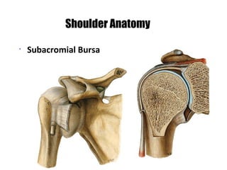
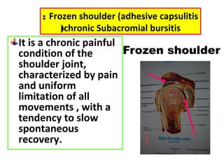
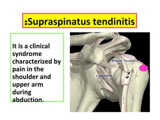
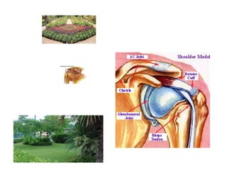
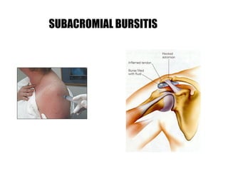
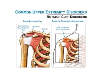
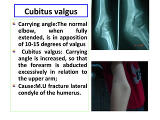
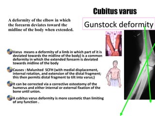
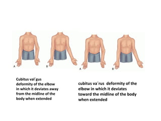
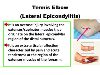
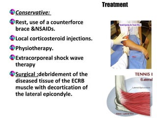
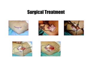
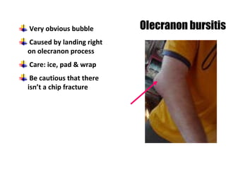
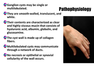
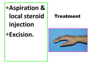
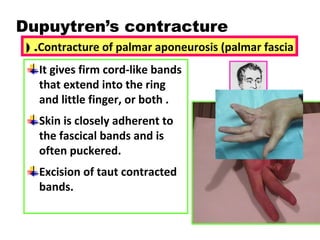
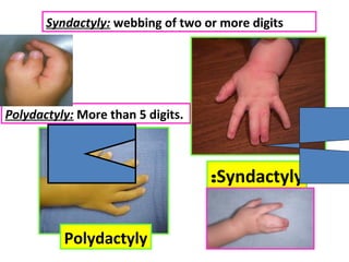
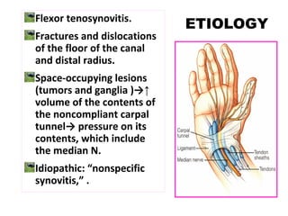
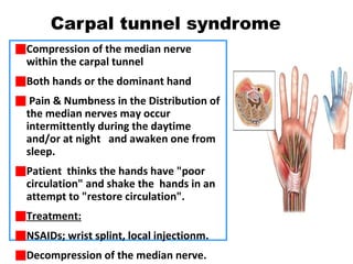
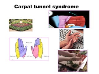
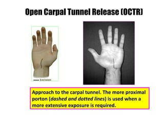
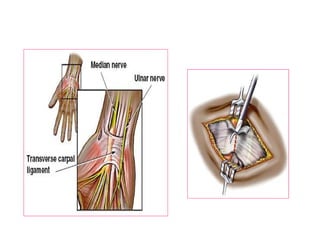
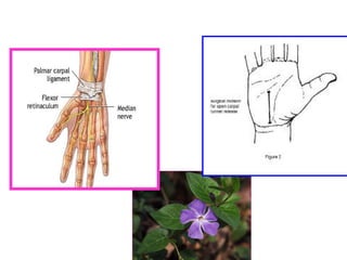
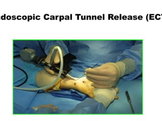
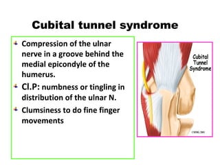
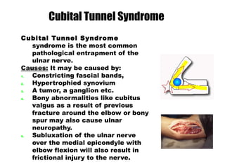
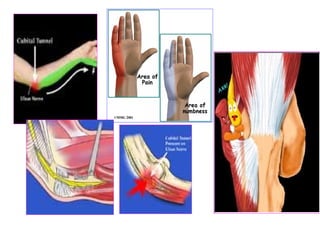
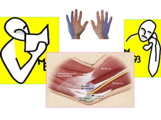
The document discusses disorders of the upper limb, including the shoulder, elbow, wrist, and hand. It provides details on anatomy, common conditions such as frozen shoulder, lateral epicondylitis, carpal tunnel syndrome, and treatments including injections, physical therapy, and surgery. It comprehensively covers the assessment and management of various musculoskeletal issues in the upper extremity.
this PPT contain detailed kinetics & kinematics of ankle joint & all joints of foot complex, muscles of ankle & foot complex, plantar arches & weight distribution during standing.
rotator cuff injuries, its causes, types,symptoms, special test and its pt management
special test for every injury types which includes rotator cuff tear, tendonitis, impingement syndrome,painfull arc syndrome, frozen shoulder.....special test includes neer's impingent, empty cane, full cane, speed test
As part of a class presentation, we attempted to make this to briefly explain what Torticollis meas, the Types of presentation of Torticollis, and Management strategies for a Physiotherapist for Congenital Torticollis especially.
I hope this helps. :)
The pictures and information had been taken from internet, complied to make a brief presentation for the purpose of class presentation.
I do not own any content.
Femoral Head (Superiorly, Medially, Anteriorly).
Acetabulum (Inferiorly, Laterally, Anteriorly).
Horseshoe-shaped (Acetabular Notch).
The deepest portion (Acetabular Fossa).
Labrum Acetabular:
Is a wedged fibrocartilaginous ring inserted into the acetabular rim to increase the acetabular concavity.
Mch Program Offered by Texila American UniversityTexila141
Texila American University has developed "Knowledge Based Assessment" program leading to M.Ch Certification. The aim and objectives of the "Magister Chirurgiae" M.Ch Trauma and Orthopedic Surgery & Surgery ( Two different Courses) course are to provide a clinically based Masters Degree, designed to gain super specialty experience in Trauma and Orthopedics.
Visit my Blog for more on Dentistry and Medicine :
http://dentistryandmedicine.blogspot.com/
Free Download e Books,PPT's and Lecture notes
Contents
Knee Joint anatomy with Diagrams
Anatomical Components of Knee
Anatomy of Patella
Innervations of the Knee
Knee Movements
Osteoarthritis in the Knee
Management of Disorders in Knee joint
paediatric injuries around the elbow
supracondylar elbow injuries
pulled elbow in paediatric age r
radiological signs around elbow in supracondylar fracture humerus
Anti ulcer drugs and their Advance pharmacology ||
Anti-ulcer drugs are medications used to prevent and treat ulcers in the stomach and upper part of the small intestine (duodenal ulcers). These ulcers are often caused by an imbalance between stomach acid and the mucosal lining, which protects the stomach lining.
||Scope: Overview of various classes of anti-ulcer drugs, their mechanisms of action, indications, side effects, and clinical considerations.
Flu Vaccine Alert in Bangalore Karnatakaaddon Scans
As flu season approaches, health officials in Bangalore, Karnataka, are urging residents to get their flu vaccinations. The seasonal flu, while common, can lead to severe health complications, particularly for vulnerable populations such as young children, the elderly, and those with underlying health conditions.
Dr. Vidisha Kumari, a leading epidemiologist in Bangalore, emphasizes the importance of getting vaccinated. "The flu vaccine is our best defense against the influenza virus. It not only protects individuals but also helps prevent the spread of the virus in our communities," he says.
This year, the flu season is expected to coincide with a potential increase in other respiratory illnesses. The Karnataka Health Department has launched an awareness campaign highlighting the significance of flu vaccinations. They have set up multiple vaccination centers across Bangalore, making it convenient for residents to receive their shots.
To encourage widespread vaccination, the government is also collaborating with local schools, workplaces, and community centers to facilitate vaccination drives. Special attention is being given to ensuring that the vaccine is accessible to all, including marginalized communities who may have limited access to healthcare.
Residents are reminded that the flu vaccine is safe and effective. Common side effects are mild and may include soreness at the injection site, mild fever, or muscle aches. These side effects are generally short-lived and far less severe than the flu itself.
Healthcare providers are also stressing the importance of continuing COVID-19 precautions. Wearing masks, practicing good hand hygiene, and maintaining social distancing are still crucial, especially in crowded places.
Protect yourself and your loved ones by getting vaccinated. Together, we can help keep Bangalore healthy and safe this flu season. For more information on vaccination centers and schedules, residents can visit the Karnataka Health Department’s official website or follow their social media pages.
Stay informed, stay safe, and get your flu shot today!
HOT NEW PRODUCT! BIG SALES FAST SHIPPING NOW FROM CHINA!! EU KU DB BK substit...GL Anaacs
Contact us if you are interested:
Email / Skype : kefaya1771@gmail.com
Threema: PXHY5PDH
New BATCH Ku !!! MUCH IN DEMAND FAST SALE EVERY BATCH HAPPY GOOD EFFECT BIG BATCH !
Contact me on Threema or skype to start big business!!
Hot-sale products:
NEW HOT EUTYLONE WHITE CRYSTAL!!
5cl-adba precursor (semi finished )
5cl-adba raw materials
ADBB precursor (semi finished )
ADBB raw materials
APVP powder
5fadb/4f-adb
Jwh018 / Jwh210
Eutylone crystal
Protonitazene (hydrochloride) CAS: 119276-01-6
Flubrotizolam CAS: 57801-95-3
Metonitazene CAS: 14680-51-4
Payment terms: Western Union,MoneyGram,Bitcoin or USDT.
Deliver Time: Usually 7-15days
Shipping method: FedEx, TNT, DHL,UPS etc.Our deliveries are 100% safe, fast, reliable and discreet.
Samples will be sent for your evaluation!If you are interested in, please contact me, let's talk details.
We specializes in exporting high quality Research chemical, medical intermediate, Pharmaceutical chemicals and so on. Products are exported to USA, Canada, France, Korea, Japan,Russia, Southeast Asia and other countries.
Ozempic: Preoperative Management of Patients on GLP-1 Receptor Agonists Saeid Safari
Preoperative Management of Patients on GLP-1 Receptor Agonists like Ozempic and Semiglutide
ASA GUIDELINE
NYSORA Guideline
2 Case Reports of Gastric Ultrasound
TEST BANK for Operations Management, 14th Edition by William J. Stevenson, Ve...kevinkariuki227
TEST BANK for Operations Management, 14th Edition by William J. Stevenson, Verified Chapters 1 - 19, Complete Newest Version.pdf
Lung Cancer: Artificial Intelligence, Synergetics, Complex System Analysis, S...Oleg Kshivets
RESULTS: Overall life span (LS) was 2252.1±1742.5 days and cumulative 5-year survival (5YS) reached 73.2%, 10 years – 64.8%, 20 years – 42.5%. 513 LCP lived more than 5 years (LS=3124.6±1525.6 days), 148 LCP – more than 10 years (LS=5054.4±1504.1 days).199 LCP died because of LC (LS=562.7±374.5 days). 5YS of LCP after bi/lobectomies was significantly superior in comparison with LCP after pneumonectomies (78.1% vs.63.7%, P=0.00001 by log-rank test). AT significantly improved 5YS (66.3% vs. 34.8%) (P=0.00000 by log-rank test) only for LCP with N1-2. Cox modeling displayed that 5YS of LCP significantly depended on: phase transition (PT) early-invasive LC in terms of synergetics, PT N0—N12, cell ratio factors (ratio between cancer cells- CC and blood cells subpopulations), G1-3, histology, glucose, AT, blood cell circuit, prothrombin index, heparin tolerance, recalcification time (P=0.000-0.038). Neural networks, genetic algorithm selection and bootstrap simulation revealed relationships between 5YS and PT early-invasive LC (rank=1), PT N0—N12 (rank=2), thrombocytes/CC (3), erythrocytes/CC (4), eosinophils/CC (5), healthy cells/CC (6), lymphocytes/CC (7), segmented neutrophils/CC (8), stick neutrophils/CC (9), monocytes/CC (10); leucocytes/CC (11). Correct prediction of 5YS was 100% by neural networks computing (area under ROC curve=1.0; error=0.0).
CONCLUSIONS: 5YS of LCP after radical procedures significantly depended on: 1) PT early-invasive cancer; 2) PT N0--N12; 3) cell ratio factors; 4) blood cell circuit; 5) biochemical factors; 6) hemostasis system; 7) AT; 8) LC characteristics; 9) LC cell dynamics; 10) surgery type: lobectomy/pneumonectomy; 11) anthropometric data. Optimal diagnosis and treatment strategies for LC are: 1) screening and early detection of LC; 2) availability of experienced thoracic surgeons because of complexity of radical procedures; 3) aggressive en block surgery and adequate lymph node dissection for completeness; 4) precise prediction; 5) adjuvant chemoimmunoradiotherapy for LCP with unfavorable prognosis.
Title: Sense of Smell
Presenter: Dr. Faiza, Assistant Professor of Physiology
Qualifications:
MBBS (Best Graduate, AIMC Lahore)
FCPS Physiology
ICMT, CHPE, DHPE (STMU)
MPH (GC University, Faisalabad)
MBA (Virtual University of Pakistan)
Learning Objectives:
Describe the primary categories of smells and the concept of odor blindness.
Explain the structure and location of the olfactory membrane and mucosa, including the types and roles of cells involved in olfaction.
Describe the pathway and mechanisms of olfactory signal transmission from the olfactory receptors to the brain.
Illustrate the biochemical cascade triggered by odorant binding to olfactory receptors, including the role of G-proteins and second messengers in generating an action potential.
Identify different types of olfactory disorders such as anosmia, hyposmia, hyperosmia, and dysosmia, including their potential causes.
Key Topics:
Olfactory Genes:
3% of the human genome accounts for olfactory genes.
400 genes for odorant receptors.
Olfactory Membrane:
Located in the superior part of the nasal cavity.
Medially: Folds downward along the superior septum.
Laterally: Folds over the superior turbinate and upper surface of the middle turbinate.
Total surface area: 5-10 square centimeters.
Olfactory Mucosa:
Olfactory Cells: Bipolar nerve cells derived from the CNS (100 million), with 4-25 olfactory cilia per cell.
Sustentacular Cells: Produce mucus and maintain ionic and molecular environment.
Basal Cells: Replace worn-out olfactory cells with an average lifespan of 1-2 months.
Bowman’s Gland: Secretes mucus.
Stimulation of Olfactory Cells:
Odorant dissolves in mucus and attaches to receptors on olfactory cilia.
Involves a cascade effect through G-proteins and second messengers, leading to depolarization and action potential generation in the olfactory nerve.
Quality of a Good Odorant:
Small (3-20 Carbon atoms), volatile, water-soluble, and lipid-soluble.
Facilitated by odorant-binding proteins in mucus.
Membrane Potential and Action Potential:
Resting membrane potential: -55mV.
Action potential frequency in the olfactory nerve increases with odorant strength.
Adaptation Towards the Sense of Smell:
Rapid adaptation within the first second, with further slow adaptation.
Psychological adaptation greater than receptor adaptation, involving feedback inhibition from the central nervous system.
Primary Sensations of Smell:
Camphoraceous, Musky, Floral, Pepperminty, Ethereal, Pungent, Putrid.
Odor Detection Threshold:
Examples: Hydrogen sulfide (0.0005 ppm), Methyl-mercaptan (0.002 ppm).
Some toxic substances are odorless at lethal concentrations.
Characteristics of Smell:
Odor blindness for single substances due to lack of appropriate receptor protein.
Behavioral and emotional influences of smell.
Transmission of Olfactory Signals:
From olfactory cells to glomeruli in the olfactory bulb, involving lateral inhibition.
Primitive, less old, and new olfactory systems with different path
Title: Sense of Taste
Presenter: Dr. Faiza, Assistant Professor of Physiology
Qualifications:
MBBS (Best Graduate, AIMC Lahore)
FCPS Physiology
ICMT, CHPE, DHPE (STMU)
MPH (GC University, Faisalabad)
MBA (Virtual University of Pakistan)
Learning Objectives:
Describe the structure and function of taste buds.
Describe the relationship between the taste threshold and taste index of common substances.
Explain the chemical basis and signal transduction of taste perception for each type of primary taste sensation.
Recognize different abnormalities of taste perception and their causes.
Key Topics:
Significance of Taste Sensation:
Differentiation between pleasant and harmful food
Influence on behavior
Selection of food based on metabolic needs
Receptors of Taste:
Taste buds on the tongue
Influence of sense of smell, texture of food, and pain stimulation (e.g., by pepper)
Primary and Secondary Taste Sensations:
Primary taste sensations: Sweet, Sour, Salty, Bitter, Umami
Chemical basis and signal transduction mechanisms for each taste
Taste Threshold and Index:
Taste threshold values for Sweet (sucrose), Salty (NaCl), Sour (HCl), and Bitter (Quinine)
Taste index relationship: Inversely proportional to taste threshold
Taste Blindness:
Inability to taste certain substances, particularly thiourea compounds
Example: Phenylthiocarbamide
Structure and Function of Taste Buds:
Composition: Epithelial cells, Sustentacular/Supporting cells, Taste cells, Basal cells
Features: Taste pores, Taste hairs/microvilli, and Taste nerve fibers
Location of Taste Buds:
Found in papillae of the tongue (Fungiform, Circumvallate, Foliate)
Also present on the palate, tonsillar pillars, epiglottis, and proximal esophagus
Mechanism of Taste Stimulation:
Interaction of taste substances with receptors on microvilli
Signal transduction pathways for Umami, Sweet, Bitter, Sour, and Salty tastes
Taste Sensitivity and Adaptation:
Decrease in sensitivity with age
Rapid adaptation of taste sensation
Role of Saliva in Taste:
Dissolution of tastants to reach receptors
Washing away the stimulus
Taste Preferences and Aversions:
Mechanisms behind taste preference and aversion
Influence of receptors and neural pathways
Impact of Sensory Nerve Damage:
Degeneration of taste buds if the sensory nerve fiber is cut
Abnormalities of Taste Detection:
Conditions: Ageusia, Hypogeusia, Dysgeusia (parageusia)
Causes: Nerve damage, neurological disorders, infections, poor oral hygiene, adverse drug effects, deficiencies, aging, tobacco use, altered neurotransmitter levels
Neurotransmitters and Taste Threshold:
Effects of serotonin (5-HT) and norepinephrine (NE) on taste sensitivity
Supertasters:
25% of the population with heightened sensitivity to taste, especially bitterness
Increased number of fungiform papillae
Pulmonary Thromboembolism - etilogy, types, medical- Surgical and nursing man...VarunMahajani
Disruption of blood supply to lung alveoli due to blockage of one or more pulmonary blood vessels is called as Pulmonary thromboembolism. In this presentation we will discuss its causes, types and its management in depth.
New Directions in Targeted Therapeutic Approaches for Older Adults With Mantl...i3 Health
i3 Health is pleased to make the speaker slides from this activity available for use as a non-accredited self-study or teaching resource.
This slide deck presented by Dr. Kami Maddocks, Professor-Clinical in the Division of Hematology and
Associate Division Director for Ambulatory Operations
The Ohio State University Comprehensive Cancer Center, will provide insight into new directions in targeted therapeutic approaches for older adults with mantle cell lymphoma.
STATEMENT OF NEED
Mantle cell lymphoma (MCL) is a rare, aggressive B-cell non-Hodgkin lymphoma (NHL) accounting for 5% to 7% of all lymphomas. Its prognosis ranges from indolent disease that does not require treatment for years to very aggressive disease, which is associated with poor survival (Silkenstedt et al, 2021). Typically, MCL is diagnosed at advanced stage and in older patients who cannot tolerate intensive therapy (NCCN, 2022). Although recent advances have slightly increased remission rates, recurrence and relapse remain very common, leading to a median overall survival between 3 and 6 years (LLS, 2021). Though there are several effective options, progress is still needed towards establishing an accepted frontline approach for MCL (Castellino et al, 2022). Treatment selection and management of MCL are complicated by the heterogeneity of prognosis, advanced age and comorbidities of patients, and lack of an established standard approach for treatment, making it vital that clinicians be familiar with the latest research and advances in this area. In this activity chaired by Michael Wang, MD, Professor in the Department of Lymphoma & Myeloma at MD Anderson Cancer Center, expert faculty will discuss prognostic factors informing treatment, the promising results of recent trials in new therapeutic approaches, and the implications of treatment resistance in therapeutic selection for MCL.
Target Audience
Hematology/oncology fellows, attending faculty, and other health care professionals involved in the treatment of patients with mantle cell lymphoma (MCL).
Learning Objectives
1.) Identify clinical and biological prognostic factors that can guide treatment decision making for older adults with MCL
2.) Evaluate emerging data on targeted therapeutic approaches for treatment-naive and relapsed/refractory MCL and their applicability to older adults
3.) Assess mechanisms of resistance to targeted therapies for MCL and their implications for treatment selection
MANAGEMENT OF ATRIOVENTRICULAR CONDUCTION BLOCK.pdfJim Jacob Roy
Cardiac conduction defects can occur due to various causes.
Atrioventricular conduction blocks ( AV blocks ) are classified into 3 types.
This document describes the acute management of AV block.
7. It is a chronic painful
condition of the
shoulder joint,
characterized by pain
and uniform
limitation of all
movements , with a
tendency to slow
spontaneous
recovery.
Frozen shoulder (adhesive capsulitis;
chronic Subacromial bursitis(
Frozen shoulder
8. Symptoms of primary frozen shoulder have been
divided into 3 phases:
1. Painful phase (there is a gradual onset of
diffuse shoulder pain lasting from weeks to
months. )
2. Stiffening phase (progressive loss of motion
that may last up to 1 year. Most patients lose
glenohumeral external rotation, internal
rotation, and abduction during this phase. )
3. Thawing phase (gradual motion improvement).
This phase may take up to 9 months for the
motion improvement for the patient to regain
a functional ROM.
9. It is clinical conditions, including:
1. Subacromial bursitis
2. Calcifying tendinitis
3. Partial rotator cuff tears.
Adhesive capsulitis (frozen shoulder syndrome ;FSS)
P
r
o
g
r
e
s
s
i
v
e
p
a
10. It is a clinical
syndrome
characterized by
pain in the
shoulder and
upper arm
during
abduction.
Supraspinatus tendinitis:
17. Cubitus varus: The
carrying angle is
decreased or
reversed;
Cause: malunited
SCFH.
ELBOW DISORDERS
Cubitus valgus:
Angle is increased,
so that the forearm
is abducted
excessively in
relation to the
upper arm;
Cause:M.U fracture
lateral condyle of
the humerus.
18. Cubitus valgus
Carrying angle:The normal
elbow, when fully
extended, is in apposition
of 10-15 degrees of valgus
Cubitus valgus: Carrying
angle is increased, so that
the forearm is abducted
excessively in relation to
the upper arm;
Cause:M.U fracture lateral
condyle of the humerus.
19. A deformity of the elbow in which
the forearm deviates toward the
midline of the body when extended.
Varus means a deformity of a limb in which part of it is
deviated towards the midline of the body) is a common
deformity in which the extended forearm is deviated
towards midline of the body
Causes : Malunited SCFH (with medial displacement,
internal rotation, and extension of the distal fragment;
this then permits distal fragment to tilt into varus;)
It can be corrected via a corrective osteotomy of the
humerus and either internal or external fixation of the
bone until union.
A cubitus varus deformity is more cosmetic than limiting
of any function .
Cubitus varus
Gunstock deformity
20.
21. Cubitus val´gus
deformity of the elbow
in which it deviates away
from the midline of the
body when extended
cubitus va´rus deformity of the
elbow in which it deviates
toward the midline of the body
when extended
22. It is an overuse injury involving the
extensor/supinator muscles that
originate on the lateral epicondylar
region of the distal humerus.
It is an extra-articular affection
characterized by pain and acute
tenderness at the region of the
extensor muscles of the forearm.
Tennis Elbow
(Lateral Epicondylitis)
23. Conservative:
Rest, use of a counterforce
brace &NSAIDs.
Local corticosteroid injections.
Physiotherapy.
Extracorporeal shock wave
therapy
Surgical :debridement of the
diseased tissue of the ECRB
muscle with decortication of
the lateral epicondyle.
Treatment
25. Very obvious bubble
Caused by landing right
on olecranon process
Care: ice, pad & wrap
Be cautious that there
isn’t a chip fracture
Olecranon bursitis
27. Wrist & Hand
Bones of the Wrist
Joint (Carpals)
Eight bones of the
carpus, which occur
in two rows (proximal
and distal).
The proximal row,
consists of scaphoid,
lunate, triquetrum
and pisiform, and the
first three of these
articulate with the
distal ends of the
radius or ulna.
The distal row, made
up of the trapezium,
trapezoid, capitate
and hamate,
articulates with the
bases of the
metacarpal bones.
28. An avascular necrosis of
the lunate bone due to
impairment of its blood
supply.
Softening,
fragmentation&
deformation of the
lunate .
It may give rise to
osteoarthritis of the
wrist joint.
Kienböck’s Disease
29. Tenovaginitis of the
abductor pollicis
longus & extensor
pollicis brevis.
Local tenderness at
the styloid process of
the radius.
Conservative TTT.
Release of the
sheaths of the 2
tendons.
e Quervain’s Disease
31. Women >Men
Age : 55-60 years .
The most commonly affected
digit is the thumb, followed by
the ring, long, little, and index
fingers.
More frequent in patients with
rheumatoid arthritis or
diabetes mellitus
Trigger Finger
nosing tendovaginitis of flexor tendon
snapping or jerking movements
32. Locking or catching during
active flexion-extension
activity; may need passive
manipulation to extend
the digit in later stages
Stiff digit, especially in
long-standing or neglected
cases
Pain over the distal palm
Pain radiating along the
digit
Clinical Picture
Triggering on active or passive
extension by the patient
Palpable snapping sensation or
crepitus over the A1 pulley
Tenderness over the A1 pulley
Palpable nodule in the line of the
FDS, just distal to the MCP joint in
the palm
Fixed-flexion deformity in late
presentations, especially the PIP
joint
Evidence of associated conditions
(eg, RA, gout)
Early signs of triggering in other
digits (may be bilateral)
SignsSymptoms
34. Incision marked out
in the distal palmar
crease for surgical
division of the A1
pulley.
A1 pulley is sectioned using blunt-
tipped fine scissors, keeping strictly in
the midline. Note the digit being held
hyperextended by an assistant to
displace the neurovascular bundles
away from the midline.
Surgical treatment of trigger finger
36. It is the commonest cystic
swelling at the back of the
wrist.
The swelling is soft and
cystic, but it may be tense.
Asymptomatic or minimally
symptomatic.
Symptoms such as limitation
of motion, pain,
paresthesias, and weakness.
Ganglion
Transillumination
37. Mucoid degeneration of collagen and
connective tissues.
Trauma or tissue irritation. Modified synovial
cells lining the synovial-capsular interface are
stimulated to produce mucin. Mucin dissects
along the attached joint ligament and capsule to
form capsular ducts, which function as valvelike
structures producing lakes. The ducts and lakes
of mucin eventually coalesce to form a solitary
ganglion cyst (Angelides ,1999).
Etiology
38. Ganglion cysts may be single or
multilobulated.
They are smooth-walled, translucent, and
white.
Their contents are characterized as clear
and highly viscous mucin that consists of
hyaluronic acid, albumin, globulin, and
glucosamine.
The cyst wall is made up of collagen
fibers.
Multilobulated cysts may communicate
through a network of ducts.
No necrosis or epithelial or synovial
cellularity of the wall occurs.
Pathophysiology
40. It gives firm cord-like bands
that extend into the ring
and little finger, or both .
Skin is closely adherent to
the fascical bands and is
often puckered.
Excision of taut contracted
bands.
Dupuytren’s contracture
Contracture of palmar aponeurosis (palmar fascia( .
44. CT is a fibro-
osseous tunnel at
the wrist formed
by a semi-circle of
carpal bones on
three sides. The
4th side that
forms the carpal
tunnel is the TCL.
TCL cannot
stretch. Thus the
CT is a defined
space that cannot
enlarge.
Contents of CT:
Median N.+ 9
flexor tendons
(FPL; 4 FDP; 4
FDS ).
Median N. lying
superficially and
anteroradially in
the tunnel.
Anatomy of CT
Carpal tunnel syndrome
45. The floor is formed by the carpal bones which are concave in its flexor surface. This bony gutter is converted
into a tunnel by the flexor retinacular on the volar aspect. The median nerve and the long flexor tendons
namely flexor pollicis longus, flexor digitorum profundus, and flexor digitorum superficialis together with their
synovial sheaths pass through this tunnel to the digits.
Carpal Tunnel Syndrome
46. Flexor tenosynovitis.
Fractures and dislocations
of the floor of the canal
and distal radius.
Space-occupying lesions
(tumors and ganglia )→↑
volume of the contents of
the noncompliant carpal
tunnel→ pressure on its
contents, which include
the median N.
Idiopathic: “nonspecific
synovitis,” .
ETIOLOGY
47. Compression of the median nerve
within the carpal tunnel
Both hands or the dominant hand
Pain & Numbness in the Distribution of
the median nerves may occur
intermittently during the daytime
and/or at night and awaken one from
sleep.
Patient thinks the hands have "poor
circulation" and shake the hands in an
attempt to "restore circulation".
Treatment:
NSAIDs; wrist splint, local injectionm.
Decompression of the median nerve.
Carpal tunnel syndrome
48. Tinel’s
Phalen’s
M E D IA N
N E RV E
C A R PA L
L IG A M E N T
T E N D O N SB O N E S
A: A flattened thenar eminence
indicates atrophy of the abductor
pollicis brevis.
B: Abductor pollicis brevis
A
B
50. § Nerve conduction studies show reduce nerve
conduction velocities across wrist
CTS
§
Management
§
Avoidance of precipitating activity
§
Night time splints
§
Local steroid injection
§
Surgery – division of flexor retinaculum and
decompression of carpal tunnel (80% success)
51. Approach to the carpal tunnel. The more proximal
porton (dashed and dotted lines) is used when a
more extensive exposure is required.
Open Carpal Tunnel Release (OCTR)
55. Compression of the ulnar
nerve in a groove behind the
medial epicondyle of the
humerus.
Cl.P: numbness or tingling in
distribution of the ulnar N.
Clumsiness to do fine finger
movements
Cubital tunnel syndrome
56. Cubital Tunnel Syndrome
syndrome is the most common
pathological entrapment of the
ulnar nerve.
Causes: It may be caused by:
1. Constricting fascial bands,
2. Hypertrophied synovium
3. A tumor, a ganglion etc.
4. Bony abnormalities like cubitus
valgus as a result of previous
fracture around the elbow or bony
spur may also cause ulnar
neuropathy.
5. Subluxation of the ulnar nerve
over the medial epicondyle with
elbow flexion will also result in
frictional injury to the nerve.
Cubital Tunnel Syndrome
57.
58.
59. Guyon's Canal Syndrome
is numbness and tingling
in the ring and small
fingers caused by
irritation of the ulnar
nerve in the Guyon's
canal.
Symptoms begin with a
feeling of pins and
needles in ring and little
finger.
This is followed by
decreased sensation and
eventually weakness and
clumsiness in the hand as
the small muscles of the
hand are involved.
Guyon’s Canal Compression