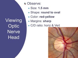
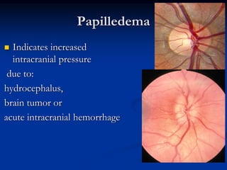
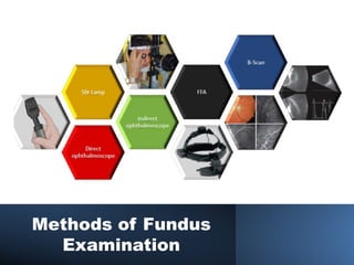
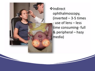
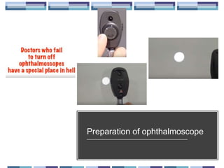
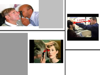
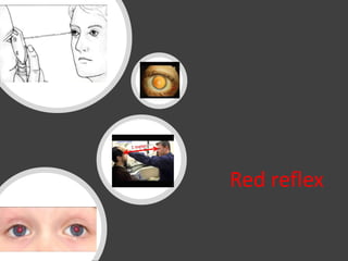
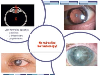
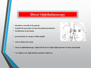
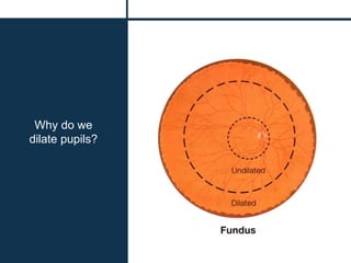
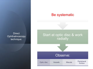
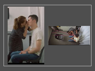
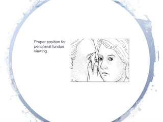
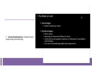
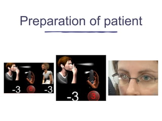
Mohamed Ahmed ELShafie
Lecturer of ophthalmology
Harvard Medical School Almuni
Director of Training Unit
01069201269
Viewing
Optic
Nerve
Head
Observe:
 Size: 1.5 mm
 Shape: round to oval
 Color: red-yellow
 Margins: sharp
 C/D ratio: horiz & Vert
Papilledema
 Indicates increased
intracranial pressure
due to:
hydrocephalus,
brain tumor or
acute intracranial hemorrhage
Optic disc
cupping
Vascular Occlusion
Methods of Fundus
Examination
Fundus Biomicroscopy
Indirect
Ophthalmoscopy
Direct
Ophthalmoscopy
Advantages
Portable
Easy to use
Upright image
Magnification  15x
Can use w/o dilation
Disadvantages
Small field of view
Lack of stereopsis
Media opacities degrade image
Direct Ophthalmoscope
Preparation of ophthalmoscope
Red reflex
• Look for media opacities
• Cataracts
• Corneal scars
• Large floaters
Basic skills
• Introduce yourself to the patient.
• Explain the procedure & take the patient permission.
• Sterilization of the hands.
• put mydriatic eye drops to dilate pupils.
• Ask to darken the room.
• Turn on ophthalmoscope, Adjust the focus & Adjust light aperture to large sized pupil.
• Use right eye & right hand for patient’s right eye.
Direct Ophthalmoscopy
Why do we
dilate pupils?
Basic skills
• Introduce yourself to the patient.
• Explain the procedure & take the patient permission.
• Sterilization of the hands.
• put mydriatic eye drops to dilate pupils.
• Ask to darken the room.
• Turn on ophthalmoscope, Adjust the focus & Adjust light aperture to large sized pupil.
• Use right eye & right hand for patient’s right eye.
Direct Ophthalmoscopy
Direct
Ophthalmoscopy
technique:
Observe:
Optic disc Vessels Macula
Peripheral
fundus
Start at optic disc & work
radially
Be systematic
Proper position for
peripheral fundus
viewing
• Contraindications: head injury
requiring monitoring
Preparation of patient
Examine Fundus
direct ophthalmoscope NMU.pptx

direct ophthalmoscope NMU.pptx