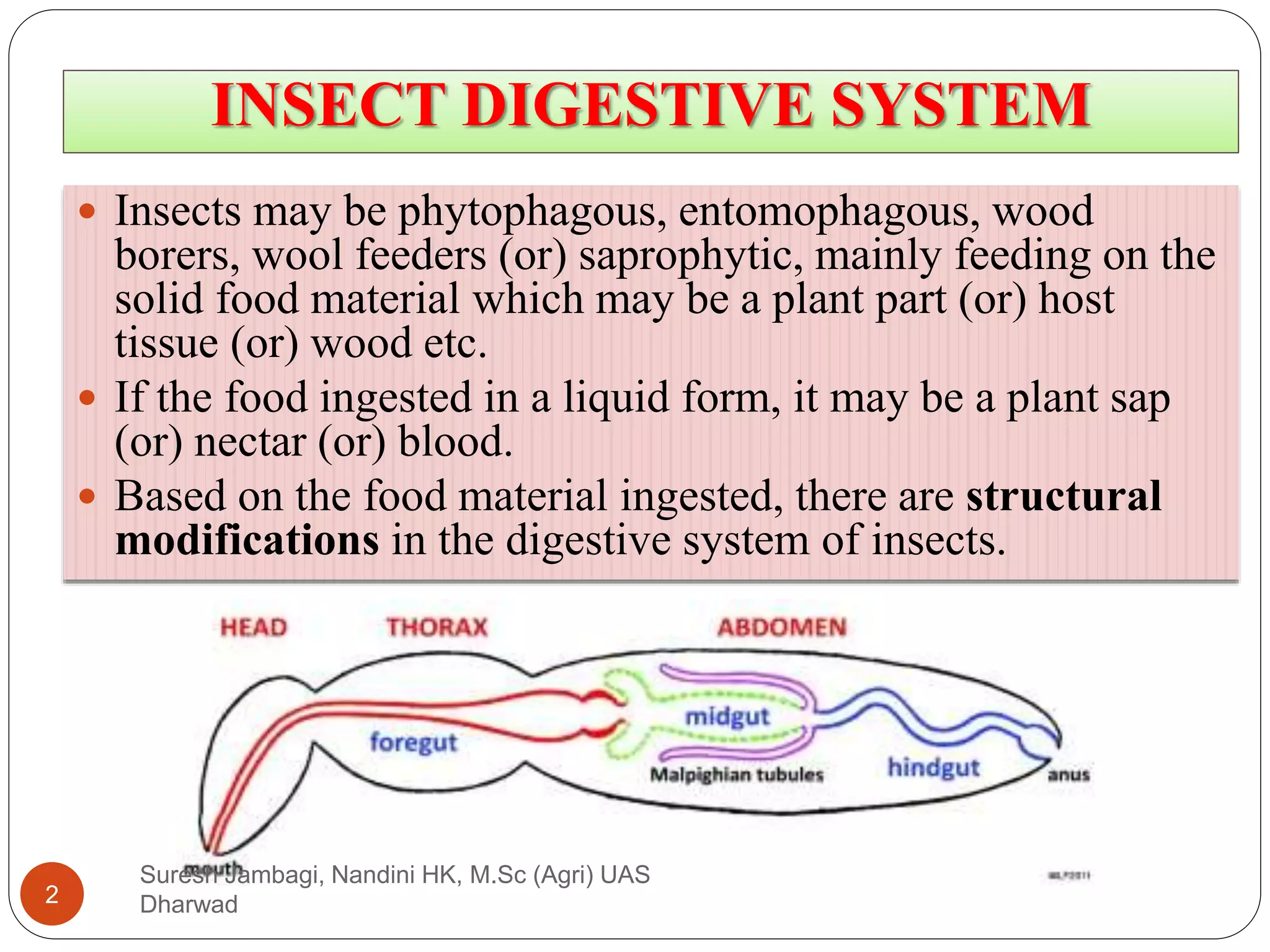
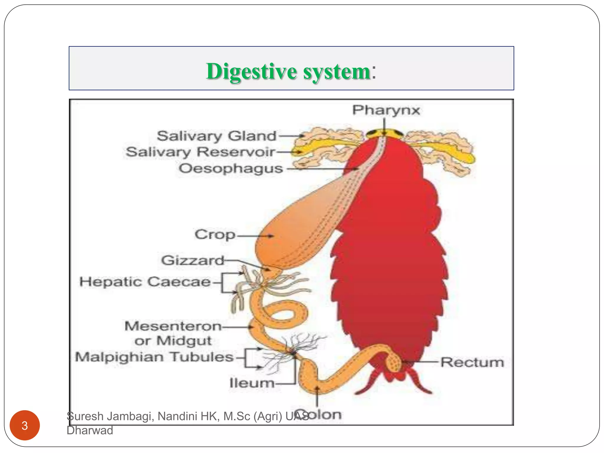
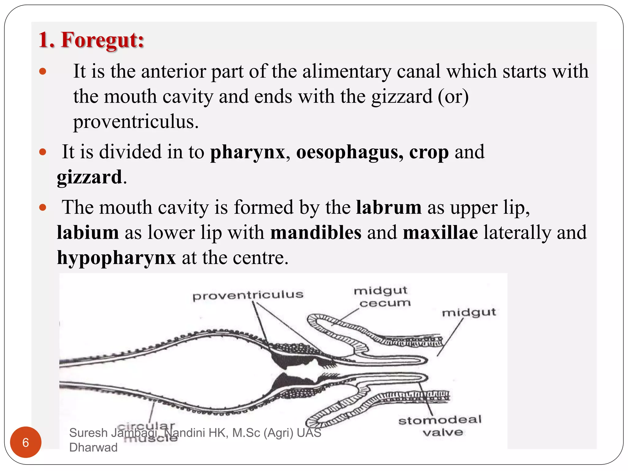
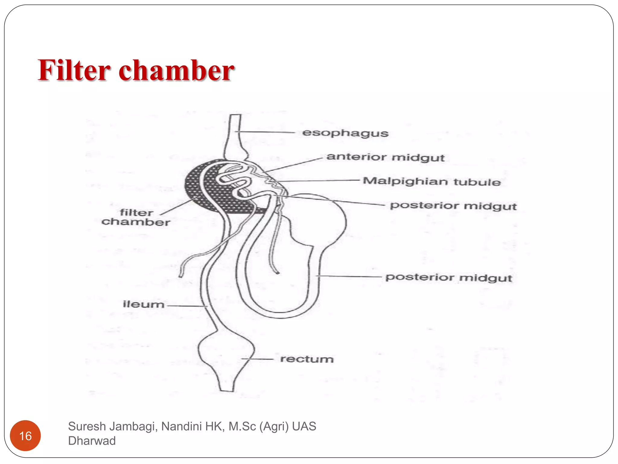
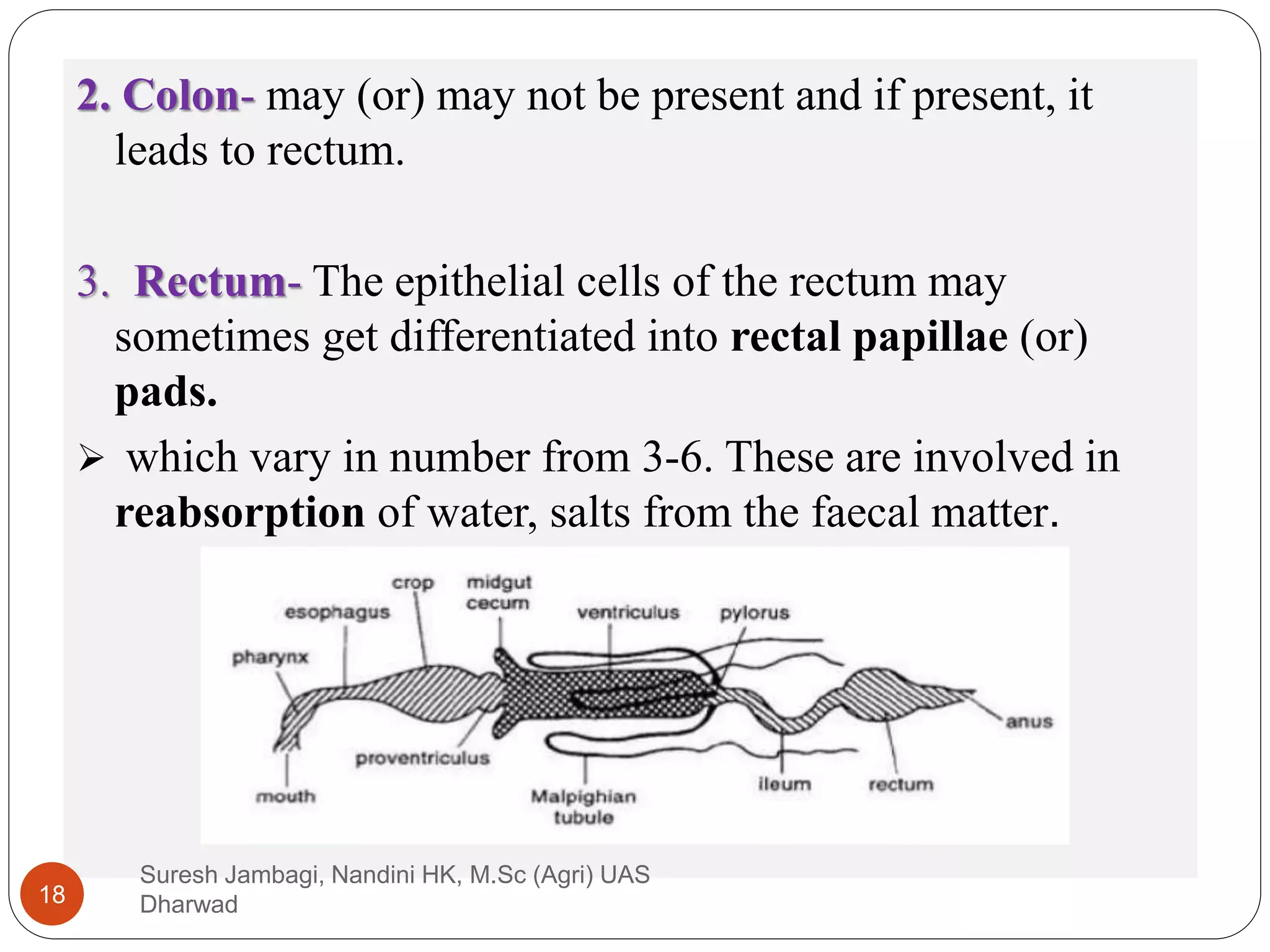
Prepared by:
Nandini HK
Suresh R. Jambagi
M.Sc. (Agri) Agril. Entomology
University of Agricultural Sciences
Dharwad, Karnataka-580005
Email: jambagisuru@gmail.com
GENERAL ENTOMOLOGY
Topics covered: i) Digestive system in insects
Presentation: 4
INSECT DIGESTIVE SYSTEM
 Insects may be phytophagous, entomophagous, wood
borers, wool feeders (or) saprophytic, mainly feeding on the
solid food material which may be a plant part (or) host
tissue (or) wood etc.
 If the food ingested in a liquid form, it may be a plant sap
(or) nectar (or) blood.
 Based on the food material ingested, there are structural
modifications in the digestive system of insects.
Suresh Jambagi, Nandini HK, M.Sc (Agri) UAS
Dharwad
2
Digestive system:
Suresh Jambagi, Nandini HK, M.Sc (Agri) UAS
Dharwad
3
 Insects having the habit of feeding on the solid food
material, possess the biting and chewing type of mouth
parts.
 Where as sap feeding ones have sucking type.
 Sap suckers possess a filter chamber and solid feeders
have well developed gizzard.
 The alimentary canal in insects extends from mouth to
anus.
Suresh Jambagi, Nandini HK, M.Sc (Agri) UAS
Dharwad
4
Which divided in to:
1. Foregut (stomodaeum)
2. Midgut (mesenteron or stomach or ventriculus)
3. Hindgut (proctodaeum).
 The foregut and hindgut are ectodermal in origin where as
the midgut is endodermal in origin.
 Compared to the carnivores (or) sap suckers, the
phytophagous solid feeders possess longer alimentary
canal.
Suresh Jambagi, Nandini HK, M.Sc (Agri) UAS
Dharwad
5
1. Foregut:
 It is the anterior part of the alimentary canal which starts with
the mouth cavity and ends with the gizzard (or)
proventriculus.
 It is divided in to pharynx, oesophagus, crop and
gizzard.
 The mouth cavity is formed by the labrum as upper lip,
labium as lower lip with mandibles and maxillae laterally and
hypopharynx at the centre.
Suresh Jambagi, Nandini HK, M.Sc (Agri) UAS
Dharwad
6
 At the base of the hypopharynx, salivary glands open into
the mouth cavity.
 Pharynx: It is the region between the mouth and
oesophagus.
 Oesophagus: it is a narrow part of the foregut through
which the food get transported from pharynx into the crop.
 Crop: It is a sac like structure which is a dilated form and
mainly serves the purpose of storage of food material.
Suresh Jambagi, Nandini HK, M.Sc (Agri) UAS
Dharwad
7
Internally foregut consists of the following layers,
(i) Inner most intima layer
(ii) Epithelial cells
(iii) Basement membrane
(iv) Longitudinal muscles
(v) Circular muscles
Suresh Jambagi, Nandini HK, M.Sc (Agri) UAS
Dharwad
8
Gizzard
 It is a small constricted part of the alimentary canal which
consists of the cuticular intima layer modified into a tooth
like denticles.
 That helps for grinding the food material.
 In some insects such as honey bees, the gizzard functions
as honey stopper (or) stainer separating the pollen from
the nectar.
 After gizzard the foregut forms into a stomodeal valve
which is surrounded by gastric (or) hepatic caecae,
which may vary from 5-6 in number.
Suresh Jambagi, Nandini HK, M.Sc (Agri) UAS
Dharwad
9
2. MIDGUT
 It is also known as mesenteron or stomach.
 Foregut opens in to midgut through stomodael /
cardiac valve.
 It is a short, straight tube in case of primitive insects
or a sac or may be pyriform or fusiform in shape in
caterpillars.
 In some insects midgut appears as a completely
separated sac like structure that gets connected with
the hindgut.
Suresh Jambagi, Nandini HK, M.Sc (Agri) UAS
Dharwad
10
Structurally midgut consists of
(i) Inner peritrophic membrane
(ii) A layer of epithelial cells
(iii) Basement membrane
(iv) Circular muscles
(v) Longitudinal muscles
 Midgut consists of an inner delicate layer called
peritrophic membrane secreted by the epithelial cells.
Suresh Jambagi, Nandini HK, M.Sc (Agri) UAS
Dharwad
11
 The peritrophic membrane protects the tender epithelial
cells of the midgut from abrasion by hard food particles as
no mucous is secreted in insects that feed on the solid food
material.
 The peritrophic membrane forms a coat over the food
particles and no damage will occur to epithelial cells of
midgut.
 This layer is said to be permeable to the digestive
enzymes and the products of digestion.
 It is absent in case of sap sucking insects.
Suresh Jambagi, Nandini HK, M.Sc (Agri) UAS
Dharwad
12
The epithelial layer of midgut consists of 3 types of cells:
1) Columnar cells: These are columnar in shape, vary in size and
release enzymes through a series of microvilli arranged in a
brush border (or) honey comb border.
2) Regenerative cells: These epithelial cells are involved in the
production (or) formation of new cells to replace the whole
columnar cells.
 Which are involved in holocrine secretion of enzymes.
 These regenerative cells may be arranged either in groups (or)
may remain scattered (or) sometimes singly.
 If they are arranged in groups (or) clusters they are called Nidi.
3) Goblet cells: Mainly serve for storage and excretion.
Suresh Jambagi, Nandini HK, M.Sc (Agri) UAS
Dharwad
13
a) Gastric caecae (Enteric caecae or Hepatic caecae):
 Finger like outgrowths found in anterior or posterior
ends of midgut.
 This structure increases the functional area of midgut
and shelter symbiotic bacteria in some insects.
b) Filter chamber:
 This is a characteristic arrangement of the midgut in
hemipteran insects (fluid feeders).
 Anterior part of midgut forms a thin-walled bladder i.e
filter chamber
Suresh Jambagi, Nandini HK, M.Sc (Agri) UAS
Dharwad
14
 Which is closely bound to either posterior part of midgut
or the anterior hindgut and Malpighian tubules.
 Filter chamber enables the excess fluids including sugar
in the food to pass directly from the anterior part of the
midgut to the hindgut without passing through the middle
portion of midgut.
 Thus preventing excessive dilution of haemolymph,
enzymes and facilitate better enzyme activity.
 In aphids, the honey dew (rich in sugars) is the substance
that is being excreted after passing through the filter
chamber.
Suresh Jambagi, Nandini HK, M.Sc (Agri) UAS
Dharwad
15
Filter chamber
Suresh Jambagi, Nandini HK, M.Sc (Agri) UAS
Dharwad
16
3. Hindgut
 It is also known as proctodaeum which is lined inside by
intima.
 It is more permeable than that of the foregut.
 Anterior end of the hindgut can be marked by the presence
of a set of malpighian tubules and a pyloric valve.
 Internally hindgut has same structure as that of the
foregut.
 Hindgut is divided into 3 regions namely ileum, colon and
rectum.
1. Ileum- is a small intestine (or) tube like structure and
appears as a pouch in scarabids.
Suresh Jambagi, Nandini HK, M.Sc (Agri) UAS
Dharwad
17
2. Colon- may (or) may not be present and if present, it
leads to rectum.
3. Rectum- The epithelial cells of the rectum may
sometimes get differentiated into rectal papillae (or)
pads.
 which vary in number from 3-6. These are involved in
reabsorption of water, salts from the faecal matter.
Suresh Jambagi, Nandini HK, M.Sc (Agri) UAS
Dharwad
18
Gut physiology
a) Salivary glands: These are a pair of glands involved in
the secretion of salivary juices.
 These glands open at the base of the hypopharynx
through small salivary ducts.
 The secretion of the glands contain the enzymes such as
amylases, lipases, proteases, but never cellulase.
 In case of silkworm (or) lepidopteran larvae, the salivary
glands produce silk which contains two proteins fibroin
and sericin
 They secrete anti coagulants in blood suckers like
mosquitoes.
Suresh Jambagi, Nandini HK, M.Sc (Agri) UAS
Dharwad
19
(b) Hepatic caecae and midgut epithelial cells: These cells
secretes most of the digestive Juices.
Two types of cells were involved in the enzyme secretion.
(i)Holocrine: Epithelial cells disintegrate in the process of
enzyme secretion.
(ii)Merocrine: Enzyme secretion occurs without cell break
down.
Suresh Jambagi, Nandini HK, M.Sc (Agri) UAS
Dharwad
20
c) Microbes in digestion: In the insect body few cells were
housing symbiotic microorganisms called as mycetocyte.
These mycetocytes aggregate to form an organ called
mycetome.
(i). Flagellate protozoa - It produces cellulase for cellulose
digestion in termites and wood cockroach.
(ii). Bacteria - It helps in wax digestion in wax moth.
Suresh Jambagi, Nandini HK, M.Sc (Agri) UAS
Dharwad
21
iii). Bed bug and cockroach obtain vitamin and aminoacids
from microbes.
 These microbes were transmitted between individuals
through food exchange (mouth to mouth feeding) called
trophallaxis, And through egg called as transovarial
transmission.
 In plant bug and ant lion grub partial digestion occurs in
the host body prior to food ingestion called as extra
intestinal digestion.
 In most of the insects digestion occurs in mid gut.
Suresh Jambagi, Nandini HK, M.Sc (Agri) UAS
Dharwad
22
Process of digestion
 The food ingested by the insects through the mouth
cavity enters into the alimentary canal, get digested and
the undigested waste material is excreted through anus.
 During this process, food material is broken down into
small particles (or) in to a form that can be readily
absorbed by the cells of the midgut and hindgut.
Suresh Jambagi, Nandini HK, M.Sc (Agri) UAS
Dharwad
23
Digestion mainly takes place in 5 steps-
1. Ingestion: Food is partially digested in the oral cavity
with the help of salivary enzymes.
 In insects like fluid feeders, carnivorous Hemiptera,
blowfly larvae etc, digestion occurs outside the intestine
by a process called extra intestinal (or) extra oral
digestion.
2. Transportation: The food material entered in to the
oesophagus is transported in to the crop by muscular
activity i.e. by peristatic movements.
 Food moves continuously from oesophagus in to the crop
where it is stored.
 From the crop, food enters in to the gizzard where it is still
broken in to very minute particles with the help of
denticles or the cuticular teeth.
Suresh Jambagi, Nandini HK, M.Sc (Agri) UAS
Dharwad
24
3. Digestion: From the gizzard through the stomodeal valve,
food passes in to the midgut where actually digestion
starts.
 The epithelial cells produce enzymes i.e. proteases which
break proteins in to amino acids
 Carbohydrases breaking carbohydrates in to mono &
disaccharides
 Lipases breaking lipids in to fatty acids and glycerol.
 In termites, digestion takes place in colon of hindgut
where mycetomes (group of cells which harbour the
micro organisms like protozoans) secrete the enzyme
cellulase which can digest the wood material rich in
cellulose.
Suresh Jambagi, Nandini HK, M.Sc (Agri) UAS
Dharwad
25
 In scarabid beetle larvae, bacteria are involved in
digestion.
 In wood feeders, keratin digestion is facilitated by
alkaline pH of midgut.
 In Tineola (cloths moth), keratinase secreted by
protozoans.
4. Absorption: Midgut epithelial cells absorb the nutrients
from the digested food and pass on the faecal matter and
undigested food material in to the hindgut.
 The Malpighian tubules maintain ionic balance by
absorbing Na and K salts from the blood.
Suresh Jambagi, Nandini HK, M.Sc (Agri) UAS
Dharwad
26
 The cells of the hindgut are also involved in the re
absorption of water, salts and other metabolites from the
faecal matter.
5. Egestion: The waste food material is discharged through
the anus due to the action of the anal muscles.
Suresh Jambagi, Nandini HK, M.Sc (Agri) UAS
Dharwad
27

Digestive system in insects

  • 1.
    Prepared by: Nandini HK SureshR. Jambagi M.Sc. (Agri) Agril. Entomology University of Agricultural Sciences Dharwad, Karnataka-580005 Email: jambagisuru@gmail.com GENERAL ENTOMOLOGY Topics covered: i) Digestive system in insects Presentation: 4
  • 2.
    INSECT DIGESTIVE SYSTEM Insects may be phytophagous, entomophagous, wood borers, wool feeders (or) saprophytic, mainly feeding on the solid food material which may be a plant part (or) host tissue (or) wood etc.  If the food ingested in a liquid form, it may be a plant sap (or) nectar (or) blood.  Based on the food material ingested, there are structural modifications in the digestive system of insects. Suresh Jambagi, Nandini HK, M.Sc (Agri) UAS Dharwad 2
  • 3.
    Digestive system: Suresh Jambagi,Nandini HK, M.Sc (Agri) UAS Dharwad 3
  • 4.
     Insects havingthe habit of feeding on the solid food material, possess the biting and chewing type of mouth parts.  Where as sap feeding ones have sucking type.  Sap suckers possess a filter chamber and solid feeders have well developed gizzard.  The alimentary canal in insects extends from mouth to anus. Suresh Jambagi, Nandini HK, M.Sc (Agri) UAS Dharwad 4
  • 5.
    Which divided into: 1. Foregut (stomodaeum) 2. Midgut (mesenteron or stomach or ventriculus) 3. Hindgut (proctodaeum).  The foregut and hindgut are ectodermal in origin where as the midgut is endodermal in origin.  Compared to the carnivores (or) sap suckers, the phytophagous solid feeders possess longer alimentary canal. Suresh Jambagi, Nandini HK, M.Sc (Agri) UAS Dharwad 5
  • 6.
    1. Foregut:  Itis the anterior part of the alimentary canal which starts with the mouth cavity and ends with the gizzard (or) proventriculus.  It is divided in to pharynx, oesophagus, crop and gizzard.  The mouth cavity is formed by the labrum as upper lip, labium as lower lip with mandibles and maxillae laterally and hypopharynx at the centre. Suresh Jambagi, Nandini HK, M.Sc (Agri) UAS Dharwad 6
  • 7.
     At thebase of the hypopharynx, salivary glands open into the mouth cavity.  Pharynx: It is the region between the mouth and oesophagus.  Oesophagus: it is a narrow part of the foregut through which the food get transported from pharynx into the crop.  Crop: It is a sac like structure which is a dilated form and mainly serves the purpose of storage of food material. Suresh Jambagi, Nandini HK, M.Sc (Agri) UAS Dharwad 7
  • 8.
    Internally foregut consistsof the following layers, (i) Inner most intima layer (ii) Epithelial cells (iii) Basement membrane (iv) Longitudinal muscles (v) Circular muscles Suresh Jambagi, Nandini HK, M.Sc (Agri) UAS Dharwad 8
  • 9.
    Gizzard  It isa small constricted part of the alimentary canal which consists of the cuticular intima layer modified into a tooth like denticles.  That helps for grinding the food material.  In some insects such as honey bees, the gizzard functions as honey stopper (or) stainer separating the pollen from the nectar.  After gizzard the foregut forms into a stomodeal valve which is surrounded by gastric (or) hepatic caecae, which may vary from 5-6 in number. Suresh Jambagi, Nandini HK, M.Sc (Agri) UAS Dharwad 9
  • 10.
    2. MIDGUT  Itis also known as mesenteron or stomach.  Foregut opens in to midgut through stomodael / cardiac valve.  It is a short, straight tube in case of primitive insects or a sac or may be pyriform or fusiform in shape in caterpillars.  In some insects midgut appears as a completely separated sac like structure that gets connected with the hindgut. Suresh Jambagi, Nandini HK, M.Sc (Agri) UAS Dharwad 10
  • 11.
    Structurally midgut consistsof (i) Inner peritrophic membrane (ii) A layer of epithelial cells (iii) Basement membrane (iv) Circular muscles (v) Longitudinal muscles  Midgut consists of an inner delicate layer called peritrophic membrane secreted by the epithelial cells. Suresh Jambagi, Nandini HK, M.Sc (Agri) UAS Dharwad 11
  • 12.
     The peritrophicmembrane protects the tender epithelial cells of the midgut from abrasion by hard food particles as no mucous is secreted in insects that feed on the solid food material.  The peritrophic membrane forms a coat over the food particles and no damage will occur to epithelial cells of midgut.  This layer is said to be permeable to the digestive enzymes and the products of digestion.  It is absent in case of sap sucking insects. Suresh Jambagi, Nandini HK, M.Sc (Agri) UAS Dharwad 12
  • 13.
    The epithelial layerof midgut consists of 3 types of cells: 1) Columnar cells: These are columnar in shape, vary in size and release enzymes through a series of microvilli arranged in a brush border (or) honey comb border. 2) Regenerative cells: These epithelial cells are involved in the production (or) formation of new cells to replace the whole columnar cells.  Which are involved in holocrine secretion of enzymes.  These regenerative cells may be arranged either in groups (or) may remain scattered (or) sometimes singly.  If they are arranged in groups (or) clusters they are called Nidi. 3) Goblet cells: Mainly serve for storage and excretion. Suresh Jambagi, Nandini HK, M.Sc (Agri) UAS Dharwad 13
  • 14.
    a) Gastric caecae(Enteric caecae or Hepatic caecae):  Finger like outgrowths found in anterior or posterior ends of midgut.  This structure increases the functional area of midgut and shelter symbiotic bacteria in some insects. b) Filter chamber:  This is a characteristic arrangement of the midgut in hemipteran insects (fluid feeders).  Anterior part of midgut forms a thin-walled bladder i.e filter chamber Suresh Jambagi, Nandini HK, M.Sc (Agri) UAS Dharwad 14
  • 15.
     Which isclosely bound to either posterior part of midgut or the anterior hindgut and Malpighian tubules.  Filter chamber enables the excess fluids including sugar in the food to pass directly from the anterior part of the midgut to the hindgut without passing through the middle portion of midgut.  Thus preventing excessive dilution of haemolymph, enzymes and facilitate better enzyme activity.  In aphids, the honey dew (rich in sugars) is the substance that is being excreted after passing through the filter chamber. Suresh Jambagi, Nandini HK, M.Sc (Agri) UAS Dharwad 15
  • 16.
    Filter chamber Suresh Jambagi,Nandini HK, M.Sc (Agri) UAS Dharwad 16
  • 17.
    3. Hindgut  Itis also known as proctodaeum which is lined inside by intima.  It is more permeable than that of the foregut.  Anterior end of the hindgut can be marked by the presence of a set of malpighian tubules and a pyloric valve.  Internally hindgut has same structure as that of the foregut.  Hindgut is divided into 3 regions namely ileum, colon and rectum. 1. Ileum- is a small intestine (or) tube like structure and appears as a pouch in scarabids. Suresh Jambagi, Nandini HK, M.Sc (Agri) UAS Dharwad 17
  • 18.
    2. Colon- may(or) may not be present and if present, it leads to rectum. 3. Rectum- The epithelial cells of the rectum may sometimes get differentiated into rectal papillae (or) pads.  which vary in number from 3-6. These are involved in reabsorption of water, salts from the faecal matter. Suresh Jambagi, Nandini HK, M.Sc (Agri) UAS Dharwad 18
  • 19.
    Gut physiology a) Salivaryglands: These are a pair of glands involved in the secretion of salivary juices.  These glands open at the base of the hypopharynx through small salivary ducts.  The secretion of the glands contain the enzymes such as amylases, lipases, proteases, but never cellulase.  In case of silkworm (or) lepidopteran larvae, the salivary glands produce silk which contains two proteins fibroin and sericin  They secrete anti coagulants in blood suckers like mosquitoes. Suresh Jambagi, Nandini HK, M.Sc (Agri) UAS Dharwad 19
  • 20.
    (b) Hepatic caecaeand midgut epithelial cells: These cells secretes most of the digestive Juices. Two types of cells were involved in the enzyme secretion. (i)Holocrine: Epithelial cells disintegrate in the process of enzyme secretion. (ii)Merocrine: Enzyme secretion occurs without cell break down. Suresh Jambagi, Nandini HK, M.Sc (Agri) UAS Dharwad 20
  • 21.
    c) Microbes indigestion: In the insect body few cells were housing symbiotic microorganisms called as mycetocyte. These mycetocytes aggregate to form an organ called mycetome. (i). Flagellate protozoa - It produces cellulase for cellulose digestion in termites and wood cockroach. (ii). Bacteria - It helps in wax digestion in wax moth. Suresh Jambagi, Nandini HK, M.Sc (Agri) UAS Dharwad 21
  • 22.
    iii). Bed bugand cockroach obtain vitamin and aminoacids from microbes.  These microbes were transmitted between individuals through food exchange (mouth to mouth feeding) called trophallaxis, And through egg called as transovarial transmission.  In plant bug and ant lion grub partial digestion occurs in the host body prior to food ingestion called as extra intestinal digestion.  In most of the insects digestion occurs in mid gut. Suresh Jambagi, Nandini HK, M.Sc (Agri) UAS Dharwad 22
  • 23.
    Process of digestion The food ingested by the insects through the mouth cavity enters into the alimentary canal, get digested and the undigested waste material is excreted through anus.  During this process, food material is broken down into small particles (or) in to a form that can be readily absorbed by the cells of the midgut and hindgut. Suresh Jambagi, Nandini HK, M.Sc (Agri) UAS Dharwad 23
  • 24.
    Digestion mainly takesplace in 5 steps- 1. Ingestion: Food is partially digested in the oral cavity with the help of salivary enzymes.  In insects like fluid feeders, carnivorous Hemiptera, blowfly larvae etc, digestion occurs outside the intestine by a process called extra intestinal (or) extra oral digestion. 2. Transportation: The food material entered in to the oesophagus is transported in to the crop by muscular activity i.e. by peristatic movements.  Food moves continuously from oesophagus in to the crop where it is stored.  From the crop, food enters in to the gizzard where it is still broken in to very minute particles with the help of denticles or the cuticular teeth. Suresh Jambagi, Nandini HK, M.Sc (Agri) UAS Dharwad 24
  • 25.
    3. Digestion: Fromthe gizzard through the stomodeal valve, food passes in to the midgut where actually digestion starts.  The epithelial cells produce enzymes i.e. proteases which break proteins in to amino acids  Carbohydrases breaking carbohydrates in to mono & disaccharides  Lipases breaking lipids in to fatty acids and glycerol.  In termites, digestion takes place in colon of hindgut where mycetomes (group of cells which harbour the micro organisms like protozoans) secrete the enzyme cellulase which can digest the wood material rich in cellulose. Suresh Jambagi, Nandini HK, M.Sc (Agri) UAS Dharwad 25
  • 26.
     In scarabidbeetle larvae, bacteria are involved in digestion.  In wood feeders, keratin digestion is facilitated by alkaline pH of midgut.  In Tineola (cloths moth), keratinase secreted by protozoans. 4. Absorption: Midgut epithelial cells absorb the nutrients from the digested food and pass on the faecal matter and undigested food material in to the hindgut.  The Malpighian tubules maintain ionic balance by absorbing Na and K salts from the blood. Suresh Jambagi, Nandini HK, M.Sc (Agri) UAS Dharwad 26
  • 27.
     The cellsof the hindgut are also involved in the re absorption of water, salts and other metabolites from the faecal matter. 5. Egestion: The waste food material is discharged through the anus due to the action of the anal muscles. Suresh Jambagi, Nandini HK, M.Sc (Agri) UAS Dharwad 27