
Recommended
More Related Content
Similar to insect head.pptx
Similar to insect head.pptx (20)
More from nowsheranss185151
More from nowsheranss185151 (8)
Recently uploaded
Recently uploaded (20)
insect head.pptx
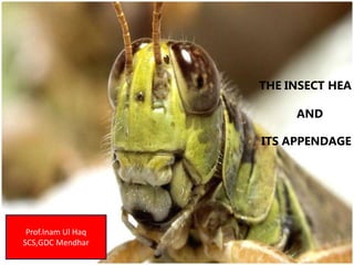
- 1. 5 THE INSECT HEA AND ITS APPENDAGE Prof.Inam Ul Haq SCS,GDC Mendhar
- 2. TAGMOSIS-Segmentation • Insect body is divided in to a series of segments, which in primitive arthropods are known as “somites” or “metameres”. • During the process of evolution, these somites gets fused with each other in different ways forming the body parts of the existing arthropods. • embryonic stage -primary segmentation & adult insects -secondary segmentation sclerotized membranous inter-segmental region. • Three regions or tagmata namely head, thorax and abdomen. • This grouping of body segments in to regions is known as tagmosis. Head-mouthparts, compound eyes, simple eyes (ocelli) and a pair of antennae. Thorax -3 segments i.e. prothorax, mesothorax and metathorax Meso+metathorax= pterothorax. All the three thoracic segments possess a pair of legs and meso and meta thorax possess one pair of wings Abdomen has 7-11 segments with genital appendages on 8th and 9th segments.
- 3. S. No. Segment Region Appendages 1 Pre Antennary Pro cephalon No 2 Antennary Antennae 3 Intercalary No 4 Mandibulary Gnatho cephalon Mandibles 5 Maxillary Maxillae 6 Labial Labium Insect head • Hard and highly sclerotized • 6 segments form a head capsule •can be divided into 2 regions i.e. 1. Procephalon 2. gnathocephalon (mouth).
- 4. Head Capsule Mouthparts orientation Neck Head orientation
- 5. Types of head According to the position /projection of mouth parts, the Insect head can be classified as (a) Hypognathous (Hypo – Below: Gnathous – Jaw ) head is vertical and at right angle to the long axis of body mouth parts are ventrally placed and projected downwards This is also kwown as Orthopteroid type Eg: Grass hopper, Cockroach (b) Prognathous : (Pro – infront: Gnathous – Jaw ) head remains in the same axis to body mouth parts are projected forward This is also known as Coleopteroid type Eg: beetles (c) Opisthognathous : (Opistho – behind: Gnathous – Jaw ) Head orientation is same as prognathous mouthparts are directed back ward held in b/w the fore legs Hemipteroid or Opisthorhynchous type Eg: bugs
- 6. Sclerites and sutures of head • Head capsule is formed by the union of number of cuticular plates i.e. sclerites • Sclerites are joined together by cuticular lines or ridges known as sutures • Sutures provide mechanical support to the cranial wall. Sclerites 1. Labrum: small sclerite forms the upper lip of the mouth cavity It is freely attached or suspended from the lower margin of the clypeus 2. Clypeus: It is situated above the labrum divided in to anterior ante-clypeus and posterior post-clypeus. 3. Frons: It is the facial part of the insect consisting of median ocellus. 4.Vertex: It is the top portion of the head behind the frons or the area between the two compound eyes. 5. Epicranium: It is the upper part of the head extending from vertex to occipital suture.
- 7. Insect Head Frons Gena Clypeus Mandible Labrum 1st Maxilla Labium (2nd Maxilla) Occiput Compound Eyes Epicranium Ocelli Vertex Antennal PostOcciput Occular
- 8. 6. Occiput: It is an inverted “U” shaped structure representing the area between the epicranium and post occiput 7. Post occiput : It is the extreme posterior part of the insect head that remains before the neck region. 8. Gena: It is the area extending below the compound eyes to just above mandibles 9. Occular sclerites: cuticular ring like structures around each compound eye 10. Antennal sclerites: These form the basis for the antennae and present around the scape which are well developed in Plecoptera (stone flies) All the above sclerites gets attached through cuticular ridges or sutures to provide the attachment for the muscles inside.
- 9. Sutures 1. Clypeolabral suture : It is the suture present between clypeus and labrum. It remains in the lower margin of the clypeus from which the labrum hangs down 2. Clypeofrontal suture or epistomal suture: The suture present between clypeus and frons 3. Epicranial: It is an inverted ‘Y’ shaped suture distributed above the facial region extending up to the epicranial part of the head. Two arms 1.frontal suture occupying the frons and stem called as coronal suture. It also known as line of weakness or ecdysial suture because the exuvial membrane splits along this suture during the process of ecdysis. 4. Occipital : It is ‘U’shaped or horseshoe shaped suture between epicranium and occiput. 5. Post occipital: It is the only real suture in insect head. Posterior end of the head is marked by this suture to which the sclerites are attached. As this suture separates the head from the neck, hence named as real suture. 6. Genal: It is the sutures present on the lateral side of the head i.e. gena. 7. Occular suture: It is circular suture present around each compound eye. 8. Antennal suture: It is a marginal depressed ring around the antennal socket.
- 10. Tentorium (plural tentoria) is a term used to refer to the framework of internal supports within an arthropod head. The tentorium is formed by ingrowths of the exoskeleton, called apophyses, which fuse in various ways to provide rigid support for the muscles of the head. Cut away view of the head capsule Anterior arms Posterior arms Dorsal arms
- 12. Insect Antenna & its Modifications 12
- 13. uniramous (unbranched), segmented and mobile structures basally fixed in to deep antennal socket (antennifer) Antennal socket is provided with an antennal suture The base of socket is connected to the edge of the socket by an articulatory membrane For free movement of antennae
- 14. Functions sense organ responding to touch, smell, odour, humidity, temperature & air currents or wind speed. Jhonston’s organ on pedicel -auditory organ responding to sound and measuring the speed of air currents. Help the mandibles for holding prey and for mastication of food material Helps in sexual dimorphism Useful for clasping the female during copulation Aid in respiration by forming an air funnel in aquatic insects. Exceptions Absent in Protura and ClassArachnida and 2 Pairs in Crustacea In Chalcidoids, Flagellum is divided into basal Ring, middle Funicle and upper Club In Collembola & Diplura,Antennae are muscular, Hence Segmented type Johnstons organs are absent in Collembola & Diplura or Ring
- 15. FILIFORM (thread-like) The segments of flagellum are of same thickness and thread-like. Example : Grass hopper
- 16. SETACEOUS (whip-like) The segments of flagellum are taper towards apex. Example : Dragon fly, Cockroach
- 17. MONILIFORM (bead-like) The segments of flagellum are globular and bead-like with clear constrictions. Example : Termites
- 18. PECTINATE (comb) The segments of flagellum have long stiff projections on one side. Example : FemaleArctiid moths
- 19. BIPECTINATE (Double comb) The segments of flagellum have long stiff projections on both sides. Example : Male Lymantrids, Mulberry silk moth
- 20. SERRATE OR DENTATE (tooth-like) The segments of flagellum have tooth-like projections on one side. Example : Pulse Beetles
- 21. CLAVATE (club-like) The segments of flagellum are gradually broaden towards apex. Example :Butterfly
- 22. Clavate with Hook The segments of flagellum are gradually broaden towards apex structure with a terminal hook like curve Example : Skipper Butterfly
- 23. CAPITATE (knob-like) The segments of flagellum Suddenly thickened to form a knob-like structure. Example : Red Flour Beetle
- 24. GENICULATE (elbow-like) In these the scape is very long and flagellum forms a sharp bend like a flexed arm. Example : Ant, Honey bee
- 25. LAMMELLATE (Plate-like) 25 enna sha The terminal segment of flagellum are extended into leaf-like plates on one side. Example : Rhinoceros beetle Dung roller
- 26. FLABELLATE (tongue-like) The segments of flagellum are produced into long and thick tongue-like projections. Example : Male stylopid
- 27. PLUMOSE (feather-like) The segments of flagellum have thick whorls of long hairs. Example : Male mosquito
- 28. PILOSE (hairy) The segments of flagellum have very thin whorls of hairs . Example :Female mosquito
- 29. ARISTATE (arista-like) The segments of flagellum form an arista or bristle like structure on dorsal surface Example : House fly
- 30. STYLATE (styliform) The flagellum forms a long unsegmented , terminal hair. Example : Robber fly
- 31. Thorax • consisting of three segments, Prothorax, Mesothorax and Metathorax • Each possess a pair of legs and a pair of wings on meso and meta thoracic segment • Meso and meta thoracic segments together known as pterothorax • Sclerite of dorsal region of thorax is tergum or notum • ventral region is called sternum and lateral region is called pleuron
- 32. • All the three thoracic segments of an insect possess a pair of legs - hexapods and class insecta as hexapoda • Insect leg consists of 5 parts viz. Coxa, Trochanter, Femur, Tibia and Tarsus. • In primitive insects, a small sclerite known as subcoxa occur before the coxa which form the true basal segment Insect Leg and its Modifications
- 33. 1. Coxa: It is the functional basal segment rigidly fixed to thorax or weakly articulated 2. Trochanter : It is very small is articulated with coxa and more or less fixed to femur 3. Femur : It is the largest, strongest segment and is articulated with the tibia 4. Tibia : It is equal or more than the length of the femur, articulated with tarsus 5. Tarsus : largest segment of the leg divided into tarsomeres, no. vary from 1-5 and are movable one on the other • 1st segment is large, big or broad in size known as basitarsus • Tarsus at it’s end consists of pretarsus with a pair of claws and cushion like pulvilli • In between the claws, if there is lobe like structure, it is known as “aroleum” as in Orthoptera and if it is bristle like, it is called “embodium” as in Diptera • In some insects, the ventral surface of pretarsus consist of a median circular plate between the claws known as unguitractor where as the claws are known as ungues
- 34. Modifications of Leg Type Leg modified Example purpose Modification Cursorial All legs Blister beetle, wasp Walking All the legs uniformly well developed without any special modification Ambulatory All legs Cockroach Running All legs are normal. coxa widely separated Saltatorial Hind legs Grasshopper , gryllids Leaping & jumping Femur and tibia elongated Fossorial Front legs Mole crickets, dung rollers Digging Tibia and tarsus short and broad with teeth like projections
- 36. Type Leg modified Example purpose Modification Raptorial (grasping ) Front legs Preying mantids Preying femur spinose and possess a central longitudinal groove. Tibia narrow, blade like spinose and fits into the groove of femur Natatorial Hind legs Water beetle, water bugs Swimming Hind legs pad like. Tibia and tarsus short and broad having dense long marginal hairs. Scansorial All legs Head louse clinging Tibia possess tibial thumb. Tarsus single segmented and pretarsus with a single long curved claw Raptorial
- 38. Type Leg modified Example purpose Modification Prehensile All legs together Dragon flies Catching prey, basket forming type Thoracic segments obliquely arranged . Tergal plates are pulled backwards and Sternal plates pushed forward, resulting that all the legs pushed forward and seen below the head, together from a basket like structure useful for catching the prey even in flight
- 39. Type Leg Example purpose Modification Antennal cleaning legs Front legs Honey bee For cleaning antennae Tibia possess a movable spine, and the first tarsal segment with a semicircular notch Wax pick type Middle legs Honey bee For picking wax plates Tibia possess a spine called wax pick for removing the wax plates from the ventral side of the abdomen Pollen basket and brush type Hind legs Honey bee For collecting pollen and cleaning the body Inner surface of large tibia has a groove and is used as pollen basket or ‘Corbicula’ for temporary storage of pollen grains. First tarsal segment enlarged and possess short stiff hairs ‘Pecten’ all over the surface called pollen brush.
- 41. Legs of immature stages The immature stage of exopterygotes i.e. nymph consist of only thoracic legs similar to its adult The endopterygote i.e. larva possess two types of legs. 1.Thoracic legs or true legs: Jointed, on 3 thoracic segments 2.Abdominal/prolegs: Unjointed sucker like legs, having flat, fleshy surfaced tip known as Planta. Planta consists of hook like structures known as Crochets ,used for clinging to the substrate The number of prolegs vary from 1-5 pairs distributed on 3rd, 4th, 5th, 6th and 10th abdominal segments For example, sawfly larva has 6-8 pairs of abdominal prolegs. In some insects leg are degenerated e.g.: Coccidae; Endoparasitic hymenopterans.