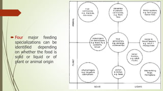
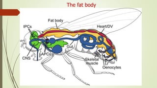
Lec7
The digestive system and nutrition
Introduction
 Insects of different groups consume a variety of foods including:
1. Watery sap (e.g.nymphs of spittle bugs)
2. Vertebrate blood (e.g. bed bugs and female mosquitoes).
3. Dry wood (e.g. some termites)
4. Bacteria and algae (e.g. black fly and many caddisfly larvae).
5. The internal tissues of other insects (e.g. endoparasitic wasp larvae).
 The diverse range of mouthpart types correlates with the diets of different
insects, but gut structure and function also reflect the mechanical properties
and the nutrient composition of the food eaten.
 Four major feeding
specializations can be
identified depending
on whether the food is
solid or liquid or of
plant or animal origin
The Gut
 Gut morphology and physiology relate to these dietary differences in the
following ways:
1. Insects that take solid food typically have a wide, straight, short gut with strong
musculature and obvious protection from abrasion.
 These features are most obvious in solid-feeders with rapid throughput of food
as in plant-feeding.
 most plant-feeding insects need to process large amounts of food because
nutrient levels in leaves and stems are often low. The gut is usually short and
without storage areas, as food is available.
 A diet of animal tissue is nutrient-rich and, at least for predators, well balanced.
However, the food may be available only intermittently and the gut normally has
large storage capacity.
Cont, the gut
2. Insects feeding on blood, sap, or nectar usually have long, narrow,
convoluted guts to allow maximal contact with the liquid food; here,
protection from abrasion is unnecessary.
 The most obvious gut specialization of liquid feeders is a mechanism for
removing excess water to concentrate nutrient substances prior to digestion,
as seen in hemipterans
Structure of the Gut
 There are three main regions to the insect gut (or alimentary canal), with
sphincters (valves) controlling food/fluid movement between regions.
1. The foregut (stomodeum), is concerned with ingestion, storage, grinding, and
transport of food to the next region
2. The midgut (mesenteron), Here digestive enzymes are produced and secreted and
absorption of the products of digestion occurs. The material remaining in the gut
lumen together with urine from the Malpighian tubules.
3. The hindgut (proctodeum), where absorption of water, salts, and other valuable
molecules occurs prior to elimination of the feces through the anus.
 The gut epithelium is one cell layer thick throughout the length of the canal and
rests on a basement membrane surrounded by a variably developed muscle layer.
Both the foregut and hindgut have a cuticular lining, whereas the midgut does not.
Structure of the Gut
1- The foregut (stomodeum)
• The foregut is subdivided into a pharynx, an
oesophagus (esophagus), and a crop (food storage
area).
• In insects that ingest solid food there is often a
grinding organ, the proventriculus (or gizzard).
• The proventriculus is especially well developed in
orthopteroid insects, such as cockroaches in which
the epithelium is folded longitudinally to form ridges
on which the cuticle is armed with spines or teeth.
1- The foregut (stomodeum)
 At the anterior end of the foregut the mouth
opens into a preoral cavity bounded by the bases
of the mouthparts and often divided into:
1. An upper area, or cibarium.
2. A lower part, or salivarium.
 The paired labial or salivary glands vary in size
and arrangement from simple elongated tubes to
complex branched or lobed structures.
 In Lepidoptera, the labial glands produce silk,
whereas mandibular glands secrete the saliva.
 In insects that store meals in their foregut, the
crop may take up the greater portion of the food
and often is capable of extreme distension, with
a posterior sphincter controlling food retention.
2- The mid gut (mesenteron)
 The generalized midgut has two main areas:
1. The tubular ventriculus
2. The blind-ending lateral diverticula called caeca (ceca).
 Most cells of the midgut are structurally similar, being
columnar with microvilli (finger-like protrusions)
covering the inner surface.
 The midgut epithelium mostly is separated from the
food by a thin sheath called the peritrophic membrane,
consisting of a network of chitin fibrils in a protein–
glycoprotein matrix.
 The peritrophic membrane either is delaminated from
the whole midgut or produced by cells in the anterior
region of the midgut.
3- The hind Gut (proctodeum)
 The beginning of the hindgut is defined by
the entry point of the Malpighian tubules,
often into a distinct pylorus forming a
muscular pyloric sphincter.
 Followed by the ileum, colon, and rectum.
 The main functions of the hindgut are the
absorption of water, salts, and other useful
substances from the feces and urine.
Saliva and food ingestion
 Salivary secretions dilute the ingested food and adjust its pH and ionic content.
 The saliva often contains digestive enzymes and, in blood-feeding insects,
anticoagulants and thinning agents are present.
 In insects with extra-intestinal digestion, such as predatory Hemiptera,
digestive enzymes are exported into the food and the resulting liquid is
ingested.
 Most Hemiptera produce an alkaline watery saliva that is a vehicle for enzymes
(either digestive or lytic), and a proteinaceous solidifying saliva that either
forms a complete sheath around the mouthparts (stylets) as they pierce and
penetrate the food or just a securing flange at the point of entry.
Digestion of food
 Most digestion occurs in the midgut, where the epithelial cells produce and
secrete digestive enzymes and also absorb the resultant food breakdown
products.
 Insect food consists principally of polymers of carbohydrates and proteins.
which are digested by enzymatically breaking these large molecules into small
monomers.
 The midgut pH usually is 6.0–7.5, and very alkaline values (pH 9–12) occur in
many plant-feeding insects that extract hemicelluloses from plant cell walls.
 In most insects, the midgut epithelium is separated from the food bolus by the
peritrophic membrane.
Digestion of food
 The peritrophic membrane which constitutes a very efficient high-flux sieve. It
is perforated by pores, which allow passage of small molecules while
restricting large molecules, bacteria, and food particles from directly accessing
the midgut cells.
 The peritrophic membrane also may protect herbivorous insects from ingested
allelochemicals such as tannins.
 In some insects, all or most midgut digestion occurs inside the peritrophic
membrane in the endoperitrophic space.
 In others, only initial digestion occurs there, and smaller food molecules then
diffuse out into the ectoperitrophic space, where further digestion takes place.
 final phase of digestion usually occurs on the surface of the midgut microvilli,
where certain enzymes are either trapped in a mucopolysaccharide coating or
bound to the cell membrane. Thus the peritrophic membrane forms a
permeability barrier and helps to compartmentalize the phases of digestion.
Digestion of food
The fat body
 In many insects, especially the larvae of holometabolous groups, fat body tissue is
a conspicuous component of the internal anatomy.
 Typically, it forms a white or yellow tissue formed of loose sheets or lobes of cells.
 The fat body is an organ of multiple metabolic functions, including the:
1. metabolism of carbohydrates, lipids, and nitrogenous compounds
2. the storage of glycogen, fat, and protein;
3. the synthesis and regulation of blood sugar
4. The synthesis of major hemolymph proteins (such as hemoglobins, vitellogenins
for yolk formation, and storage proteins).
5. Fat body cells can switch their activities in response to nutritional and hormonal
signals to supply the requirements of insect growth, metamorphosis, and
reproduction
The fat body

Insect Digestive System

  • 2.
  • 3.
    Introduction  Insects ofdifferent groups consume a variety of foods including: 1. Watery sap (e.g.nymphs of spittle bugs) 2. Vertebrate blood (e.g. bed bugs and female mosquitoes). 3. Dry wood (e.g. some termites) 4. Bacteria and algae (e.g. black fly and many caddisfly larvae). 5. The internal tissues of other insects (e.g. endoparasitic wasp larvae).  The diverse range of mouthpart types correlates with the diets of different insects, but gut structure and function also reflect the mechanical properties and the nutrient composition of the food eaten.
  • 4.
     Four majorfeeding specializations can be identified depending on whether the food is solid or liquid or of plant or animal origin
  • 5.
    The Gut  Gutmorphology and physiology relate to these dietary differences in the following ways: 1. Insects that take solid food typically have a wide, straight, short gut with strong musculature and obvious protection from abrasion.  These features are most obvious in solid-feeders with rapid throughput of food as in plant-feeding.  most plant-feeding insects need to process large amounts of food because nutrient levels in leaves and stems are often low. The gut is usually short and without storage areas, as food is available.  A diet of animal tissue is nutrient-rich and, at least for predators, well balanced. However, the food may be available only intermittently and the gut normally has large storage capacity.
  • 6.
    Cont, the gut 2.Insects feeding on blood, sap, or nectar usually have long, narrow, convoluted guts to allow maximal contact with the liquid food; here, protection from abrasion is unnecessary.  The most obvious gut specialization of liquid feeders is a mechanism for removing excess water to concentrate nutrient substances prior to digestion, as seen in hemipterans
  • 7.
    Structure of theGut  There are three main regions to the insect gut (or alimentary canal), with sphincters (valves) controlling food/fluid movement between regions. 1. The foregut (stomodeum), is concerned with ingestion, storage, grinding, and transport of food to the next region 2. The midgut (mesenteron), Here digestive enzymes are produced and secreted and absorption of the products of digestion occurs. The material remaining in the gut lumen together with urine from the Malpighian tubules. 3. The hindgut (proctodeum), where absorption of water, salts, and other valuable molecules occurs prior to elimination of the feces through the anus.  The gut epithelium is one cell layer thick throughout the length of the canal and rests on a basement membrane surrounded by a variably developed muscle layer. Both the foregut and hindgut have a cuticular lining, whereas the midgut does not.
  • 8.
  • 9.
    1- The foregut(stomodeum) • The foregut is subdivided into a pharynx, an oesophagus (esophagus), and a crop (food storage area). • In insects that ingest solid food there is often a grinding organ, the proventriculus (or gizzard). • The proventriculus is especially well developed in orthopteroid insects, such as cockroaches in which the epithelium is folded longitudinally to form ridges on which the cuticle is armed with spines or teeth.
  • 10.
    1- The foregut(stomodeum)  At the anterior end of the foregut the mouth opens into a preoral cavity bounded by the bases of the mouthparts and often divided into: 1. An upper area, or cibarium. 2. A lower part, or salivarium.  The paired labial or salivary glands vary in size and arrangement from simple elongated tubes to complex branched or lobed structures.  In Lepidoptera, the labial glands produce silk, whereas mandibular glands secrete the saliva.  In insects that store meals in their foregut, the crop may take up the greater portion of the food and often is capable of extreme distension, with a posterior sphincter controlling food retention.
  • 11.
    2- The midgut (mesenteron)  The generalized midgut has two main areas: 1. The tubular ventriculus 2. The blind-ending lateral diverticula called caeca (ceca).  Most cells of the midgut are structurally similar, being columnar with microvilli (finger-like protrusions) covering the inner surface.  The midgut epithelium mostly is separated from the food by a thin sheath called the peritrophic membrane, consisting of a network of chitin fibrils in a protein– glycoprotein matrix.  The peritrophic membrane either is delaminated from the whole midgut or produced by cells in the anterior region of the midgut.
  • 12.
    3- The hindGut (proctodeum)  The beginning of the hindgut is defined by the entry point of the Malpighian tubules, often into a distinct pylorus forming a muscular pyloric sphincter.  Followed by the ileum, colon, and rectum.  The main functions of the hindgut are the absorption of water, salts, and other useful substances from the feces and urine.
  • 13.
    Saliva and foodingestion  Salivary secretions dilute the ingested food and adjust its pH and ionic content.  The saliva often contains digestive enzymes and, in blood-feeding insects, anticoagulants and thinning agents are present.  In insects with extra-intestinal digestion, such as predatory Hemiptera, digestive enzymes are exported into the food and the resulting liquid is ingested.  Most Hemiptera produce an alkaline watery saliva that is a vehicle for enzymes (either digestive or lytic), and a proteinaceous solidifying saliva that either forms a complete sheath around the mouthparts (stylets) as they pierce and penetrate the food or just a securing flange at the point of entry.
  • 14.
    Digestion of food Most digestion occurs in the midgut, where the epithelial cells produce and secrete digestive enzymes and also absorb the resultant food breakdown products.  Insect food consists principally of polymers of carbohydrates and proteins. which are digested by enzymatically breaking these large molecules into small monomers.  The midgut pH usually is 6.0–7.5, and very alkaline values (pH 9–12) occur in many plant-feeding insects that extract hemicelluloses from plant cell walls.  In most insects, the midgut epithelium is separated from the food bolus by the peritrophic membrane.
  • 15.
    Digestion of food The peritrophic membrane which constitutes a very efficient high-flux sieve. It is perforated by pores, which allow passage of small molecules while restricting large molecules, bacteria, and food particles from directly accessing the midgut cells.  The peritrophic membrane also may protect herbivorous insects from ingested allelochemicals such as tannins.  In some insects, all or most midgut digestion occurs inside the peritrophic membrane in the endoperitrophic space.  In others, only initial digestion occurs there, and smaller food molecules then diffuse out into the ectoperitrophic space, where further digestion takes place.  final phase of digestion usually occurs on the surface of the midgut microvilli, where certain enzymes are either trapped in a mucopolysaccharide coating or bound to the cell membrane. Thus the peritrophic membrane forms a permeability barrier and helps to compartmentalize the phases of digestion.
  • 16.
  • 17.
    The fat body In many insects, especially the larvae of holometabolous groups, fat body tissue is a conspicuous component of the internal anatomy.  Typically, it forms a white or yellow tissue formed of loose sheets or lobes of cells.  The fat body is an organ of multiple metabolic functions, including the: 1. metabolism of carbohydrates, lipids, and nitrogenous compounds 2. the storage of glycogen, fat, and protein; 3. the synthesis and regulation of blood sugar 4. The synthesis of major hemolymph proteins (such as hemoglobins, vitellogenins for yolk formation, and storage proteins). 5. Fat body cells can switch their activities in response to nutritional and hormonal signals to supply the requirements of insect growth, metamorphosis, and reproduction
  • 18.

Editor's Notes

  • #5 Some insect species clearly fall into a single category, but others with generalized diet may fall between two or more of them, and most endopterygotes will occupy different categories at differe stages of their life (e.g. moths and butterflies switch from solid-plant as larvae to liquid-plant adults).
  • #11 للفهم
  • #17 للفهم
  • #19 للفهم