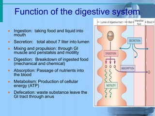
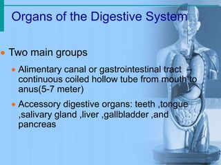
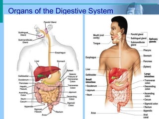
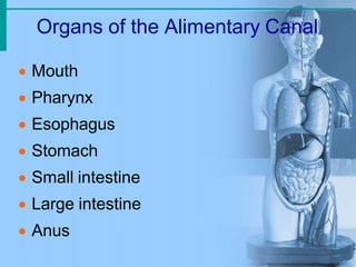
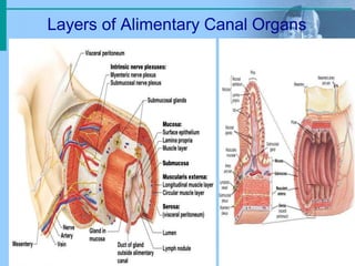
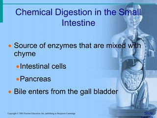
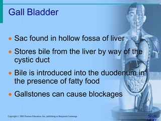
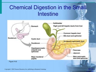
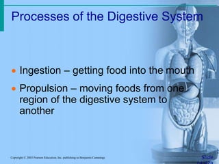
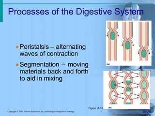
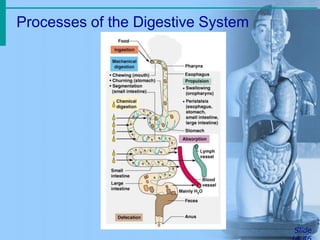
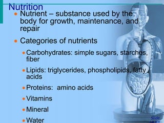
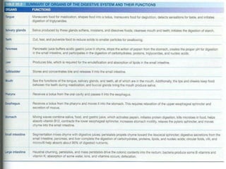
Digestive system
Anil N Lawrence
Lecturer
LNH CON
Function of the digestive system





 Ingestion: taking food and liquid into
mouth
Secretion: total about 7 liter into lumen
Mixing and propulsion: through GI
muscle and peristalsis and motility
Digestion: Breakdown of ingested food
(mechanical and chemical)
Absorption: Passage of nutrients into
the blood
 Metabolism: Production of cellular
energy (ATP)
Defecation: waste substance leave the
GI tract through anus
Organs of the Digestive System
 Two main groups
 Alimentary canal or gastrointestinal tract –
continuous coiled hollow tube from mouth to
anus(5-7 meter)
 Accessory digestive organs: teeth ,tongue
,salivary gland ,liver ,gallbladder ,and
pancreas
Organs of the Digestive System
Organs of the Alimentary Canal
 Mouth
 Pharynx
 Esophagus
 Stomach
 Small intestine
 Large intestine
 Anus
Mouth (Oral Cavity) Anatomy
 Lips (labia) – protect
the anterior opening
 Cheeks – form the
lateral walls
 Hard palate – forms
the anterior roof
 Soft palate – forms
the posterior roof
 Uvula – fleshy
projection of the
soft palate
Mouth (Oral Cavity) Anatomy
 Vestibule – space
between lips
externally and teeth
and gums internally
 Oral cavity – area
contained by the
teeth
 Tongue – attached
at hyoid bone and
styloid processes of
the skull, and by the
lingual frenulum
Tongue

 Dorsum (upper part of
tongue covered with
papillae taste receptor and
buds)
filiform papillae( griping
food)
Fungiform papillae
(The fungiform papillae are involved
in the sensations of taste and have
taste buds)
 circumvallate papillae
( particularly sensitive to sour
and bitter tastes)
Cont.
Palatine tonsil: play a role in protecting the body against respiratory
and gastrointestinal infections.
lingual tonsil: production of antibodies in response to invading
pathogenic bacteria or viruses.
Teeth
• Teeth
(mechanical breakdown)
– Incisors used for cutting
– Canines used for stabbing
and holding
– Molars large surface area
used for grinding
• Primary or deciduous
teeth 20
• Secondary or permanent
teeth 32
Structure of Teeth
Crown - exposed surface of tooth
Neck - boundary between root and crown
Enamel - outer surface (the hardest substance in the
body 95% calcium salts)
Dentin – bone-like, but noncellular(70% calcium
salts)
Pulp cavity - hollow with blood vessels and nerves
Root canal - canal length of root
gingival sulcus - where gum and tooth meet
Processes of the Mouth
 Mastication (chewing) of food
 Mixing masticated food with saliva to
produce easy digestied food called
bolus
 Saliva contain 2 enzyme,
salivary amylase(hydrolysis of starch into
sugars.) and lingual lipase(breakdown of
fats to fatty acids)
 Initiation of swallowing by the tongue
 Allowing for the sense of taste
Layers of Alimentary Canal Organs
 Mucosa
 Submucosa
 Muscularis propria
 serosa
Layers of Alimentary Canal Organs
Pharynx Anatomy
 Nasopharynx –
not part of the
digestive system
 Oropharynx –
posterior to oral
cavity
 Laryngopharynx –
below the oropharynx
and connected to
the esophagus
Pharynx Function
 Serves as a passageway for air and
food
 Food is propelled to the esophagus by
two muscle layers
Longitudinal outer layer
Circular inner layer
 Food movement is by alternating
contractions of the muscle layers
(peristalsis)
Esophagus
 Runs from pharynx to stomach through
the diaphragm( 25 cm)
 Conducts food by peristalsis
(slow rhythmic squeezing): contraction
of circular layer above the food and
contraction of longitudinal below the
food
 Passageway for food only (respiratory
system branches off after the pharynx)
Peristalsis in Esophagus
Bolus of
food
Muscles relax,
allowing
passageway
to open
Stomach
Muscles
contract,
constricting
passageway
and pushing
bolus down
Muscles
relax
Muscles contract
Muscles relax
Muscles contract
Stomach Anatomy
 Located on the left side of the
abdominal cavity
 Food enters at the
cardioesophageal sphincter
Site where food is churned into chyme
Protein digestion begins
Stomach Anatomy
 Regions of the stomach
Cardiac region – near the heart
Fundus
Body
Phylorus – funnel-shaped terminal end
 Food empties into the small intestine at
the pyloric sphincter
Stomach
Stomach Anatomy
 Rugae – internal folds of the mucosa
 External regions
Lesser curvature
Greater curvature
Stomach
Stomach Functions
 Acts as a storage tank for food
 Site of food breakdown and mixing
 Chemical breakdown of protein begins
 Delivers chyme (processed food) to the
small intestine
Specialized Mucosa of the
Stomach
 Simple columnar epithelium




 Mucous neck cells – produce a sticky
alkaline mucus
Gastric glands – secrete gastric juice
Chief cells – produce protein-digesting
enzymes (pepsinogens)
Parietal cells – produce hydrochloric acid
and Intrinsic factor(B12 absorption)
Endocrine cells (G cell) – produce gastrin
which stimulates both parietal and chief cells)
Peritoneum
• Is the largest serous membrane of the body consist of
mesothelium
• Divide into
1. Parietal peritoneum: lines the wall of abdominopelvic
cavity internally
2. Visceral peritoneum: cover some oh the organs in the
cavity
3. The space between them contain fluid and called
peritoneal cavity this cavity may be accumulated by
several liters of fluid state called ascites
Small Intestine
 The body’s major digestive organ
 Site of nutrient absorption into the blood
 Muscular tube extending form the
pyloric sphincter to the ileocecal valve
Subdivisions of the Small Intestine
 Duodenum(25cm)
Attached to the stomach
Curves around the head of the pancreas
Fixed retroperitoneal structure
 Jejunum (2.5m)
Attaches anteriorly to the duodenum
 Ileum (3.5m)
Extends from jejunum to large intestine
Regions of Small Intestine
Small intestine
Chemical Digestion in the Small
Intestine
SlideCopyright © 2003 Pearson Education, Inc. publishing as Benjamin Cummings
 Source of enzymes that are mixed with
chyme
Intestinal cells
Pancreas
 Bile enters from the gall bladder
Structures Involved in Absorption
of Nutrients
Copyright © 2003 Pearson Education, Inc. publishing as Benjamin Cummings
 Absorptive
cells(absorption of nutrients)
 Blood capillaries
(transport nutrients)
 Lacteals (specialized
lymphatic capillaries)
absorb digested fats
Figure 14.7b
Large Intestine
SlideCopyright © 2003 Pearson Education, Inc. publishing as Benjamin Cummings
 Larger in diameter, but shorter than the
small intestine
 Frames the internal abdomen
Large Intestine
SlideCopyright © 2003 Pearson Education, Inc. publishing as Benjamin Cummings
Figure 14.8
Regions of Large Intestine
Cecum – pocket at proximal end with
Appendix
Colon
Ascending colon - on right
Transverse colon - horizontal
portion
Descending colon - left side
Sigmoid colon - S bend near terminal
end
Rectum – terminal end is anal canal - ending at the anus -
which has internal involuntary sphincter and external voluntary
sphincter
Functions of the Large Intestine
SlideCopyright © 2003 Pearson Education, Inc. publishing as Benjamin Cummings
 Absorption of water
 Eliminates indigestible food from the
body as feces
 Does not participate in digestion of food
 Goblet cells produce mucus to act as a
lubricant
Structures of the Large Intestine
SlideCopyright © 2003 Pearson Education, Inc. publishing as Benjamin Cummings
 Cecum – saclike first part of the large
intestine
 Appendix
Accumulation of lymphatic tissue that
sometimes becomes inflamed
(appendicitis)
Hangs from the cecum
Structures of the Large Intestine
SlideCopyright © 2003 Pearson Education, Inc. publishing as Benjamin Cummings
 Colon
Ascending
Transverse
Descending
S-shaped sigmoidal
 Rectum
 Anus – external body opening
Food Breakdown and Absorption in
the Large Intestine
SlideCopyright © 2003 Pearson Education, Inc. publishing as Benjamin Cummings
 No digestive enzymes are produced
 Resident bacteria digest remaining
nutrients
Produce some vitamin K and B
Release gases
 Water and vitamins K and B are absorbed
 Remaining materials are eliminated via
feces
Propulsion in the Large Intestine
SlideCopyright © 2003 Pearson Education, Inc. publishing as Benjamin Cummings
 Sluggish peristalsis
 Mass movements
Slow, powerful movements
Occur three to four times per day
 Presence of feces in the rectum causes
a defecation reflex
Internal anal sphincter is relaxed
Defecation occurs with relaxation of the
voluntary (external) anal sphincter
Liver
SlideCopyright © 2003 Pearson Education, Inc. publishing as Benjamin Cummings
 Largest gland in the body
 Located on the right side of the body
under the diaphragm
 Consists of four lobes suspended from
the diaphragm and abdominal wall by
the falciform ligament
 Connected to the gall bladder via the
common hepatic duct
Liver
e
,
On right under diaphragm,
largest organ made up of 4
lobes (left and right, caudat
and quadrate)
Hilus (porta hepatis) –
underside "entry" point
Gall bladder
Role of the Liver in Metabolism
SlideCopyright © 2003 Pearson Education, Inc. publishing as Benjamin Cummings
 Several roles in digestion
 Detoxifies drugs and alcohol
 Degrades hormones
 Produce cholesterol, blood proteins
(albumin and clotting proteins)
 Plays a central role in metabolism
Bile
SlideCopyright © 2003 Pearson Education, Inc. publishing as Benjamin Cummings
 Produced by cells in the liver
 Composition
Bile salts
Bile pigment (mostly bilirubin from the
breakdown of hemoglobin)
Cholesterol
Phospholipids
Electrolytes
Gall Bladder
SlideCopyright © 2003 Pearson Education, Inc. publishing as Benjamin Cummings
 Sac found in hollow fossa of liver
 Stores bile from the liver by way of the
cystic duct
 Bile is introduced into the duodenum in
the presence of fatty food
 Gallstones can cause blockages
Chemical Digestion in the Small
Intestine
SlideCopyright © 2003 Pearson Education, Inc. publishing as Benjamin Cummings
Figure 14.6
Processes of the Digestive System
SlideCopyright © 2003 Pearson Education, Inc. publishing as Benjamin Cummings
 Ingestion – getting food into the mouth
 Propulsion – moving foods from one
region of the digestive system to
another
Processes of the Digestive System
SlideCopyright © 2003 Pearson Education, Inc. publishing as Benjamin Cummings
Peristalsis – alternating
waves of contraction
Segmentation – moving
materials back and forth
to aid in mixing
Figure 14.12
Processes of the Digestive System
SlideCopyright © 2003 Pearson Education, Inc. publishing as Benjamin Cummings
 Mechanical digestion
Mixing of food in the mouth by the tongue
Churning of food in the stomach
Segmentation in the small intestine
Processes of the Digestive System
SlideCopyright © 2003 Pearson Education, Inc. publishing as Benjamin Cummings
 Chemical Digestion
Enzymes break down food molecules into
their building blocks
Each major food group uses different
enzymes
Carbohydrates are broken to simple sugars
Proteins are broken to amino acids
Fats are broken to fatty acids and alcohols
Processes of the Digestive System
SlideCopyright © 2003 Pearson Education, Inc. publishing as Benjamin Cummings
 Absorption
End products of digestion are absorbed in
the blood or lymph
Food must enter mucosal cells and then
into blood or lymph capillaries
 Defecation
Elimination of indigestible substances as
feces
Processes of the Digestive System
Slide
Nutrition
Slide
 Nutrient – substance used by the
body for growth, maintenance, and
repair
 Categories of nutrients
Carbohydrates: simple sugars, starches,
fiber
Lipids: triglycerides, phospholipids, fatty
acids
Proteins: amino acids
Vitamins
Mineral
Water
Git system Anatomy & Physiology

Git system Anatomy & Physiology

  • 1.
    Digestive system Anil NLawrence Lecturer LNH CON
  • 2.
    Function of thedigestive system       Ingestion: taking food and liquid into mouth Secretion: total about 7 liter into lumen Mixing and propulsion: through GI muscle and peristalsis and motility Digestion: Breakdown of ingested food (mechanical and chemical) Absorption: Passage of nutrients into the blood  Metabolism: Production of cellular energy (ATP) Defecation: waste substance leave the GI tract through anus
  • 3.
    Organs of theDigestive System  Two main groups  Alimentary canal or gastrointestinal tract – continuous coiled hollow tube from mouth to anus(5-7 meter)  Accessory digestive organs: teeth ,tongue ,salivary gland ,liver ,gallbladder ,and pancreas
  • 4.
    Organs of theDigestive System
  • 5.
    Organs of theAlimentary Canal  Mouth  Pharynx  Esophagus  Stomach  Small intestine  Large intestine  Anus
  • 6.
    Mouth (Oral Cavity)Anatomy  Lips (labia) – protect the anterior opening  Cheeks – form the lateral walls  Hard palate – forms the anterior roof  Soft palate – forms the posterior roof  Uvula – fleshy projection of the soft palate
  • 7.
    Mouth (Oral Cavity)Anatomy  Vestibule – space between lips externally and teeth and gums internally  Oral cavity – area contained by the teeth  Tongue – attached at hyoid bone and styloid processes of the skull, and by the lingual frenulum
  • 8.
    Tongue   Dorsum (upperpart of tongue covered with papillae taste receptor and buds) filiform papillae( griping food) Fungiform papillae (The fungiform papillae are involved in the sensations of taste and have taste buds)  circumvallate papillae ( particularly sensitive to sour and bitter tastes)
  • 9.
    Cont. Palatine tonsil: playa role in protecting the body against respiratory and gastrointestinal infections. lingual tonsil: production of antibodies in response to invading pathogenic bacteria or viruses.
  • 10.
    Teeth • Teeth (mechanical breakdown) –Incisors used for cutting – Canines used for stabbing and holding – Molars large surface area used for grinding • Primary or deciduous teeth 20 • Secondary or permanent teeth 32
  • 11.
    Structure of Teeth Crown- exposed surface of tooth Neck - boundary between root and crown Enamel - outer surface (the hardest substance in the body 95% calcium salts) Dentin – bone-like, but noncellular(70% calcium salts) Pulp cavity - hollow with blood vessels and nerves Root canal - canal length of root gingival sulcus - where gum and tooth meet
  • 12.
    Processes of theMouth  Mastication (chewing) of food  Mixing masticated food with saliva to produce easy digestied food called bolus  Saliva contain 2 enzyme, salivary amylase(hydrolysis of starch into sugars.) and lingual lipase(breakdown of fats to fatty acids)  Initiation of swallowing by the tongue  Allowing for the sense of taste
  • 13.
    Layers of AlimentaryCanal Organs  Mucosa  Submucosa  Muscularis propria  serosa
  • 14.
  • 15.
    Pharynx Anatomy  Nasopharynx– not part of the digestive system  Oropharynx – posterior to oral cavity  Laryngopharynx – below the oropharynx and connected to the esophagus
  • 16.
    Pharynx Function  Servesas a passageway for air and food  Food is propelled to the esophagus by two muscle layers Longitudinal outer layer Circular inner layer  Food movement is by alternating contractions of the muscle layers (peristalsis)
  • 17.
    Esophagus  Runs frompharynx to stomach through the diaphragm( 25 cm)  Conducts food by peristalsis (slow rhythmic squeezing): contraction of circular layer above the food and contraction of longitudinal below the food  Passageway for food only (respiratory system branches off after the pharynx)
  • 18.
    Peristalsis in Esophagus Bolusof food Muscles relax, allowing passageway to open Stomach Muscles contract, constricting passageway and pushing bolus down Muscles relax Muscles contract Muscles relax Muscles contract
  • 19.
    Stomach Anatomy  Locatedon the left side of the abdominal cavity  Food enters at the cardioesophageal sphincter Site where food is churned into chyme Protein digestion begins
  • 20.
    Stomach Anatomy  Regionsof the stomach Cardiac region – near the heart Fundus Body Phylorus – funnel-shaped terminal end  Food empties into the small intestine at the pyloric sphincter
  • 21.
  • 22.
    Stomach Anatomy  Rugae– internal folds of the mucosa  External regions Lesser curvature Greater curvature
  • 23.
  • 24.
    Stomach Functions  Actsas a storage tank for food  Site of food breakdown and mixing  Chemical breakdown of protein begins  Delivers chyme (processed food) to the small intestine
  • 25.
    Specialized Mucosa ofthe Stomach  Simple columnar epithelium      Mucous neck cells – produce a sticky alkaline mucus Gastric glands – secrete gastric juice Chief cells – produce protein-digesting enzymes (pepsinogens) Parietal cells – produce hydrochloric acid and Intrinsic factor(B12 absorption) Endocrine cells (G cell) – produce gastrin which stimulates both parietal and chief cells)
  • 26.
    Peritoneum • Is thelargest serous membrane of the body consist of mesothelium • Divide into 1. Parietal peritoneum: lines the wall of abdominopelvic cavity internally 2. Visceral peritoneum: cover some oh the organs in the cavity 3. The space between them contain fluid and called peritoneal cavity this cavity may be accumulated by several liters of fluid state called ascites
  • 28.
    Small Intestine  Thebody’s major digestive organ  Site of nutrient absorption into the blood  Muscular tube extending form the pyloric sphincter to the ileocecal valve
  • 29.
    Subdivisions of theSmall Intestine  Duodenum(25cm) Attached to the stomach Curves around the head of the pancreas Fixed retroperitoneal structure  Jejunum (2.5m) Attaches anteriorly to the duodenum  Ileum (3.5m) Extends from jejunum to large intestine
  • 30.
  • 31.
  • 32.
    Chemical Digestion inthe Small Intestine SlideCopyright © 2003 Pearson Education, Inc. publishing as Benjamin Cummings  Source of enzymes that are mixed with chyme Intestinal cells Pancreas  Bile enters from the gall bladder
  • 33.
    Structures Involved inAbsorption of Nutrients Copyright © 2003 Pearson Education, Inc. publishing as Benjamin Cummings  Absorptive cells(absorption of nutrients)  Blood capillaries (transport nutrients)  Lacteals (specialized lymphatic capillaries) absorb digested fats Figure 14.7b
  • 34.
    Large Intestine SlideCopyright ©2003 Pearson Education, Inc. publishing as Benjamin Cummings  Larger in diameter, but shorter than the small intestine  Frames the internal abdomen
  • 35.
    Large Intestine SlideCopyright ©2003 Pearson Education, Inc. publishing as Benjamin Cummings Figure 14.8
  • 36.
    Regions of LargeIntestine Cecum – pocket at proximal end with Appendix Colon Ascending colon - on right Transverse colon - horizontal portion Descending colon - left side Sigmoid colon - S bend near terminal end Rectum – terminal end is anal canal - ending at the anus - which has internal involuntary sphincter and external voluntary sphincter
  • 37.
    Functions of theLarge Intestine SlideCopyright © 2003 Pearson Education, Inc. publishing as Benjamin Cummings  Absorption of water  Eliminates indigestible food from the body as feces  Does not participate in digestion of food  Goblet cells produce mucus to act as a lubricant
  • 38.
    Structures of theLarge Intestine SlideCopyright © 2003 Pearson Education, Inc. publishing as Benjamin Cummings  Cecum – saclike first part of the large intestine  Appendix Accumulation of lymphatic tissue that sometimes becomes inflamed (appendicitis) Hangs from the cecum
  • 39.
    Structures of theLarge Intestine SlideCopyright © 2003 Pearson Education, Inc. publishing as Benjamin Cummings  Colon Ascending Transverse Descending S-shaped sigmoidal  Rectum  Anus – external body opening
  • 40.
    Food Breakdown andAbsorption in the Large Intestine SlideCopyright © 2003 Pearson Education, Inc. publishing as Benjamin Cummings  No digestive enzymes are produced  Resident bacteria digest remaining nutrients Produce some vitamin K and B Release gases  Water and vitamins K and B are absorbed  Remaining materials are eliminated via feces
  • 41.
    Propulsion in theLarge Intestine SlideCopyright © 2003 Pearson Education, Inc. publishing as Benjamin Cummings  Sluggish peristalsis  Mass movements Slow, powerful movements Occur three to four times per day  Presence of feces in the rectum causes a defecation reflex Internal anal sphincter is relaxed Defecation occurs with relaxation of the voluntary (external) anal sphincter
  • 42.
    Liver SlideCopyright © 2003Pearson Education, Inc. publishing as Benjamin Cummings  Largest gland in the body  Located on the right side of the body under the diaphragm  Consists of four lobes suspended from the diaphragm and abdominal wall by the falciform ligament  Connected to the gall bladder via the common hepatic duct
  • 43.
    Liver e , On right underdiaphragm, largest organ made up of 4 lobes (left and right, caudat and quadrate) Hilus (porta hepatis) – underside "entry" point Gall bladder
  • 44.
    Role of theLiver in Metabolism SlideCopyright © 2003 Pearson Education, Inc. publishing as Benjamin Cummings  Several roles in digestion  Detoxifies drugs and alcohol  Degrades hormones  Produce cholesterol, blood proteins (albumin and clotting proteins)  Plays a central role in metabolism
  • 45.
    Bile SlideCopyright © 2003Pearson Education, Inc. publishing as Benjamin Cummings  Produced by cells in the liver  Composition Bile salts Bile pigment (mostly bilirubin from the breakdown of hemoglobin) Cholesterol Phospholipids Electrolytes
  • 46.
    Gall Bladder SlideCopyright ©2003 Pearson Education, Inc. publishing as Benjamin Cummings  Sac found in hollow fossa of liver  Stores bile from the liver by way of the cystic duct  Bile is introduced into the duodenum in the presence of fatty food  Gallstones can cause blockages
  • 47.
    Chemical Digestion inthe Small Intestine SlideCopyright © 2003 Pearson Education, Inc. publishing as Benjamin Cummings Figure 14.6
  • 48.
    Processes of theDigestive System SlideCopyright © 2003 Pearson Education, Inc. publishing as Benjamin Cummings  Ingestion – getting food into the mouth  Propulsion – moving foods from one region of the digestive system to another
  • 49.
    Processes of theDigestive System SlideCopyright © 2003 Pearson Education, Inc. publishing as Benjamin Cummings Peristalsis – alternating waves of contraction Segmentation – moving materials back and forth to aid in mixing Figure 14.12
  • 50.
    Processes of theDigestive System SlideCopyright © 2003 Pearson Education, Inc. publishing as Benjamin Cummings  Mechanical digestion Mixing of food in the mouth by the tongue Churning of food in the stomach Segmentation in the small intestine
  • 51.
    Processes of theDigestive System SlideCopyright © 2003 Pearson Education, Inc. publishing as Benjamin Cummings  Chemical Digestion Enzymes break down food molecules into their building blocks Each major food group uses different enzymes Carbohydrates are broken to simple sugars Proteins are broken to amino acids Fats are broken to fatty acids and alcohols
  • 52.
    Processes of theDigestive System SlideCopyright © 2003 Pearson Education, Inc. publishing as Benjamin Cummings  Absorption End products of digestion are absorbed in the blood or lymph Food must enter mucosal cells and then into blood or lymph capillaries  Defecation Elimination of indigestible substances as feces
  • 53.
    Processes of theDigestive System Slide
  • 54.
    Nutrition Slide  Nutrient –substance used by the body for growth, maintenance, and repair  Categories of nutrients Carbohydrates: simple sugars, starches, fiber Lipids: triglycerides, phospholipids, fatty acids Proteins: amino acids Vitamins Mineral Water