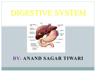
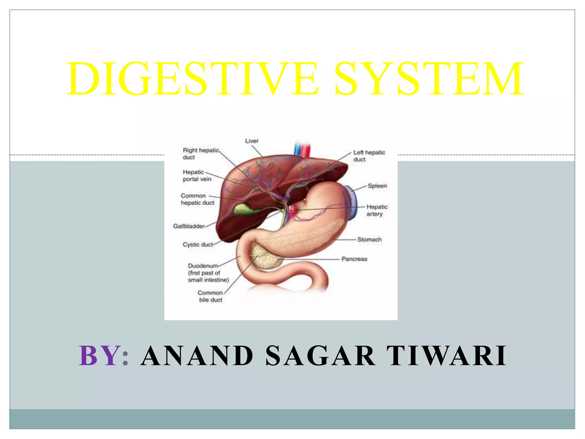
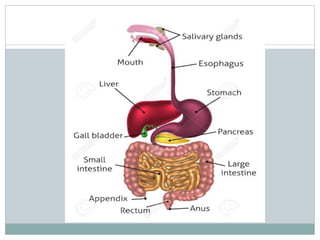
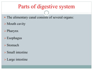
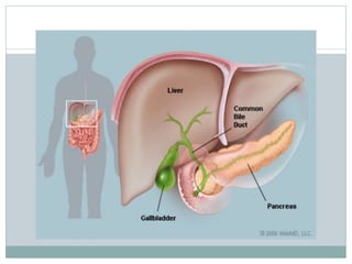
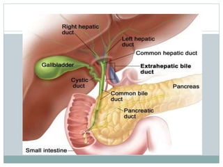
BY: ANAND SAGAR TIWARI
DIGESTIVE SYSTEM
Introduction
 The human digestive system consists of the
gastrointestinal tract plus the accessory organs.
 Human alimentary canal is a long hollow muscular tube
with a length of 9 meters.
 Digestion involves the conversion of food into smaller
and smaller substances, until they can be absorbed and
assimilated by the body.
 The movement of food substance in the gastrointestinal
tract is carried out by peristalsis.
 Peristaltic movement, a rhythmic contraction of muscles
begins in the esophagus and continues throughout the
GIT.
Contd…
 The wall of alimentary canal is formed by four layers:
i. Visceral peritoneum
ii. Muscular layer
iii. Submucous layer
iv. Mucous layer
• Nerve fibres called myoentric plexus present in between
two muscular layer controls the muscular contraction
and movement of alimentary canal.
• Plexus of Meissner in between submucous and mucous
layer controls the secretion of juices.
Parts of digestive system
 The alimentary canal consists of several organs:
Mouth cavity
Pharynx
Esophagus
Stomach
Small intestine
Large intestine
Accessory organs
 Teeth
 Tongue
 Glands
Gastric glands
Pancreatic glands
Intestinal glands
Liver glands
Salivary glands
Mouth cavity
Teeth
Teeth
Salivary glands
Salivary glands
 Salivary glands secrete saliva which help in the digestion of
polysaccharides, starch, glycogen, dextrin.
 This is done by the hydrolyzing action of enzyme ptyalin.
 Also this is done by the disaccharide maltose by enzyme
maltase.
 Saliva moistens and lubricates the oral cavity, tongue, lips for
speech, lubrication and swallowing.
 The lysozyme secreted by the salivary glands prevents the
decay of teeth and thus maintain oral hygiene.
Tongue
Pharynx
Pharynx
 It is located just behind the soft palate and about 12cm long.
 It serves as the common pathway for both digestive and
respiratory systems.
 The pharynx is made of three parts: Nasopharynx, Oropharynx
and Laryngopharynx.
 Eustachian tubes originating from the sides of middle ear open
at the side walls of the nasopharynx.
 Oropharynx is connected to the oral cavity.
Contd…
 The posterior part of laryngopharynx is connected with
the proximal part of esophagus and the anterior part is
connected with pharynx.
 The pharynx is made with three layers: outer muscular,
middle fibrous and inner mucous.
Function: Pharynx performs dual function by sending the
food particles into the esophagus and inhaled air into the
larynx as well as respiratory tract.
Esophagus
Esophagus
 The esophagus is about 25 cm long hollow musculo-
membranous canal.
 It descends through the neck and thorax behind the
trachea and heart.
 It pierces into the diaphragm and finally ends at the
cardiac orifice of the stomach.
Function: The esophagus helps in transporting the food
downward to the stomach from pharynx by the peristaltic
movement.
Stomach
Stomach
 It is located just below the diaphragm in the left portion of the
abdomen and is J shaped.
 Length is about 25-30 cm and width is about 13-15 cm.
 The stomach can be divided into fundus, body and pylorus.
 Like other parts of alimentary canal the stomach is made up of
the four layers.
 The gastric glands present in the stomach are made up of
different cells.
Contd….
 The cells are : Chief cells or peptic cells, parietal cells or
oxyntic cells and mucous cells.
 Chief cells secrete pepsinogen, oxyntic cells secrete
hydrochloric acid and the mucous cells secrete mucus.
Function:
Mechanical function
Secretory function
Digestive function
Absorptive function
Excretory function
Small & Large intestine
Small intestine
 It is a narrow coiled tube, which extends from the pyloric
sphincter of the stomach to the ileocecal valve.
 At that point it joins the large intestine.
 The small intestine is made up of three main parts i.e.
duodenum, jejunum and ileum.
 The luminal surface of the small intestine remains covered
with numerous micro shaped projection called villi which
helps in the absorption of food.
Contd…
 The small intestine contains numerous tubular glands for
the secretion of enzyme and mucus called crypts of
Liberkuhn.
 The inner layer of small intestine contains several spiral
and circular folds called folds of Kerckring.
 This increases the surface area of the lumen of small
intestine when food enters.
Function
 Small intestine is concerned with the digestion of all
foodstuffs and absorption of the digested food materials.
i. Digested products of lipids are absorbed in the villi of
small intestine.
ii. Mucous membrane of small intestine absorbs all the
digested products of carbohydrates.
iii. Blood capillaries of villi absorb the digested proteins.
 The small intestine also contain endocrine cells that
secrete local hormones such as secretin, cholecystokinin
pancreozymin.
Large intestine
 The large intestine extends from the end of ileum to the
anus.
 The length of large intestine is 1.5 meters.
 It is composed of three main parts: caecum, colon and
rectum.
 Caecum is the first part and is about 6 cm wide.
 The caecum contains a long 8 cm worm shaped blind
tubular projection known as vermiform appendix. It is a
vestigial organ in humans.
Contd…
 Colon is the largest part of the large intestine.
 It is made of four parts i.e. ascending, transverse,
descending and sigmoid colon.
 The blends in between the ascending and transverse colon
is called hepatic flexure.
 The blend in between transverse and descending colon is
called splenic flexure.
 Rectum is the last part and 15 cm long and “S” shaped.
 It is wide in the middle and narrow at the end. The
terminal part is called anal canal and about 2.5 cm long.
Function
 Large intestine stores undigested food material
temporarily before eliminating as faeces.
 The mucus produced by the goblet cells helps in the
lubrication and easy elimination of faeces.
 It also absorbs vitamins, minerals, glucose, vitamins,
amino acids from the food residues.
 It also excretes some elements such as arsenic, bismuth,
lead, mercury etc.
 The vitamin B- Complex and vitamin K produced by the
colon bacteria are absorbed by the large intestine.
Functions of digestive system
 Digestion and absorption
 Defection
 Synthesis of vitamins
 Erythropoiesis
 Regulation of blood sugar
 Maintenance of water balance
Some concepts
 Pepsin: Active form of proteolytic enzyme of gastric juice. It
breaks protein into peptone.
 Pepsinogen: Inactive precursor of the enzyme pepsin.
 Conversion of pepsinogen to pepsin takes place under the
influence of HCl of gastric juice.
 Trypsin: Secreted from exocrine cells of the pancreas which is
an active proteolytic enzyme.
 Trypsinogen: Inactive form of trypsin.
 Conversion of trypsinogen to trypsin takes place by the action
of enteropeptidase of intestinal juice in the duodenum.
DIGESTIVE SYSTEM
DIGESTIVE SYSTEM
DIGESTIVE SYSTEM

DIGESTIVE SYSTEM

  • 1.
    BY: ANAND SAGARTIWARI DIGESTIVE SYSTEM
  • 2.
    Introduction  The humandigestive system consists of the gastrointestinal tract plus the accessory organs.  Human alimentary canal is a long hollow muscular tube with a length of 9 meters.  Digestion involves the conversion of food into smaller and smaller substances, until they can be absorbed and assimilated by the body.  The movement of food substance in the gastrointestinal tract is carried out by peristalsis.  Peristaltic movement, a rhythmic contraction of muscles begins in the esophagus and continues throughout the GIT.
  • 3.
    Contd…  The wallof alimentary canal is formed by four layers: i. Visceral peritoneum ii. Muscular layer iii. Submucous layer iv. Mucous layer • Nerve fibres called myoentric plexus present in between two muscular layer controls the muscular contraction and movement of alimentary canal. • Plexus of Meissner in between submucous and mucous layer controls the secretion of juices.
  • 5.
    Parts of digestivesystem  The alimentary canal consists of several organs: Mouth cavity Pharynx Esophagus Stomach Small intestine Large intestine
  • 6.
    Accessory organs  Teeth Tongue  Glands Gastric glands Pancreatic glands Intestinal glands Liver glands Salivary glands
  • 7.
  • 8.
  • 9.
  • 10.
  • 11.
    Salivary glands  Salivaryglands secrete saliva which help in the digestion of polysaccharides, starch, glycogen, dextrin.  This is done by the hydrolyzing action of enzyme ptyalin.  Also this is done by the disaccharide maltose by enzyme maltase.  Saliva moistens and lubricates the oral cavity, tongue, lips for speech, lubrication and swallowing.  The lysozyme secreted by the salivary glands prevents the decay of teeth and thus maintain oral hygiene.
  • 12.
  • 13.
  • 14.
    Pharynx  It islocated just behind the soft palate and about 12cm long.  It serves as the common pathway for both digestive and respiratory systems.  The pharynx is made of three parts: Nasopharynx, Oropharynx and Laryngopharynx.  Eustachian tubes originating from the sides of middle ear open at the side walls of the nasopharynx.  Oropharynx is connected to the oral cavity.
  • 15.
    Contd…  The posteriorpart of laryngopharynx is connected with the proximal part of esophagus and the anterior part is connected with pharynx.  The pharynx is made with three layers: outer muscular, middle fibrous and inner mucous. Function: Pharynx performs dual function by sending the food particles into the esophagus and inhaled air into the larynx as well as respiratory tract.
  • 16.
  • 17.
    Esophagus  The esophagusis about 25 cm long hollow musculo- membranous canal.  It descends through the neck and thorax behind the trachea and heart.  It pierces into the diaphragm and finally ends at the cardiac orifice of the stomach. Function: The esophagus helps in transporting the food downward to the stomach from pharynx by the peristaltic movement.
  • 18.
  • 19.
    Stomach  It islocated just below the diaphragm in the left portion of the abdomen and is J shaped.  Length is about 25-30 cm and width is about 13-15 cm.  The stomach can be divided into fundus, body and pylorus.  Like other parts of alimentary canal the stomach is made up of the four layers.  The gastric glands present in the stomach are made up of different cells.
  • 20.
    Contd….  The cellsare : Chief cells or peptic cells, parietal cells or oxyntic cells and mucous cells.  Chief cells secrete pepsinogen, oxyntic cells secrete hydrochloric acid and the mucous cells secrete mucus. Function: Mechanical function Secretory function Digestive function Absorptive function Excretory function
  • 21.
    Small & Largeintestine
  • 22.
    Small intestine  Itis a narrow coiled tube, which extends from the pyloric sphincter of the stomach to the ileocecal valve.  At that point it joins the large intestine.  The small intestine is made up of three main parts i.e. duodenum, jejunum and ileum.  The luminal surface of the small intestine remains covered with numerous micro shaped projection called villi which helps in the absorption of food.
  • 23.
    Contd…  The smallintestine contains numerous tubular glands for the secretion of enzyme and mucus called crypts of Liberkuhn.  The inner layer of small intestine contains several spiral and circular folds called folds of Kerckring.  This increases the surface area of the lumen of small intestine when food enters.
  • 24.
    Function  Small intestineis concerned with the digestion of all foodstuffs and absorption of the digested food materials. i. Digested products of lipids are absorbed in the villi of small intestine. ii. Mucous membrane of small intestine absorbs all the digested products of carbohydrates. iii. Blood capillaries of villi absorb the digested proteins.  The small intestine also contain endocrine cells that secrete local hormones such as secretin, cholecystokinin pancreozymin.
  • 25.
    Large intestine  Thelarge intestine extends from the end of ileum to the anus.  The length of large intestine is 1.5 meters.  It is composed of three main parts: caecum, colon and rectum.  Caecum is the first part and is about 6 cm wide.  The caecum contains a long 8 cm worm shaped blind tubular projection known as vermiform appendix. It is a vestigial organ in humans.
  • 26.
    Contd…  Colon isthe largest part of the large intestine.  It is made of four parts i.e. ascending, transverse, descending and sigmoid colon.  The blends in between the ascending and transverse colon is called hepatic flexure.  The blend in between transverse and descending colon is called splenic flexure.  Rectum is the last part and 15 cm long and “S” shaped.  It is wide in the middle and narrow at the end. The terminal part is called anal canal and about 2.5 cm long.
  • 27.
    Function  Large intestinestores undigested food material temporarily before eliminating as faeces.  The mucus produced by the goblet cells helps in the lubrication and easy elimination of faeces.  It also absorbs vitamins, minerals, glucose, vitamins, amino acids from the food residues.  It also excretes some elements such as arsenic, bismuth, lead, mercury etc.  The vitamin B- Complex and vitamin K produced by the colon bacteria are absorbed by the large intestine.
  • 28.
    Functions of digestivesystem  Digestion and absorption  Defection  Synthesis of vitamins  Erythropoiesis  Regulation of blood sugar  Maintenance of water balance
  • 29.
    Some concepts  Pepsin:Active form of proteolytic enzyme of gastric juice. It breaks protein into peptone.  Pepsinogen: Inactive precursor of the enzyme pepsin.  Conversion of pepsinogen to pepsin takes place under the influence of HCl of gastric juice.  Trypsin: Secreted from exocrine cells of the pancreas which is an active proteolytic enzyme.  Trypsinogen: Inactive form of trypsin.  Conversion of trypsinogen to trypsin takes place by the action of enteropeptidase of intestinal juice in the duodenum.